. 2014 Dec;7(Suppl 1):S059-67.
doi: 10.1055/s-0034-1389560.

The Comprehensive AOCMF Classification System: Midface Fractures - Level 2 Tutorial

Affiliations

The Comprehensive AOCMF Classification System: Midface Fractures - Level 2 Tutorial

Christoph Kunz et al. Craniomaxillofac Trauma Reconstr. 2014 Dec.

Abstract

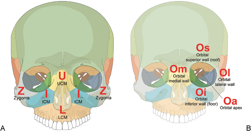
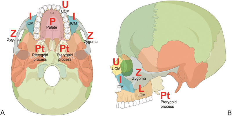
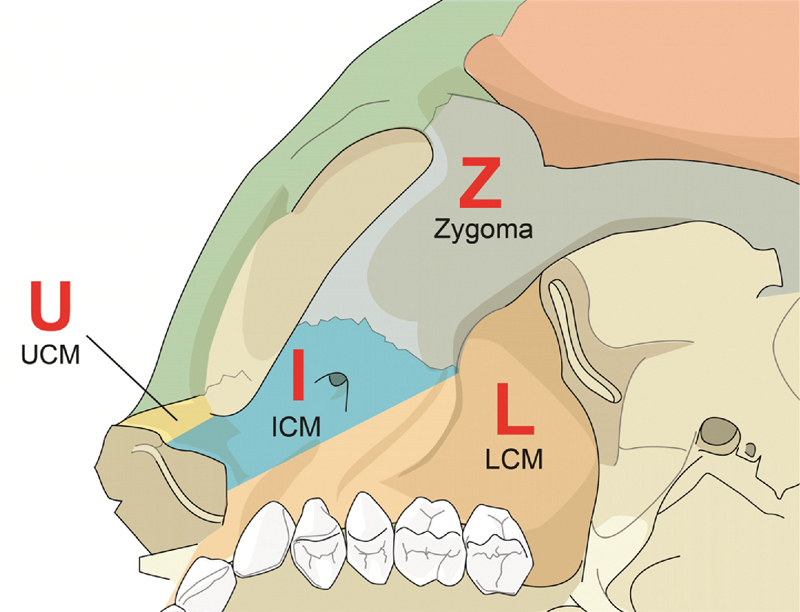
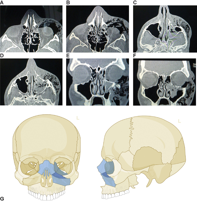
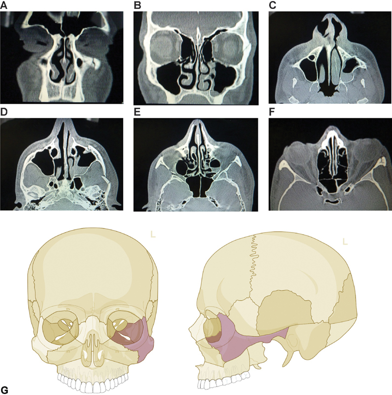
The AOCMF Classification Group developed a hierarchical three-level craniomaxillofacial classification system with increasing level of complexity and details. The highest level 1 system distinguish four major anatomical units including the mandible (code 91), midface (code 92), skull base (code 93), and cranial vault (code 94). This tutorial presents the level 2 system for the midface unit that concentrates on the location of the fractures within defined regions in the central (upper, intermediate, and lower) and lateral (zygoma, pterygoid) midface, as well as the internal orbit and palate. The level 2 midface fracture location outlines the topographic boundaries of the anatomical regions. The common nasoorbitoethmoidal and zygoma en bloc fracture patterns, as well as the time-honored Le Fort classification are taken into account. This tutorial is organized in a sequence of sections dealing with the description of the classification system with illustrations of the topographical cranial midface regions along with rules for fracture location and coding, a series of case examples with clinical imaging and a general discussion on the design of this classification. Individual fracture mapping in these regions regarding severity, fragmentation, displacement of the fragment or bone defect is addressed in a more detailed level 3 system in the subsequent articles.

Keywords: anatomic regions; fracture classification; midface.


Figures

Figure 1
Figure 1
Midface (A) and orbit (B) mapping of anatomical subdivisions on the anterior skull. UCM, upper central midface–nasal skeleton including bone and nasofrontal maxilla; ICM, intermediate central midface–parapiriform maxilla and infraorbital maxilla; LCM, lower central midface–maxillary bodies including infrazygomatic maxilla; Z, zygoma/zygomatic arch.
Figure 2
Figure 2
Midface mapping of anatomical subdivisions on the caudal (A) and lateral (B) skull. UCM, upper central midface–nasal skeleton including bone and nasofrontal maxilla; ICM, intermediate central midface–parapiriform maxilla and infraorbital maxilla; LCM, lower central midface–maxillary bodies including infrazygomatic maxilla; Z, zygoma/zygomatic arch.
Figure 3
Figure 3
Lower central midface extension in the maxillary tuberosity region. UCM, upper central midface; ICM, intermediate central midface; LCM, lower central midface; Z, zygoma/zygomatic arch.
Figure 4
Figure 4
Fracture of the origin of the zygomatic arch. CT scan axial view showing a fracture of the origin of the zygomatic arch, passing through the temporal bony base of the glenoid fossa (arrow) that is added to the zygomatic region.
Figure 5
Figure 5
Fracture of the upper central midface. Imaging: CT scan axial view (A–D) and coronal view (E, F). Description: Nasoorbitoethmoidal (NOE) fracture with involvement of nasal bones bilaterally (UCM), medial orbital and inferior orbital rim left side (UCM and ICM), the anterior medial wall (Om) and the orbital floor (Oi) are fractured. (G) Level 2 Code: 92 U.Omi.Ii. This case example CMTR-92-001 is made available electronically for viewing using the AOCOIAC software at www.aocmf.org/classification.
Figure 6
Figure 6
Bilateral Le Fort I fracture associated with nasal bones and zygoma fracture on the left side. Imaging: CT scan coronal view (A–F) and axial view (G–M). Description: Bilateral Le Fort I fracture (ICM and LCM) and nasal bones (UCM), with fracture of the right pterygoid bone (Pt) as well as a zygoma fracture on the left side (Z) bilateral orbital floor fracture (Om) and involvement of the left lateral orbital wall (Ol). There is no clearly visible fracture of the medial orbital wall fracture (Om is not coded).(N) Level 2 Code: 92 I.L.Pt.Oi.U.Oil.L.I.Z. This case example CMTR-92-002 is made available electronically for viewing using the AOCOIAC software at www.aocmf.org/classification.
Figure 7
Figure 7
Zygoma fracture on the left side. Imaging: CT scan coronal view (A, B) and axial view (C–F). Description: En bloc zygoma fracture on the left side, fracture of the arch with intermediate fragment (Z); the lateral orbital wall (Ol) is slightly displaced. (G) Level 2 Code: 92 m.Oil.Z (m.ZEB). Orbit (left): R(li).W1(li). This case example CMTR-92-003 is made available electronically for viewing using the AOCOIAC software at www.aocmf.org/classification.

References

    1. Audigé L, Cornelius C P, Di Ieva A, Prein J. CMF Classification Group . The first AO classification system for fractures of the craniomaxillofaxial skeleton: rationale, methodological background, developmental process and objectives. Craniomaxillofac Trauma Reconstr. 2014;7 01:S6–S14. - PMC - PubMed
    1. Cornelius C P, Audigé L, Kunz C. et al.The comprehensive AOCMF classification system: midface fractures - level 3 tutorial. Craniomaxillofac Trauma Reconstr. 2014;7 01:S68–S91. - PMC - PubMed
    1. Kunz C, Cornelius C P, Audigé L. et al.The comprehensive AOCMF classification system: orbital fractures - level 3 tutorial. Craniomaxillofac Trauma Reconstr. 2014;7 01:S92–S102. - PMC - PubMed
    1. Di Ieva A, Audigé L, Kellman R M. et al.The comprehensive AOCMF classification system: skull base and cranial vault fractures level 2 and 3 tutorial. Craniomaxillofac Trauma Reconstr. 2014;7 01:S103–S113. - PMC - PubMed
    1. Marsh J L Slongo T F Agel J et al.Fracture and dislocation classification compendium - 2007: Orthopaedic Trauma Association classification, database and outcomes committee J Orthop Trauma 200721(10, Suppl)S1–S133. - PubMed

LinkOut - more resources