. 2014 Dec;7(Suppl 1):S092-102.
doi: 10.1055/s-0034-1389562.

The Comprehensive AOCMF Classification System: Orbital Fractures - Level 3 Tutorial

Affiliations

The Comprehensive AOCMF Classification System: Orbital Fractures - Level 3 Tutorial

Christoph Kunz et al. Craniomaxillofac Trauma Reconstr. 2014 Dec.

Abstract

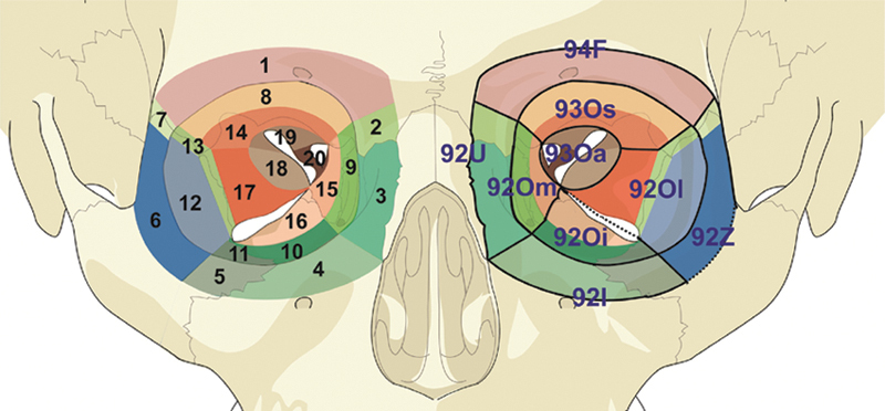
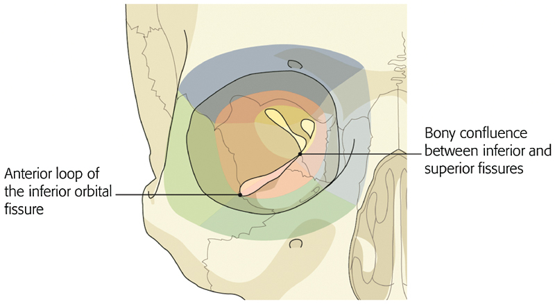
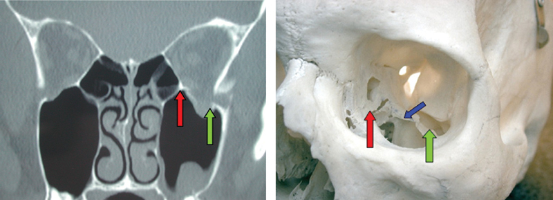
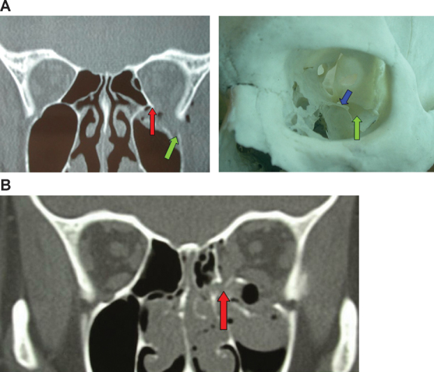
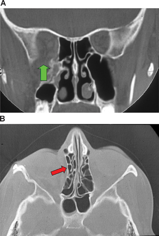
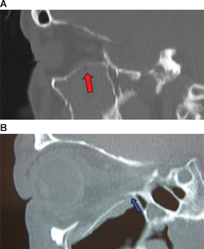
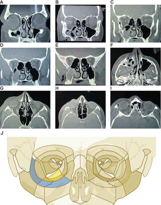
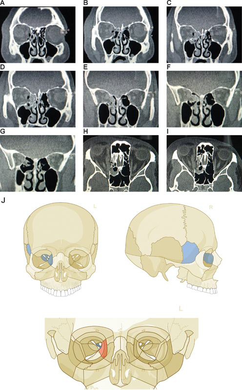
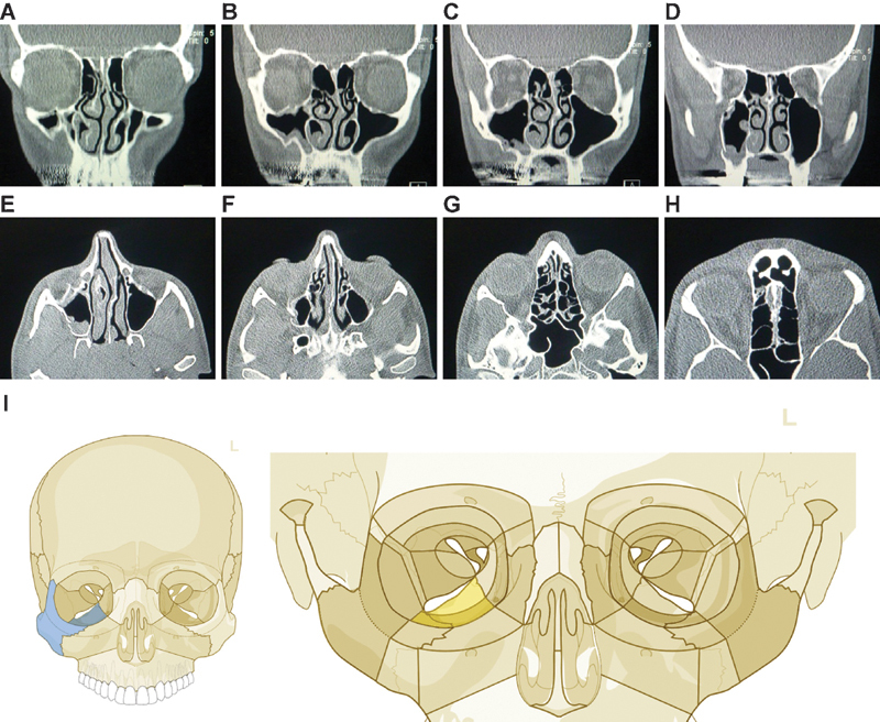
The AOCMF Classification Group developed a hierarchical three-level craniomaxillofacial classification system with increasing level of complexity and details. Within the midface (level 1 code 92), the level 2 system describes the location of the fractures within defined regions in the central and lateral midface including the internal orbit. This tutorial outlines the level 3 detailed classification system for fractures of the orbit. It depicts the orbital fractures according to the subregions defined as orbital rims, anterior orbital walls, midorbit, and apex. The system allows documentation of the involvement of specific orbital structures such as inferior orbital fissure, internal orbital buttress, the greater wing of sphenoid, lacrimal bone, superior orbital fissure, and optic canal. The classification system is presented along with rules for fracture location and coding, a series of case examples with clinical imaging and a general discussion on the design of this classification.

Keywords: anatomic regions; fracture classification; midface; orbit.


Figures

Figure 1
Figure 1
Shape of the anterior section (A) and the apex (B) of the orbit.
Figure 2
Figure 2
Orbital structure and fracture mapping. The left and right orbits show level 2 regions and level 3 subregions, respectively. The numbering on the right orbit (orbital structure ID from 1 to 20) is described in Table 2. Orbital rims: Subregions 1–7 contribute to the orbital rims; orbital walls: subregions 8–12 contribute to anterior section (anterior “third”), subregions 13–17 correspond to midorbit (middle “third”), and subregions 18–20 correspond to the apex (posterior “third”).
Figure 3
Figure 3
Orbital landmarks. The anterior loop of the inferior orbital fissure marks the boundary of the anterior section of the orbital cavity, the confluence of the superior and the inferior orbital fissure defines the posterior border of the midorbit and the entrance to the apex of the orbit.
Figure 4
Figure 4
Intact inferior orbital fissure and internal orbital buttress. CT scan and anatomic specimen showing an intact inferior orbital fissure (green arrow) without widening (the posterior ledge is marked with a blue arrow) and an intact internal orbital buttress (red arrow).
Figure 5
Figure 5
Involvement of inferior orbital fissure and internal orbital buttress. (A) Involvement of inferior orbital fissure with widening (green arrow), and intact internal orbital buttress (red arrow) is intact. The posterior ledge is marked with a blue arrow. (B) The internal orbital buttress is displaced an no longer a reliable landmark.
Figure 6
Figure 6
Lack of landmarks after involvement of inferior orbital fissure and internal orbital buttress. (A) The inferior orbital fissure shows severe widening (green arrow) resulting in an increased orbital volume. (B) Coronal aspect of a secondary orbital deformity due to malalignement of the lateral orbital wall and displacement of the internal orbital buttress (red arrow).
Figure 7
Figure 7
Assessment of intraorbital landmarks. (A) The intact retrobulbar constriction (red arrow) has important impact on the position of the globes, (B) Blow out fracture with displacement of the orbital floor (the posterior ledge is marked with a blue arrow).
Figure 8
Figure 8
Orbital floor fracture with intraorbital buttress involvement. Imaging: CT scan coronal view (A–E) and axial view (F–I). Description: Fracture of orbital floor anterior and middle section (W1i, W2i). Fracture of lateral wall; involvement of inferior rim and lateral rim IOB stable, IOF not widened. (J) Level 3 code: 92 Zli.Ii.Oi.m, Orbit (right): R(li).W1(i)2(i). This case example CMTR-92-201 is made available electronically for viewing using the AOCOIAC software at www.aocmf.org/classification.
Figure 9
Figure 9
Isolated medial orbital wall fracture with apex involvement. Imaging: CT scan coronal view (A–G), axial view (H–I). Description: Isolated medial orbital wall fracture middle (W2m) and posterior section (Am), IOB stable, IOF not widened. (J) Level 3 code: 92 Om.m- 93 S.Oa.m, Orbit (right): W1(m)2(m).A(m). This case example CMTR-92-202 is made available electronically for viewing using the AOCOIAC software at www.aocmf.org/classification.
Figure 10
Figure 10
Zygoma fracture on the right side. Imaging: CT scan coronal view (A–D), axial view (E–H). Description: Zygoma fracture (Z) on the right side with fracture of the orbital floor in the anterior and middle portion (W1i, W2i). The internal orbital fissure (IOF) is widened, and internal orbital buttress (IOB) is stable. (I) Level 3 code: 92 Z.Oi.m, Orbit (right): W1(i)2(i). This case example CMTR-92-203 is made available electronically for viewing using the AOCOIAC software at www.aocmf.org/classification.

References

    1. Audigé L, Cornelius C P, Di Ieva A, Prein J. CMF Classification Group . The first AO classification system for fractures of the craniomaxillofaxial skeleton: rationale, methodological background, developmental process and objectives. Craniomaxillofac Trauma Reconstr. 2014;7 01:S6–S14. - PMC - PubMed
    1. Kunz C, Cornelius C P, Prein J. et al.The comprehensive AOCMF classification system: midface fractures - level 2 tutorial. Craniomaxillofac Trauma Reconstr. 2014;7 01:S59–S67. - PMC - PubMed
    1. Cornelius C P, Audigé L, Kunz C. et al.The comprehensive AOCMF classification system: midface fractures - level 3 tutorial. Craniomaxillofac Trauma Reconstr. 2014;7 01:S68–S91. - PMC - PubMed
    1. Di Ieva A, Audigé L, Kellman R M. et al.The comprehensive AOCMF classification system: skull base and cranial vault fractures - level 2 and 3 tutorial. Craniomaxillofac Trauma Reconstr. 2014;7 01:S103–S113. - PMC - PubMed
    1. Audigé L, Cornelius C P, Buitrago-Téllez C H. et al.The comprehensive AOCMF classification system: classification and documentation within AOCOIAC software. Craniomaxillofac Trauma Reconstr. 2014;7 01:S114–S122. - PMC - PubMed

LinkOut - more resources