

Review
Doppler assessment of maternal central venous hemodynamics in uncomplicated pregnancy: a comprehensive review
Affiliations
- PMID: 25489462
- PMCID: PMC4255508
Item in Clipboard
Review
Doppler assessment of maternal central venous hemodynamics in uncomplicated pregnancy: a comprehensive review
Facts Views Vis Obgyn.
2009.
No abstract available
Keywords: Cardiovascular adaptation; doppler; gestational physiology; maternal veins; venous hemodynamics.
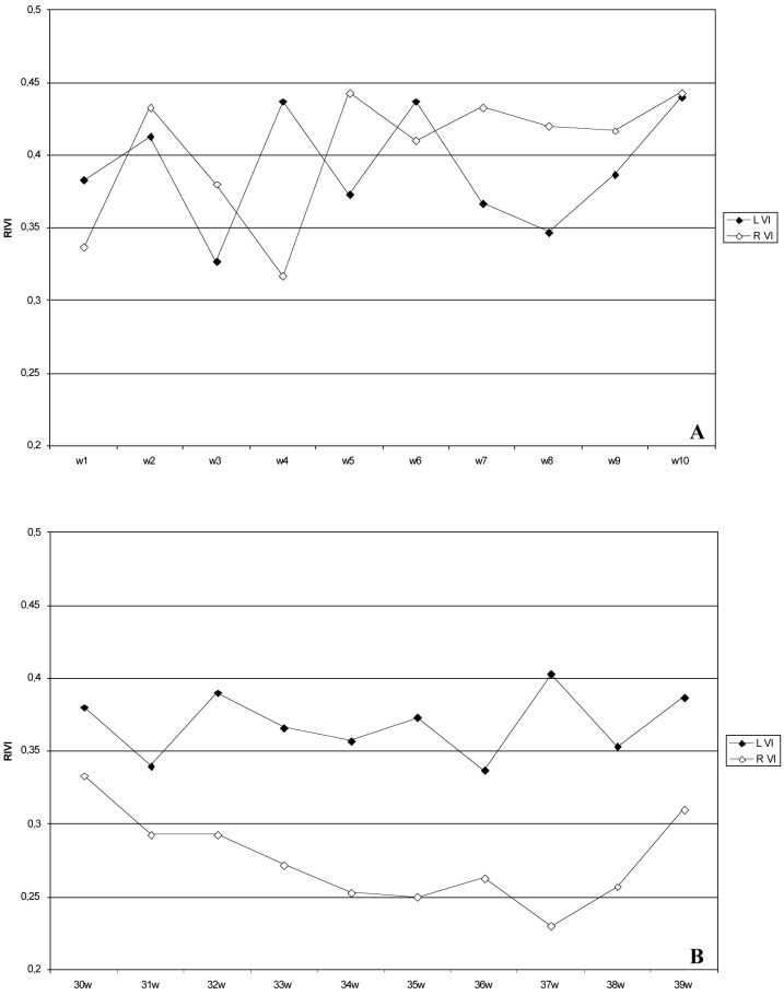
Figures
Similar articles
-
Maternal Venous Hemodynamic Dysfunction in Proteinuric Gestational Hypertension: Evidence and Implications.J Clin Med. 2019 Mar 11;8(3):335. doi: 10.3390/jcm8030335. J Clin Med. 2019. PMID: 30862007 Free PMC article. Review.
-
Role of dysfunctional maternal venous hemodynamics in the pathophysiology of pre-eclampsia: a review.Ultrasound Obstet Gynecol. 2011 Aug;38(2):123-9. doi: 10.1002/uog.9061. Ultrasound Obstet Gynecol. 2011. PMID: 21611996 Review.
-
Time interval between maternal electrocardiogram and venous Doppler waves in normal pregnancy and preeclampsia: a pilot study.Ultraschall Med. 2012 Dec;33(7):E119-E125. doi: 10.1055/s-0029-1245698. Epub 2010 Oct 11. Ultraschall Med. 2012. PMID: 20938893
-
Maternal venous Doppler characteristics are abnormal in pre-eclampsia but not in gestational hypertension.Ultrasound Obstet Gynecol. 2015 Apr;45(4):421-6. doi: 10.1002/uog.13427. Epub 2015 Mar 9. Ultrasound Obstet Gynecol. 2015. PMID: 24890401
-
Maternal venous hemodynamics assessment for prediction of preeclampsia should be longitudinal.J Matern Fetal Neonatal Med. 2015 Feb;28(3):311-5. doi: 10.3109/14767058.2014.916673. Epub 2014 May 20. J Matern Fetal Neonatal Med. 2015. PMID: 24846698
Cited by
-
Why non-invasive maternal hemodynamics assessment is clinically relevant in early pregnancy: a literature review.BMC Pregnancy Childbirth. 2016 Oct 12;16(1):302. doi: 10.1186/s12884-016-1091-9. BMC Pregnancy Childbirth. 2016. PMID: 27729024 Free PMC article. Review.
-
Early-onset preeclampsia is characterised by an increased vascular tone in internal jugular veins.Front Cardiovasc Med. 2022 Aug 12;9:911059. doi: 10.3389/fcvm.2022.911059. eCollection 2022. Front Cardiovasc Med. 2022. PMID: 36035962 Free PMC article.
-
Placental protein 13 (PP13) stimulates rat uterine vessels after slow subcutaneous administration.Int J Womens Health. 2019 Mar 27;11:213-222. doi: 10.2147/IJWH.S188303. eCollection 2019. Int J Womens Health. 2019. PMID: 30988643 Free PMC article.
-
Maternal Venous Hemodynamic Dysfunction in Proteinuric Gestational Hypertension: Evidence and Implications.J Clin Med. 2019 Mar 11;8(3):335. doi: 10.3390/jcm8030335. J Clin Med. 2019. PMID: 30862007 Free PMC article. Review.
-
Changes in lower extremity blood flow during advancing phases of pregnancy and the effects of special footwear.J Vasc Bras. 2017 Jul-Sep;16(3):214-219. doi: 10.1590/1677-5449.002617. J Vasc Bras. 2017. PMID: 29930649 Free PMC article.
References
-
- Abramowicz JS, Sheiner E. Ultrasound of the placenta: a systematic approach. Part II: functional assessment (Doppler) Placenta. 2008;29:921–929. - PubMed
-
- Ahmed K, Sampath R, Khan MS. Current trends in the diagnosis and management of renal nutcracker syndrome: a review. Eur J Vasc Endovasc Surg. 2006;31:410–416. - PubMed
-
- Bateman GA, Cuganesan R. Renal vein Doppler sonography of obstructive uropathy. AJR Am J Roentgenol. 2002;178:921–925. - PubMed
-
- Bateman GA, Giles W, England SL. Renal venous Doppler sonography in preeclampsia. J Ultrasound Med. 2004;23:1607–1611. - PubMed
-
- Berne R, Levy M. Berne R, Levy M, editors. Cardiovascular physiology. London: The C.V. Mosby Company; 2001. Control of cardiac output: coupling of heart and blood vessels; pp. 199–226.
Publication types
LinkOut - more resources
Full Text Sources