. 2014 Nov 17;4(3):285-91.
eCollection 2014 Jul.

Thyroid hormones increase collagen I and cartilage oligomeric matrix protein (COMP) expression in vitro human tenocytes

Affiliations

Thyroid hormones increase collagen I and cartilage oligomeric matrix protein (COMP) expression in vitro human tenocytes

Anna C Berardi et al. Muscles Ligaments Tendons J. .

Abstract

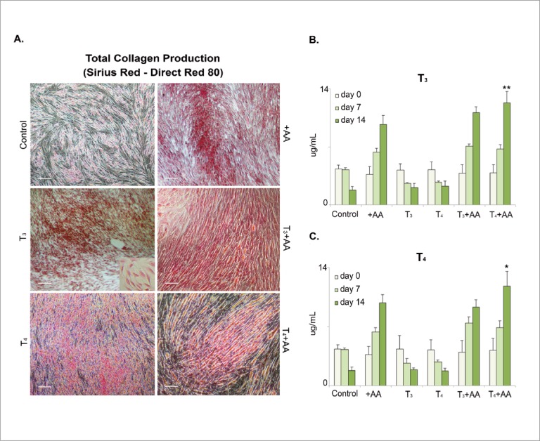
Background: we previously demonstrated the presence of high levels of thyroid hormones (THs) receptors isoforms in healthy tendons, their protective action during tenocyte apoptosis, and the capability to enhance tenocyte proliferation in vitro. In the present study we tested the ability of THs to influence ECM protein tenocyte secretion in an in vitro system.

Methods: primary tenocyte-like cells were cultivated for 1, 7 and 14 days in the presence of T3 or T4 individually or in combination with ascorbic acid (AA).

Results: THs (T3 or T4) in synergism with AA increase significantly the total collagen production after 14 days. THs in synergism with AA increase significantly the expression of collagen I,biglycan and COMP, after some days.

Conclusion: THs play a role on the extra cellular matrix of tendons, enhancing in vitro the production of several proteins such as collagen I, biglycan and COMP. THs receptors are active on human tenocytes, and can play a role in tendon ailments.

Keywords: COMP; ascorbic acid; collagen I; tendons; tenocytes; thyroid hormones.


Figures

Figure 1.
Figure 1.
Total collagens production of primary tenoyte-like cells in vitro culture. A) primary tenocyte-like cells isolated from 5 healthy patients, were cultivated for 1, 7 and 14 days in the presence of T3 or T4 or T3+ AA and/or T4+AA as control, we used cells without any treatment and cells in the presence of AA were used as positive control for the secretion and accumulation of collagen from primary tenocyte-like cells. Red color show the accumulation of total collagen in the 24 well plates after 14 days of culture followed by Sirius red staining; B), C) quantification of total collagen after 1,7 and 14 days of in vitro culture in the presence of T3 or T4. The collagen was quantified by spectrophotometry at 540 nm. Representative images from 8 independent experiments. The data are expressed as mean ± SD for 8 independent experiments for samples run in triplicate.(n = 8, *P <0.05, **P <0.01). Scale bar(A): 50 μm.
Figure 2.
Figure 2.
Collagen I, III and V expression of primary tenoyte-like cells in vitro culture isolated from 5 healthy patients, and stained as described in material and methods, section Immunofluorescence Staining. Representative images from 4 independent experiments. A) Expression of Collagen I after 14 days of culture in green fluorescence and in natural light, showing the primary tenocyte-like cells. B) Collagen I Intensity (Total Area was quantified by anti-collagen I) it was measured by Nikon software. C) Collagen III Intensity (Total Area). D) Expression of Collagen V after 7 and 14 days of culture in red fluorescence and in regular light image showing the primary tenocyte-like cells. E) Collagen V Intensity (Total Area). F) Data are expressed as mean ± SD for 4 independent experiments for samples run in triplicate (n = 4, *P <0.05, **P <0.01). Scale bar(A, D, E): 50 μm.
Figure 3.
Figure 3.
Biglycan and COMP expression of primary tenoyte-like cells in vitro culture. A) Byglican Intensity (Total Area) after 1 and 14 days of culture. B) Expression of COMP after 14 days of culture in green fluorescence and in regular light image showing the primary tenocyte-like cells. C) COMP Intensity (Total Area). Representative results from 5 independent experiments. Data are expressed as mean ± SD for 5 independent experiments for samples run in triplicate. (*P <0.05, **P <0.01). Scale bar(B): 50 μm.

Similar articles

Cited by

References

    1. de Oliveira RR, Martins CS, Rocha YR, et al. Experimental diabetes induces structural, inflammatory and vascular changes of Achilles tendons. PLoS One. 2013;8(10) - PMC - PubMed
    1. Ji JH, Shafi M, Moon CY, Park SE, Kim YJ, Kim SE. Arthroscopic suture bridge technique for intratendinous tear of rotator cuff in chronically painful calcific tendinitis of the shoulder. Orthop Surg. 2013;5(4):289–92. - PMC - PubMed
    1. Harvie P, Pollard TC, Carr AJ. Calcific tendinitis: natural history and association with endocrine disorders. J Shoulder Elbow Surg. 2007;16(2):169–73. - PubMed
    1. Oliva F, Barisani D, Grasso A, Maffulli N. Gene expression analysis in calcific tendinopathy of the rotator cuff. Eur Cell Mater. 2011;21:548–57. - PubMed
    1. Tresoldi I, Oliva F, Benvenuto M, Fantini M, Masuelli L, Bei R, Modesti A. Tendon’s ultrastructure. Muscles Ligaments Tendons J. 2013;3(1):2–6. - PMC - PubMed

LinkOut - more resources