

Effect of divalent cation removal on the structure of gram-negative bacterial outer membrane models
- PMID: 25489959
- PMCID: PMC4295546
- DOI: 10.1021/la504407v
Effect of divalent cation removal on the structure of gram-negative bacterial outer membrane models
Abstract
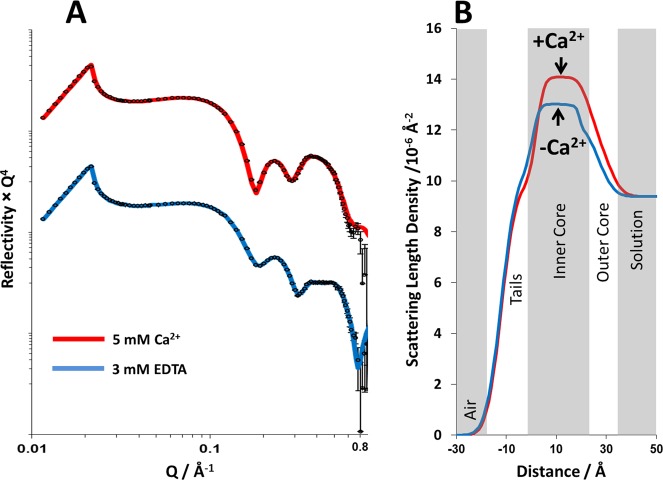
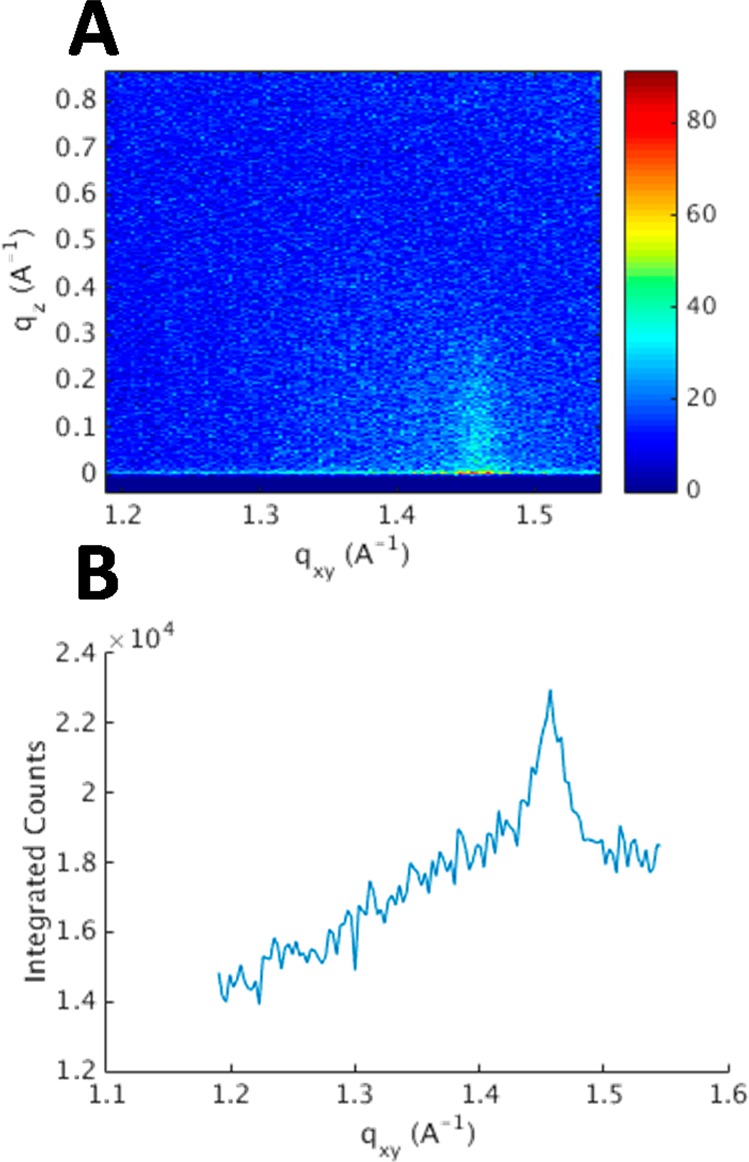
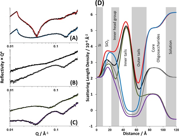
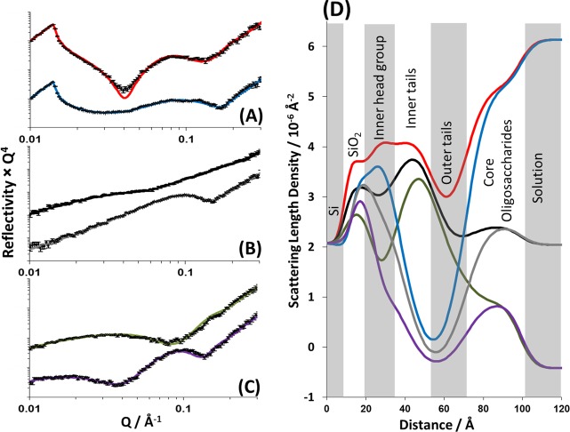
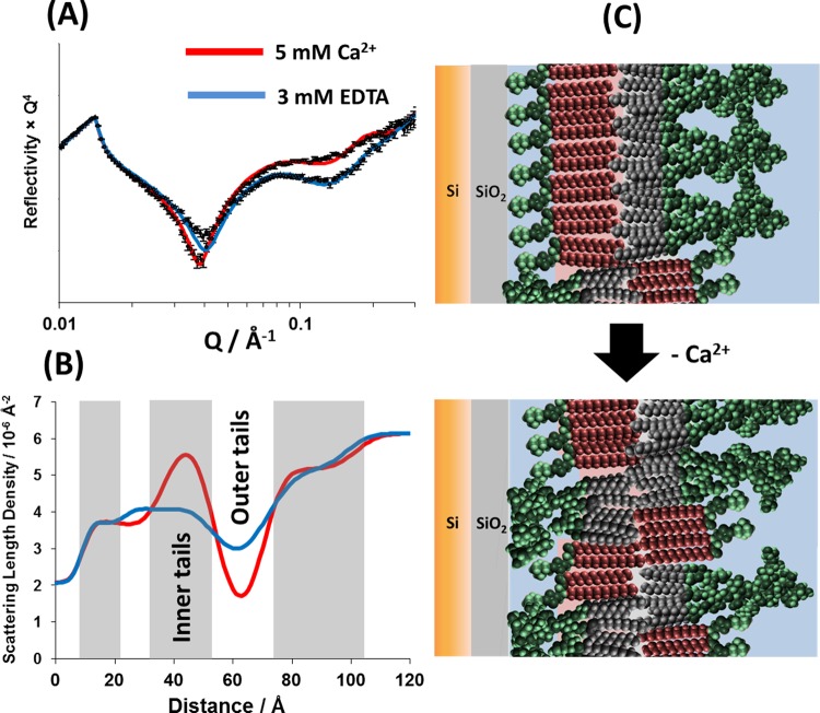
The Gram-negative bacterial outer membrane (GNB-OM) is asymmetric in its lipid composition with a phospholipid-rich inner leaflet and an outer leaflet predominantly composed of lipopolysaccharides (LPS). LPS are polyanionic molecules, with numerous phosphate groups present in the lipid A and core oligosaccharide regions. The repulsive forces due to accumulation of the negative charges are screened and bridged by the divalent cations (Mg(2+) and Ca(2+)) that are known to be crucial for the integrity of the bacterial OM. Indeed, chelation of divalent cations is a well-established method to permeabilize Gram-negative bacteria such as Escherichia coli. Here, we use X-ray and neutron reflectivity (XRR and NR, respectively) techniques to examine the role of calcium ions in the stability of a model GNB-OM. Using XRR we show that Ca(2+) binds to the core region of the rough mutant LPS (RaLPS) films, producing more ordered structures in comparison to divalent cation free monolayers. Using recently developed solid-supported models of the GNB-OM, we study the effect of calcium removal on the asymmetry of DPPC:RaLPS bilayers. We show that without the charge screening effect of divalent cations, the LPS is forced to overcome the thermodynamically unfavorable energy barrier and flip across the hydrophobic bilayer to minimize the repulsive electrostatic forces, resulting in about 20% mixing of LPS and DPPC between the inner and outer bilayer leaflets. These results reveal for the first time the molecular details behind the well-known mechanism of outer membrane stabilization by divalent cations. This confirms the relevance of the asymmetric models for future studies of outer membrane stability and antibiotic penetration.
Figures
References
-
- Pagès J. M.; James C. E.; Winterhalter M. The Porin and the Permeating Antibiotic: A Selective Diffusion Barrier in Gram-Negative Bacteria. Nat. Rev. Microbiol. 2008, 6, 893–903. - PubMed
-
- Stavenger R. A.; Winterhalter M. TRANSLOCATION Project: How to Get Good Drugs into Bad Bugs. Sci. Transl. Med. 2014, 6, 228ed7. - PubMed
-
- Erridge C.; Bennett-Guerrero E.; Poxton I. R. Structure and Function of Lipopolysaccharides. Microbes Infect. 2002, 4, 837–851. - PubMed
Publication types
MeSH terms
Substances
Grants and funding
LinkOut - more resources
Full Text Sources
Other Literature Sources
Miscellaneous
