. 2015 Aug;53(2):159-73.
doi: 10.1165/rcmb.2014-0107OC.

Role of Cigarette Smoke-Induced Aggresome Formation in Chronic Obstructive Pulmonary Disease-Emphysema Pathogenesis

Affiliations

Role of Cigarette Smoke-Induced Aggresome Formation in Chronic Obstructive Pulmonary Disease-Emphysema Pathogenesis

Ian Tran et al. Am J Respir Cell Mol Biol. 2015 Aug.

Abstract

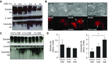
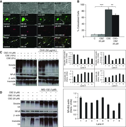
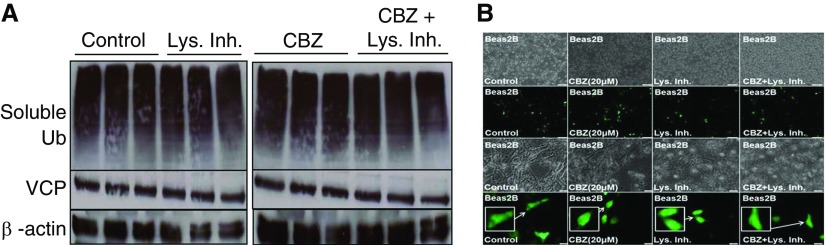
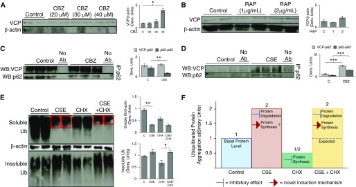
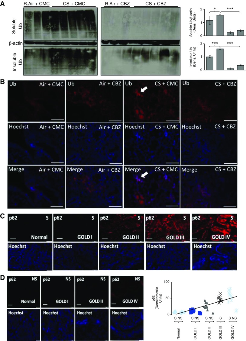
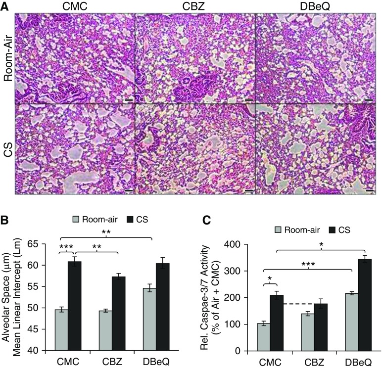
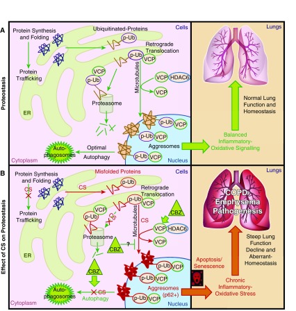
Cigarette smoke (CS) exposure is known to induce proteostasis imbalance that can initiate accumulation of ubiquitinated proteins. Therefore, the primary goal of this study was to determine if first- and secondhand CS induces localization of ubiquitinated proteins in perinuclear spaces as aggresome bodies. Furthermore, we sought to determine the mechanism by which smoke-induced aggresome formation contributes to chronic obstructive pulmonary disease (COPD)-emphysema pathogenesis. Hence, Beas2b cells were treated with CS extract (CSE) for in vitro experimental analysis of CS-induced aggresome formation by immunoblotting, microscopy, and reporter assays, whereas chronic CS-exposed murine model and human COPD-emphysema lung tissues were used for validation. In preliminary analysis, we observed a significant (P < 0.01) increase in ubiquitinated protein aggregation in the insoluble protein fraction of CSE-treated Beas2b cells. We verified that CS-induced ubiquitin aggregrates are localized in the perinuclear spaces as aggresome bodies. These CS-induced aggresomes (P < 0.001) colocalize with autophagy protein microtubule-associated protein 1 light chain-3B(+) autophagy bodies, whereas U.S. Food and Drug Administration-approved autophagy-inducing drug (carbamazepine) significantly (P < 0.01) decreases their colocalization and expression, suggesting CS-impaired autophagy. Moreover, CSE treatment significantly increases valosin-containing protein-p62 protein-protein interaction (P < 0.0005) and p62 expression (aberrant autophagy marker; P < 0.0001), verifying CS-impaired autophagy as an aggresome formation mechanism. We also found that inhibiting protein synthesis by cycloheximide does not deplete CS-induced ubiquitinated protein aggregates, suggesting the role of CS-induced protein synthesis in aggresome formation. Next, we used an emphysema murine model to verify that chronic CS significantly (P < 0.0005) induces aggresome formation. Moreover, we observed that autophagy induction by carbamazepine inhibits CS-induced aggresome formation and alveolar space enlargement (P < 0.001), confirming involvement of aggresome bodies in COPD-emphysema pathogenesis. Finally, significantly higher p62 accumulation in smokers and severe COPD-emphysema lungs (Global Initiative for Chronic Obstructive Lung Disease Stage III/IV) as compared with normal nonsmokers (Global Initiative for Chronic Obstructive Lung Disease Stage 0) substantiates the pathogenic role of autophagy impairment in aggresome formation and COPD-emphysema progression. In conclusion, CS-induced aggresome formation is a novel mechanism involved in COPD-emphysema pathogenesis.

Keywords: aggresomes; autophagy; chronic obstructive pulmonary disease; cigarette smoke; ubiquitin.


Figures

Figure 1.
Figure 1.
Cigarette smoke (CS) extract (CSE) induces perinuclear ubiquitin accumulation in Beas2b cells. (A) Beas2b cells were treated with the indicated doses of freshly prepared CSE for 6 hours. Increasing CSE dose leads to greater aggregation of ubiquitinated proteins in the insoluble protein fraction (aggresome; ubiquitin [Ub] pellet, bottom panel) and decreased accumulation of ubiquitinated proteins in the soluble protein fraction. More specifically, in cells treated with low doses of CSE (lanes 1–3), ubiquitinated protein is mainly in the soluble fraction (top panel; potentially in process of homeostatic proteasomal degradation), rather than in the insoluble fraction (pellet/aggresomes). In cells treated with high-dose CSE (lanes 4–6), we observed a decrease in accumulation of ubiquitinated proteins in the soluble fraction (top panel), whereas aggregation of ubiquitinated proteins was elevated in the insoluble fraction (aggresome; bottom panel; lanes 4–6), suggesting translocation of ubiquitinated proteins from soluble to insoluble fractions. β-actin was used as a loading control for soluble fractions. Data suggest that, as CSE treatment strength increases, a distinct change in the localization and aggregation of ubiquitinated proteins is apparent. (B) Beas2b cells were transfected for 48 hours with Ub–red fluorescent protein (RFP) and treated with CSE (10%) or MG-132 (1 μM) for the final 6 hours of transfection. CSE treatment induced accumulation of ubiquitinated proteins in perinuclear spaces (red perinuclear bodies; aggresomes, white arrows) similar to MG-132 (positive control), suggesting aggresome formation. Scale bars, 20 μm. (C) Beas2b cells were treated with indicated low doses of CSE for 6 hours. A significant (P < 0.05) translocation of ubiquitinated protein initiated at 0.1% CSE treatment. β-actin was used as a loading control for soluble fractions. (D) Densitometric analysis for C shows a trend toward a decrease in soluble ubiquitin. Even at small doses, 1% CSE shows a significant (P < 0.05) increase over 0.01% CSE of ubiquitin in the insoluble cellular fraction. CSE induces ubiquitination, translocation, and aggregation of proteins in aggresome bodies. Data are presented as mean (±SEM). *P < 0.05.
Figure 2.
Figure 2.
CSE induces aggresome formation by interfering with proteostasis and autophagy processes. (A) An equal number of Beas2b cells were transfected for 24 hours with Ub-RFP and autophagy protein microtubule–associated protein 1 light chain-3B (LC3)–green fluorescent protein (GFP) to quantify autophagy-mediated Ub accumulation. Carbamazepine (CBZ; 20 μM) and freshly prepared CSE (10%) treatments were added for the final 6 hours before capturing images. Scale bars, 100 μm. (B) Based on plating of an equal number of Beas2b cells (row 1 in A), the percentage of colocalized cells was calculated as displayed in the merged image (row 4 in A). CSE treatment significantly (∼11-fold; P < 0.001) induced Ub accumulation (red) in LC3+ (green; magnified inset provided) autophagy bodies (LC3 and Ub colocalization [yellow] merged images of same cellular field), indicating aggresome formation. Short, 6-hour treatment with CBZ shows a decrease in Ub-RFP puntas (red perinuclear bodies), Ub-LC3+ yellow autophagy bodies (∼1.2-fold; P < 0.01), and increased LC3-GFP (autophagy reporter) expression, suggesting that CSE-induced aggresome formation can be controlled by autophagy induction. (C) Beas2b cells were treated with freshly prepared CSE (5 or 10%), cycloheximide (CHX) (50 μg/ml), and/or CBZ (10 μM) for 6 hours. We observed greater accumulation of ubiquitinated proteins in the insoluble protein fraction of CSE-treated Beas2b cells. Moreover, levels of ubiquitinated proteins in the soluble protein fraction were decreased in samples that had increased aggregation in the insoluble fraction, suggesting translocation and aggregation of ubiquitinated proteins as aggresome bodies. The magnitude of protein movement from the soluble fraction to the insoluble aggresomes tended to correlate to the CSE dose. In CSE samples treated with the low-dose autophagy–inducing drug, CBZ (lane 5 versus 6), soluble ubiquitinated and NF-κB protein levels were reduced, although the effect on accumulation of ubiquitinated proteins in the insoluble fraction was insignificant with the selected dose (10 μM). Moreover, when protein synthesis was halted by CHX, similar effects were observed, suggesting that CSE induces protein synthesis to override the effect of CHX treatment to exacerbate CSE-induced aggresome formation. β-actin was used as a loading control for soluble fractions. (D) We used proteasome inhibitor (MG-132) treatment as a positive control to induce aggresome formation (ubiquitinated protein aggregation) in Beas2b cells, which were treated overnight with MG-132 (1 μM) and/or CBZ (5, 10, 20 μM). Overnight treatment of MG-132 (1 μM) caused accumulation of ubiquitinated proteins in both the soluble and insoluble fractions, as anticipated. Multiple overnight treatment doses of CBZ were tested to investigate their effectiveness in reducing MG-132–induced aggresome bodies (Ub pellet, insoluble fraction) and total NF-κB protein levels. In MG-132–treated cells, even a low dose (10 μM) of CBZ was effective in decreasing NF-κB levels. As anticipated, CBZ (20 μM) was effective in inhibiting MG-132–induced aggresome bodies (Ub pellet, insoluble fraction). β-actin was used as a loading control for soluble fractions. CBZ has the potential to alleviate CSE-induced accumulation of ubiquitinated proteins by modulating autophagy and proteostasis mechanisms. Data are presented as mean (±SEM). **P < 0.01; ***P < 0.001.
Figure 3.
Figure 3.
CBZ treatment induces valosin-containing protein (VCP) relocalization and expression to aggresome bodies. (A) Beas2b cells were treated with CBZ (20 μM, 48 h) and/or lysosome inhibitor ([Lys. Inh.] E64 and pepstatin A, 20 μg/ml each, final 6 h). Treatment with lysosome inhibitor in the presence of CBZ did not affect basal ubiquitin or VCP protein levels, suggesting that CBZ does not influence lysosomal degradation machinery to induce autophagy. β-actin was used as a loading control for soluble fractions. (B) Beas2b cells were transfected for 48 hours with VCP-GFP and treated with CBZ (20 μM, 36 h) and/or lysosome inhibitor (E64 and pepstatin A, 20 μg/ml each, final 6 h). CBZ treatment induced VCP-GFP relocalization (magnified inset included) to the perinuclear spaces and slightly modulated VCP-GFP expression, suggesting the involvement of VCP as a mechanism for autophagy induction and aggresome formation. Scale bars, 100 μm for rows 1 and 2 and 20 μm for rows 3 and 4. CBZ induced VCP relocalization to perinuclear spaces (aggresomes bodies), suggesting its participation in CBZ induced autophagy.
Figure 4.
Figure 4.
The mechanism for CSE-induced aggresome formation and CBZ-mediated autophagy induction. (A) Beas2B cells were treated with CBZ, a known autophagy inducer, at indicated doses for 24 hours. Cells treated with a high dosage of CBZ (40 μM, lanes 7 and 8) showed significant (P < 0.05) induction of VCP as compared with untreated cells, verifying the ability of CBZ in modulating a VCP autophagy pathway, in addition to VCP relocalization to perinuclear spaces (Figure 3B), suggesting its role in elevating VCP-mediated autophagy to control aggresome bodies. (B) Beas2B cells were treated with indicated doses of another autophagy inducer, rapamycin (RAP) (24 h), as a positive control. As anticipated, autophagy induction by RAP induced VCP expression, suggesting its role in autophagy. (C) Beas2B cells were treated with CBZ for 6 hours, and equal amounts of total proteins were used to immunoprecipitate p62, a known autophagy marker, followed by immunoblotting for VCP and p62. Significant (P < 0.01) inhibition of p62 expression (immunoprecipitation [IP] and Western blot [WB]: p62) in CBZ-treated samples confirmed autophagy induction. Moreover, VCP–p62 protein–protein interaction (IP, p62; WB, VCP) during CBZ treatment remained constant, whereas p62 expression decreased, providing further evidence that CBZ induces VCP relocalization to autophagy bodies. (D) Beas2B cells were similarly treated with CSE for 6 hours, and equal amounts of total proteins were used to immunoprecipitate p62, followed by immunoblotting for VCP and p62. CSE-treated cells displayed a significant (P < 0.0001) increase in p62 expression, indicating CSE-induced aberrant autophagy. CSE treatment also significantly (P < 0.0005) induced VCP–p62 protein–protein interaction, confirming that VCP relocalizes to p62+ aggresome bodies as a cellular response to CSE-induced aberrant autophagy. (E) Beas2B cells were next treated with CHX for 24 hours and/or CSE for the last 6 hours. CSE treatment significantly (P < 0.01) inhibited accumulation of ubiquitinated proteins in the soluble fraction, which corresponds to induction of ubiquitinated protein aggregation in the insoluble fraction (bottom panel, lanes 3 and 4). Treatment with a protein synthesis inhibitor, CHX, decreased accumulation of ubiquitinated proteins in both the soluble and insoluble fractions. Halting protein synthesis by CHX treatment in the presence of CSE, however, did not inhibit aggregation of ubiquitinated proteins in the insoluble fraction (bottom panel, lanes 7 and 8), suggesting that CSE treatment induces protein synthesis to overcome the inhibitory effect of CHX. β-actin was used as a loading control for soluble fractions for A to E. (F) Statistical analysis of insoluble protein fractions in binary units used aggregation of ubiquitinated proteins in the insoluble protein fractions shown in (E). CSE is known to inhibit protein degradation, but CHX halts protein synthesis. Therefore, cotreatment with CHX should persuade inhibition of CSE-induced ubiquitinated protein aggregation. However, CSE+CHX–cotreated cells show approximately equal amounts of ubiquitinated proteins in the insoluble fraction, similar to cells treated with only CSE. Data suggest that CSE induces protein synthesis to overcome the inhibitory effect of CHX, thus retaining ubiquitinated protein levels in the insoluble fraction. CSE treatment induced protein synthesis and VCP expression/localization to aggravate aggresome formation, whereas CBZ could induce VCP-mediated autophagy for clearance of these aggresome bodies. Data are presented as mean (±SEM). Ab, antibody. *P < 0.05, **P < 0.01, ***P < 0.001.
Figure 5.
Figure 5.
Chronic CS–induced aggresome bodies and chronic obstructive pulmonary disease (COPD)–emphysema can be controlled by autophagy induction. (A) Total protein extracts from room air (R.Air)– and chronic CS–exposed C57BL/6 mouse lungs were sonicated and immunoblotted for Ub and β-actin. Chronic CS–exposed mice showed a significant (P < 0.01, soluble; P < 0.0005, insoluble) increase in aggresome bodies (ubiquitinated proteins), especially in the insoluble fractions, demonstrating CS-induced aggresome formation in murine COPD–emphysema lungs (left panel). Moreover, in CBZ-treated mice (right panel), CS-induced accumulation of ubiquitinated proteins (soluble/insoluble protein fractions) is significantly (P < 0.0001, soluble; P < 0.0001, insoluble) inhibited compared with levels seen in carrier, carboxymethylcellulose (CMC)-treated room air– and chronic CS–exposed mice, showing the potential of autophagy induction in controlling CS-induced aggresome bodies. β-actin was used as a loading control for soluble fractions. (B) Immunofluorescence microscopy of longitudinal lung tissue sections from chronic CS–exposed mice shows a significant accumulation of ubiquitinated protein (red; top panel), confirming aggresome formation (top row, third column, white arrow). In addition, CBZ inhibited CS-induced aggresome bodies (top row, fourth column). The middle row uses nuclear staining (Hoechst, blue) to show uniform lung tissue sections. Merged images of the same tissue section field are provided (bottom row) to indicate CS-induced perinuclear ubiquitin accumulation (white arrow). Scale bars, 20 μm. (C and D) Paraffin-embedded longitudinal human lung tissue sections from nonsmokers (NS) and smokers (S) classified as GOLD (Global Initiative for Chronic Obstructive Lung Disease) I–IV emphysema (n = 40[S] + 5[NS] = 45) and GOLD 0 nonemphysema control (n = 5[S] + 10[NS] = 15) were immunostained for aberrant autophagy marker p62. Lung tissue sections from smokers with COPD exhibited significant p62 accumulation as lung function declined in subjects with severe emphysema–COPD (S, GOLD III–IV versus S and NS, GOLD 0–I), demonstrating increase in CS-induced autophagy impairment with COPD–emphysema progression. The bottom panel uses nuclear staining (Hoechst, blue) showing uniform lung tissue sections. Chronic CS–induced aggresome formation and aberrant autophagy in COPD–emphysema can be controlled by autophagy induction. Scale bars, 50 μM. Data are presented as mean (±SEM). *P < 0.05, ***P < 0.001.
Figure 6.
Figure 6.
CBZ controls CS-induced chronic emphysema in a murine model. (A and B) Chronic CS– or room air–exposed C57BL/6 (n = 3–5, 1.5-mo-old + 28-wk air/CS exposure) were used as a COPD–emphysema murine model to standardize and evaluate the therapeutic efficacy of CBZ in controlling autophagy and resulting emphysema. Control groups were subcutaneously injected with 0.5% CMC as the drug carrier, whereas the two other groups were subcutaneously injected with autophagy inducer CBZ (subcutaneous 20 mg/kg body weight/d for the last 10 d = 200 mg/kg body weight) and inhibitor N2,N4-dibenzylquinazoline-2,4-diamine (DBeQ; subcutaneous 5 mg/kg body weight/d for the last 10 d = 50 mg/kg body weight), the latter acting as a negative control. The longitudinal lung tissue sections were fixed in neutral buffer formalin (10%), stained (hematoxylin and eosin), and analyzed for changes in alveolar spaces (4 × 3 = 12 section measurements from n = 3–5 replicates) by microscopy. Autophagy induction (CBZ) significantly (P < 0.01) inhibited chronic CS–induced alveolar space enlargement (mean linear intercept [Lm]), whereas autophagy inhibition (DBeQ, P < 0.01) induced alveolar space enlargement similar to the CS exposure treatment groups, verifying the role of autophagy impairment in alveolar wall destruction and emphysema pathogenesis. Scale bar, 100 μm. (C) Quantification of caspase-3/7 activity in murine lung tissue extracts shows that autophagy induction by CBZ treatment decreases chronic CS–induced apoptosis. In addition, autophagy inhibition by DBeQ significantly increased caspase-3/7 activity (air, P < 0.0005; CS, P < 0.05) in murine lungs. Autophagy induction (CBZ) controls CS-induced chronic emphysema, whereas autophagy inhibition (DBeQ) induced apoptosis, which can exacerbate emphysema pathogenesis. Data are presented as mean (±SEM). *P < 0.05, **P < 0.01, ***P < 0.001.
Figure 7.
Figure 7.
Schematic showing cellular compartmentalization of CS-induced aggresome formation mechanisms in COPD–emphysema. (A) Functional cellular compartments showing the specific steps of homeostatic protein processing mechanism (proteostasis). The optimal autophagy and proteasomal degradation mechanisms can clear off aggresome bodies in a healthy cell, maintaining balanced inflammatory oxidative responses for normal lung function and homeostasis. (B) CS exposure induced protein synthesis, misfolding, and polyubiquitination (p-Ub), initiating microtubule-mediated translocation of ubiquitinated proteins to perinuclear spaces (aggresomes). Histone deacetylase 6 (HDAC6) and VCP facilitated this translocation and aggresome formation. The CS impaired autophagy to inhibit degradation of aggresome bodies, inducing chronic inflammatory apoptotic responses, leading to disordered alveolarization, steep lung function decline, and severe COPD–emphysema pathogenesis. Autophagy induction by CBZ induced VCP translocation and expression in p62+ aggresome bodies to assist with autophagy-mediated protein degradation, potentially by inducing its segregase/unfoldase activity to process and present aggregated proteins to autophagosomes. CS induced pathologic p62+ aggresome bodies by inducing protein synthesis, misfolding, ubiquitination, translocation, and aggregation, while restraining autophagy that activates chronic inflammatory oxidative–apoptotic responses and senescence to accelerate COPD–emphysema pathogenesis. ER, endoplasmic reticulum.

Similar articles

Cited by

References

    1. Barnes PJ. Chronic obstructive pulmonary disease: a growing but neglected global epidemic. PLoS Med. 2007;4:e112. - PMC - PubMed
    1. Yoshida T, Tuder RM. Pathobiology of cigarette smoke–induced chronic obstructive pulmonary disease. Physiol Rev. 2007;87:1047–1082. - PubMed
    1. Lopez AD, Shibuya K, Rao C, Mathers CD, Hansell AL, Held LS, Schmid V, Buist S. Chronic obstructive pulmonary disease: current burden and future projections. Eur Respir J. 2006;27:397–412. - PubMed
    1. Ito K, Barnes PJ. COPD as a disease of accelerated lung aging. Chest. 2009;135:173–180. - PubMed
    1. Rahman I, Adcock IM. Oxidative stress and redox regulation of lung inflammation in COPD. Eur Respir J. 2006;28:219–242. - PubMed

Publication types

MeSH terms