. 2014 Dec 9;9(12):e114182.
doi: 10.1371/journal.pone.0114182. eCollection 2014.

Key regulatory role of dermal fibroblasts in pigmentation as demonstrated using a reconstructed skin model: impact of photo-aging

Affiliations

Key regulatory role of dermal fibroblasts in pigmentation as demonstrated using a reconstructed skin model: impact of photo-aging

Christine Duval et al. PLoS One. .

Abstract

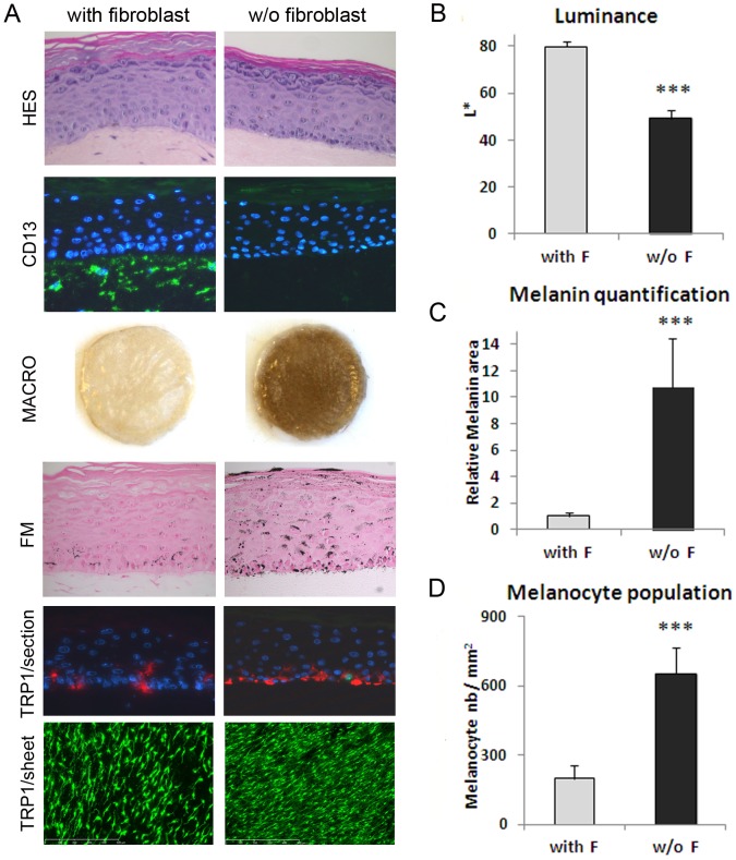
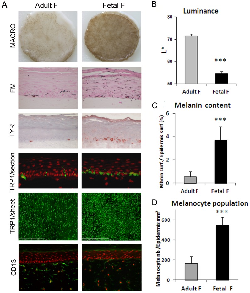
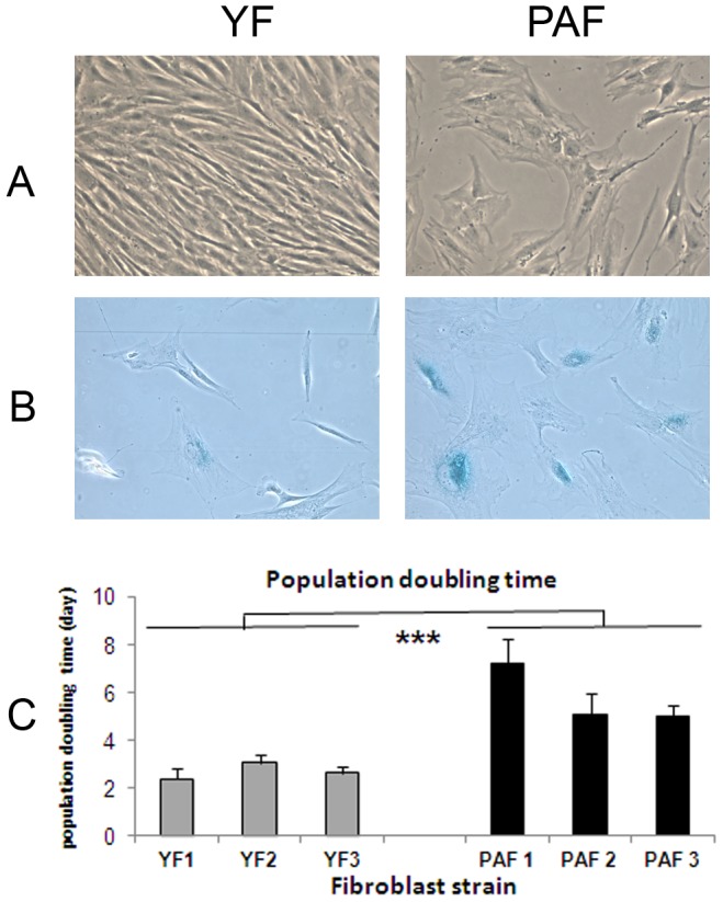
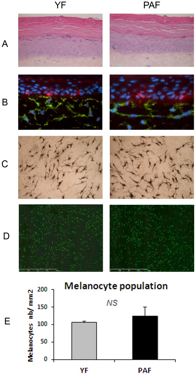
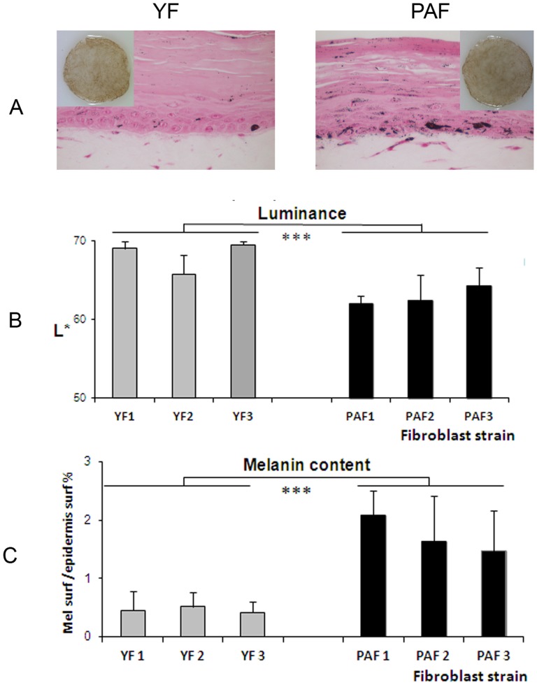
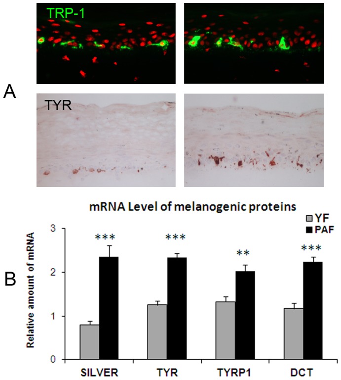
To study cutaneous pigmentation in a physiological context, we have previously developed a functional pigmented reconstructed skin model composed of a melanocyte-containing epidermis grown on a dermal equivalent comprising living fibroblasts. The present studies, using the same model, aimed to demonstrate that dermal fibroblasts influence skin pigmentation up to the macroscopic level. The proof of principle was performed with pigmented skins differing only in the fibroblast component. First, the in vitro system was reconstructed with or without fibroblasts in order to test the global influence of the presence of this cell type. We then assessed the impact of the origin of the fibroblast strain on the degree of pigmentation using fetal versus adult fibroblasts. In both experiments, impressive variation in skin pigmentation at the macroscopic level was observed and confirmed by quantitative parameters related to skin color, melanin content and melanocyte numbers. These data confirmed the responsiveness of the model and demonstrated that dermal fibroblasts do indeed impact the degree of skin pigmentation. We then hypothesized that a physiological state associated with pigmentary alterations such as photo-aging could be linked to dermal fibroblasts modifications that accumulate over time. Pigmentation of skin reconstructed using young unexposed fibroblasts (n = 3) was compared to that of tissues containing natural photo-aged fibroblasts (n = 3) which express a senescent phenotype. A stimulation of pigmentation in the presence of the natural photo-aged fibroblasts was revealed by a significant increase in the skin color (decrease in Luminance) and an increase in both epidermal melanin content and melanogenic gene expression, thus confirming our hypothesis. Altogether, these data demonstrate that the level of pigmentation of the skin model is influenced by dermal fibroblasts and that natural photo-aged fibroblasts can contribute to the hyperpigmentation that is associated with photo-aging.


Conflict of interest statement

Competing Interests: The authors have the following interests. All authors are employed by L'Oréal Research & Innovation. C.D. and F.B. are inventors on the filed patents applications numbered FR298654 and FR2930644 entitled "Equivalent de peau pigmentée fonctionnelle” and “Procédé d'évaluation de la pigmentation” respectively, and claiming a functional pigmented skin model, the method of preparation and the use of the model to assess modulation of pigmentation induced by chemical compounds. This does not alter the authors' adherence to all the PLOS ONE policies on sharing data and materials, as detailed online in the guide for authors. All the other authors declare no conflict of interest. There are no products in development or marketed products to declare related to this study.

Figures

Figure 1
Figure 1. Microscopic examination and measurement of pigmentation in the in vitro pigmented skin model reconstructed in the presence or absence of fibroblasts in the dermal compartment.
A) Microscopic examination of HES staining of cross-sections showed a correct epidermal architecture and differentiation process leading to the development of a stratum granulosum and stratum corneum in both 3D systems. The CD13 staining (green) of cross-sections confirmed the presence or absence of fibroblasts (from adult 21 yr-old donor) in the dermal equivalent. Macroscopic pictures of PRS illustrate the resulting drastic difference in pigmentation between the two conditions which was confirmed by B) Luminance measurement (mean of 3 experiments). Fontana-Masson staining for melanin granules detection (black points), and C) graph of melanin quantification (mean of 3 experiments) performed by image analysis of Fontana-Masson (FM) staining on cross-sections. To compare the two conditions, results on melanin content were normalized to the control condition (with fibroblasts). TRP-1 staining (red) of cross-sections shows melanocytes positioned at the basal layer of the epidermis in both 3D systems. TRP-1 staining (green) of PRS epidermal sheets and D) corresponding graph of melanocyte quantification (mean of 3 experiments) performed by image analysis. The values represent means ± standard deviation (SD). Statistical significance was evaluated by the Student's t-test; *** p-value <0.001. Magnification: A HES, FM  =  x400, CD13, TRP1/section  =  x200, TRP1/sheet  =  x50.
Figure 2
Figure 2. Comparison of the effect of fibroblasts from fetal versus adult origin on reconstructed skin pigmentation.
PRS samples were reconstructed with either fetal fibroblasts (GM10) or adult (21 yr-old donor) fibroblasts within the dermal equivalent. Identical keratinocyte and melanocyte strains were used for the epidermal reconstruction. A drastic increase in pigmentation of the PRS and activation of melanocytes in the presence of fetal fibroblasts as compared to adult fibroblasts, were noted A) macroscopically (Macro), on histological sections stained with Fontana-Masson (FM) and by tyrosinase staining (Tyr) of tissue sections. The hyper-pigmentation observed in fetal versus adult fibroblast condition was quantified by B) a decrease in Luminance value and C) an increase in melanin content. TRP-1 labeling of tissue sections showed that melanocytes were correctly located at the basal layer in both conditions (A). TRP-1 staining of epidermal sheets revealed an increase in melanocyte numbers in the presence of fetal fibroblasts (A) which was confirmed by image analysis (D). CD13 staining revealed no change in fibroblast morphology or density (A). Values are expressed as the mean +/- SD calculated for 4 different samples in 2 independent experiments and analyzed using the two-tailed unpaired Student's t-test, *** p<0.001. Magnifications (A): FM, TYR, and TRP-1/section  =  x400, CD13/section =  x200, TRP1/sheet =  x50.
Figure 3
Figure 3. Morphology and senescence-associated β-galactosidase staining of fibroblasts from photo-aged and young skins.
Fibroblasts isolated from photo-aged skin (PAF) and from young unexposed skin (YF) were grown in DMEM + 0 % FBS at 10% CO2 and passaged until P4. As shown by A) classical microscopy, B) senescence-associated β-galactosidase (SA-β-gal) staining and C) population doubling times (time required for a two-fold increase in fibroblast number), PAF exhibited a flattened and enlarged cellular body, a reduced growth rate and an increase in SA-β-galactosidase activity, thus revealing a senescent phenotype. Values are expressed as the mean +/- SD calculated in 3 independent experiments and analyzed using the two-tailed unpaired Student's t-test; *** p<0.001. A and B magnification  =  x200.
Figure 4
Figure 4. Reconstruction of pigmented skin with fibroblasts from photo-aged or young skin.
Pigmented skins reconstructed with a dermal equivalent containing either photo-aged fibroblasts (PAF) or young fibroblasts (YF) were analyzed after 14 days of emersion. The same keratinocyte and melanocyte strains were used for epidermal reconstruction. In both photo-aged and young fibroblast conditions, HES colorations showed that the histological quality of the epidermis was correct (A). TRP-1 (red) and CD13 (green) co-staining of skin sections indicated the correct location of melanocytes in the basal layer and the presence of fibroblasts within the dermal equivalent (B) and Dopa reaction (C) and TRP1 staining (D) of epidermal sheets revealed the correct morphology of melanocytes in both conditions. E) Graph reporting the number of melanocytes as estimated by counting TRP1-positive cells/mm2 epidermal surface shows no significant difference between the number of melanocytes in the photo-aged versus young fibroblast condition. Values are expressed as the mean +/- SD calculated for 6 samples and analyzed using the two-tailed unpaired Student's t-test; NS: non-significant. Magnification: A, B =  x 400, C =  x200, D =  x50.
Figure 5
Figure 5. Pigmentation levels in skin reconstructed with natural photo-aged fibroblasts versus young fibroblasts.
Pigmentation in the skin reconstructed with either natural photo-aged fibroblasts (PAF) or young fibroblasts (YF) was analyzed macroscopically and on histological sections stained with Fontana Masson (A) and quantified by Luminance measurements (L*) (B) and melanin quantification (C) after 14 days of emersion. In the presence of the three PAF strains versus the three YF conditions, a significant increase in pigmentation was observed as shown by i) an increase in the concentration of melanin granules as measured by image analysis on histological sections, and ii) the darkening of the skin samples which correlates with a decrease in Luminance. Values are expressed as a mean +/- SD calculated for 3 samples in 3 independent experiments and analyzed using the two-tailed unpaired Student's t-test; *** p<0.001. Magnification: A =  x400.
Figure 6
Figure 6. Effect of fibroblasts from photo-aged versus young skin on the expression level of melanogenic proteins in reconstructed skin.
Analysis of melanocyte location and activity in the reconstructed skin samples containing either photo-aged fibroblasts (PAF) or young fibroblasts (YF) was analyzed by immunostaining on histological sections using TRP-1 and Tyrosinase antibodies (A) and by measuring expression levels of melanogenic genes by RT-qPCR (B). Immunostaining of tissue sections showed the correct location of melanocytes in the basal layer and increased labeling of Tyrosinase in PAF conditions as compared to YF samples. The analysis of expression levels of genes involved in melanogenesis in the epidermis revealed the increase in mRNA levels of Silver, Tyrosinase, TRP-1 and DCT (TRP-2) in the PAF-containing model as compared to the YF condition confirming that melanocytes were stimulated when photo-aged fibroblasts were present in dermal equivalent. Values are expressed as a mean +/- SD calculated for 3 samples in one experiment and analyzed using the two-tailed unpaired Student's t-test; ** p<0.01, *** p<0.001. Similar results were obtained in three independent experiments. Magnification: A =  x400.

References

    1. Abdel-Malek Z, Swope VB, Suzuki I, Akcali C, Harriger MD, et al. (1995) Mitogenic and melanogenic stimulation of normal human melanocytes by melanotropic peptides. Proc Natl Acad Sci U S A 92:1789–1793. - PMC - PubMed
    1. Slominski A, Wortsman J, Luger T, Paus R, Solomon S (2000) Corticotropin releasing hormone and proopiomelanocortin involvement in the cutaneous response to stress. Physiol Rev 80:979–1020. - PubMed
    1. Slominski A, Tobin DJ, Shibahara S, Wortsman J (2004) Melanin pigmentation in mammalian skin and its hormonal regulation. Physiol Rev 84:1155–1228. - PubMed
    1. Slominski AT, Zmijewski MA, Skobowiat C, Zbytek B, Slominski RM, et al. (2012) Sensing the environment: regulation of local and global homeostasis by the skin's neuroendocrine system. Adv Anat Embryol Cell Biol 212: v, vii, 1-v, vii115. - PMC - PubMed
    1. Yamaguchi Y, Hearing VJ (2009) Physiological factors that regulate skin pigmentation. Biofactors 35:193–199. - PMC - PubMed

Publication types