Comparative Study
. 2015 Mar;85(2):253-62.
doi: 10.2319/030514-156.1. Epub 2014 Dec 9.

Tooth-borne vs bone-borne rapid maxillary expanders in late adolescence

Affiliations
Comparative Study

Tooth-borne vs bone-borne rapid maxillary expanders in late adolescence

Lu Lin et al. Angle Orthod. 2015 Mar.

Abstract

Objective: To evaluate the immediate effects of rapid maxillary expansion (RME) on the transverse skeletal and dentoalveolar changes with bone-borne (C-expander) and tooth-borne type expanders using cone-beam computed tomography (CBCT) in late adolescents.

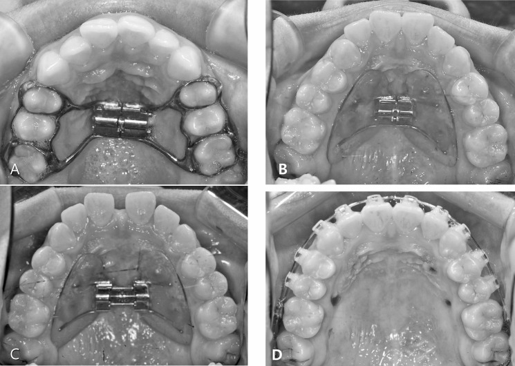
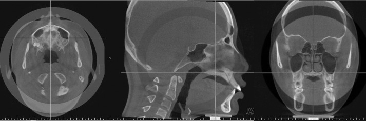
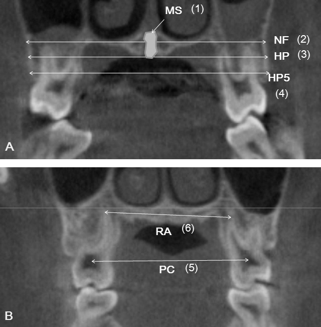
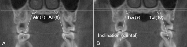
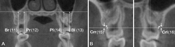
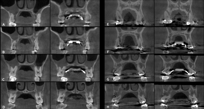
Materials and methods: A sample of 28 female late-adolescent patients was divided into two groups according to the type of expander: bone-borne (C-expander, n = 15, age = 18.1 ± 4.4 years) and tooth-borne (hyrax, bands on premolars and molars, n = 13, age = 17.4 ± 3.4 years). CBCT scans were taken at 0.2-mm voxel size before treatment (T1) and 3 months after RME (T2). Transverse skeletal and dental expansion, alveolar inclination, tooth axis, vertical height of tooth, and buccal dehiscence were evaluated on maxillary premolars and molars. Paired t-test, independent t-test, one-way analysis of variance, and Scheffé post hoc analysis were performed.

Results: The C-expander group produced greater skeletal expansion, except in the region of the first premolar (P < .05 or < .01), which showed slight buccal tipping of the alveolar bone. The Hyrax group had more buccal tipping of the alveolar bone and the tooth axes, except in the region of the second molar (P < .05 or < .01 or < .001). Dental expansion at the apex level was similar in the banded teeth (the first premolar and the first molar). Vertical height changes were apparent on the second premolar in the hyrax group (P < .05 or < .01). Significant buccal dehiscence occurred at the first premolar in the hyrax group (P < .01 or < .001). There were no significant differences between tooth types for any variables in the C-expander group.

Conclusions: For patients in late adolescence, bone-borne expanders produced greater orthopedic effects and fewer dentoalveolar side effects compared to the hyrax expanders.

Keywords: Alveolar bone; Bone-borne expander; C-implant; Cone-beam computed tomogram; Hyrax expander; Rapid maxillary expansion.


Figures

Figure 1. Expander design used in the study: tooth-borne (A) and bone-borne (B). Intraoral photos after expansion (C) and 1 week after removal of C-expander in bone-borne group (D).
Figure 1.
Expander design used in the study: tooth-borne (A) and bone-borne (B). Intraoral photos after expansion (C) and 1 week after removal of C-expander in bone-borne group (D).
Figure 2. Reorientation of CBCT scans: reoriented as perpendicular to midpalatal suture (axial section, x-plane), parallel to palatal plane (sagittal section, y-plane), and tangent to nasal floor at its most inferior level, where both of the palatal roots of the maxillary first molars are shown (coronal section, z-plane).
Figure 2.
Reorientation of CBCT scans: reoriented as perpendicular to midpalatal suture (axial section, x-plane), parallel to palatal plane (sagittal section, y-plane), and tangent to nasal floor at its most inferior level, where both of the palatal roots of the maxillary first molars are shown (coronal section, z-plane).
Figure 3. Definition of measurements. Skeletal (A) and dental (B) transverse distances. NF indicates maxillary width tangent to the nasal floor at its most inferior level; HP, maxillary width tangent to the hard palate at the most inferior level; HP5, maxillary width parallel to the line NF and 5 mm below the line HP; MS, area of midpalatal suture; RA, distance between palatal root apices; and PC, distance between pulp chambers.
Figure 3.
Definition of measurements. Skeletal (A) and dental (B) transverse distances. NF indicates maxillary width tangent to the nasal floor at its most inferior level; HP, maxillary width tangent to the hard palate at the most inferior level; HP5, maxillary width parallel to the line NF and 5 mm below the line HP; MS, area of midpalatal suture; RA, distance between palatal root apices; and PC, distance between pulp chambers.
Figure 4. Alveolar bone inclination (A) (between the palatal alveolar slop and NF. Alr, right side; All, left side) and tooth inclination (B) (between the palatal root axis and NF. Tor, right side; Tol, left side).
Figure 4.
Alveolar bone inclination (A) (between the palatal alveolar slop and NF. Alr, right side; All, left side) and tooth inclination (B) (between the palatal root axis and NF. Tor, right side; Tol, left side).
Figure 5. (A) Vertical dental heights from NF to mesiobuccal cusp (Br, right side; Bl, left side) or mesiopalatal cusp (Pr, right side; Pl, left side); (B) Alveolar bone dehiscence measured from the cemento-enamel junction (CEJ) to the alveolar crest on buccal side. Crr indicates right side; Crl, left side.
Figure 5.
(A) Vertical dental heights from NF to mesiobuccal cusp (Br, right side; Bl, left side) or mesiopalatal cusp (Pr, right side; Pl, left side); (B) Alveolar bone dehiscence measured from the cemento-enamel junction (CEJ) to the alveolar crest on buccal side. Crr indicates right side; Crl, left side.
Figure 6. The examples of rapid maxillary expansion with bone-borne (A–D) and tooth-borne (E–H) expanders, compared between pretreatment period and after expansion at the first premolar (A and E), the second premolar (B and F), the first molar (C and G), and the second molar (D and H).
Figure 6.
The examples of rapid maxillary expansion with bone-borne (A–D) and tooth-borne (E–H) expanders, compared between pretreatment period and after expansion at the first premolar (A and E), the second premolar (B and F), the first molar (C and G), and the second molar (D and H).

References

    1. Ramires T, Maia RA, Barone JR. Nasal cavity changes and the respiratory standard after maxillary expansion. Braz J Otorhinolaryngol. 2008;74:763–769. - PMC - PubMed
    1. Bishara SE, Jakobsen JR, Treder J, Nowak A. Arch width changes from 6 weeks to 45 years of age. Am J Orthod Dentofacial Orthop. 1997;111:401–409. - PubMed
    1. Baccetti T, Franchi L, Cameron CG, McNamara JA., Jr Treatment timing for rapid maxillary expansion. Angle Orthod. 2001;71:343–350. - PubMed
    1. Erverdi N, Okar I, Kucukkeles N, Arbak S. A comparison of two different rapid palatal expansion techniques from the point of root resorption. Am J Orthod Dentofacial Orthop. 1994;106:47–51. - PubMed
    1. Weissheimer A, de Menezes LM, Mezomo M, Dias DM, de Lima EM, Rizzatto SM. Immediate effects of rapid maxillary expansion with Haas-type and hyrax-type expanders: a randomized clinical trial. Am J Orthod Dentofacial Orthop. 2011;140:366–376. - PubMed

Publication types

MeSH terms