. 2014 Dec;26(12):4834-42.
doi: 10.1105/tpc.114.131193. Epub 2014 Dec 9.

The Arabidopsis cellulose synthase complex: a proposed hexamer of CESA trimers in an equimolar stoichiometry

Affiliations

The Arabidopsis cellulose synthase complex: a proposed hexamer of CESA trimers in an equimolar stoichiometry

Joseph L Hill Jr et al. Plant Cell. 2014 Dec.

Abstract

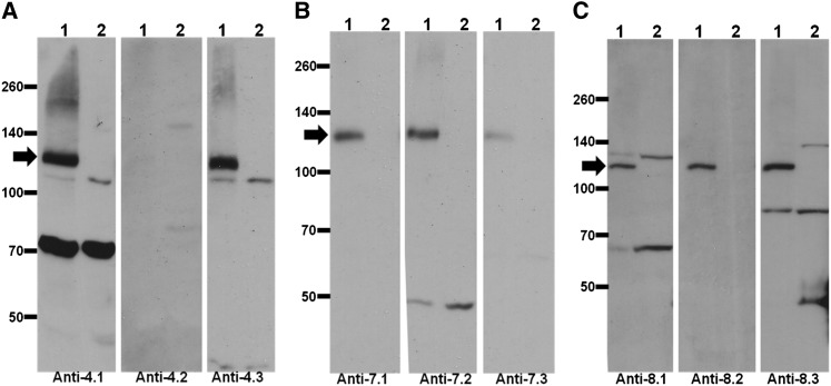
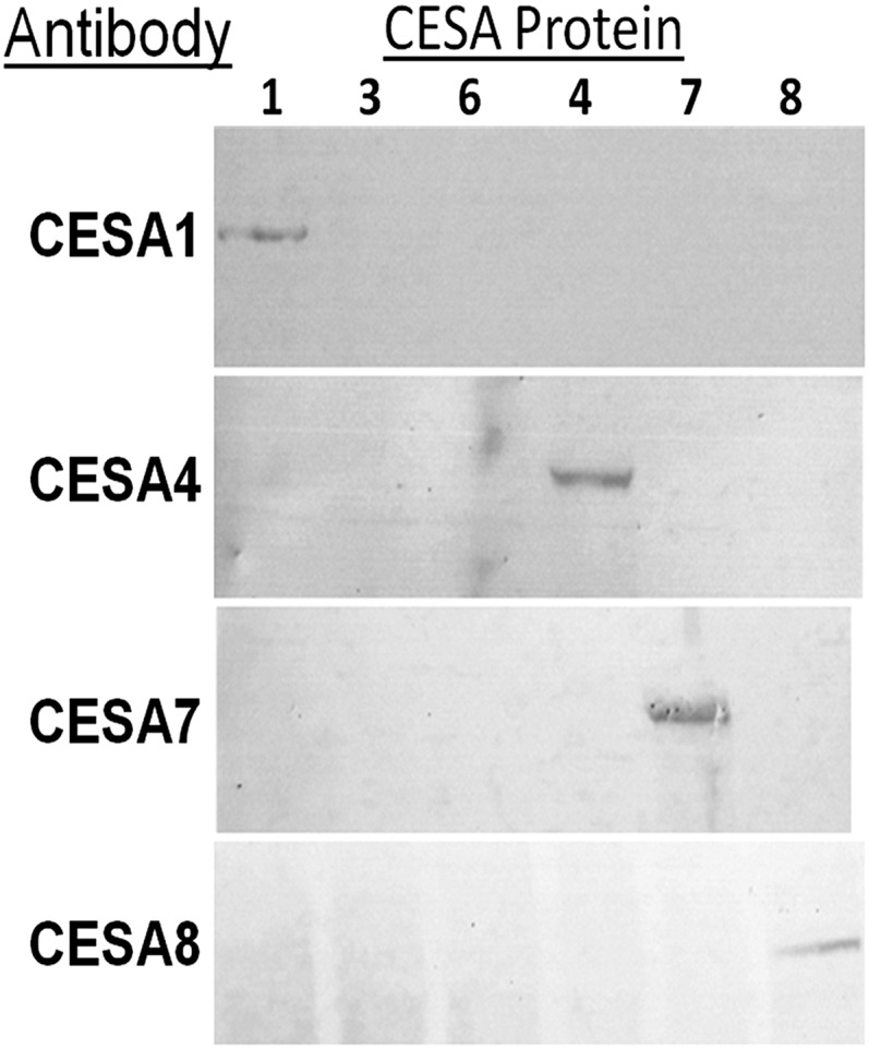
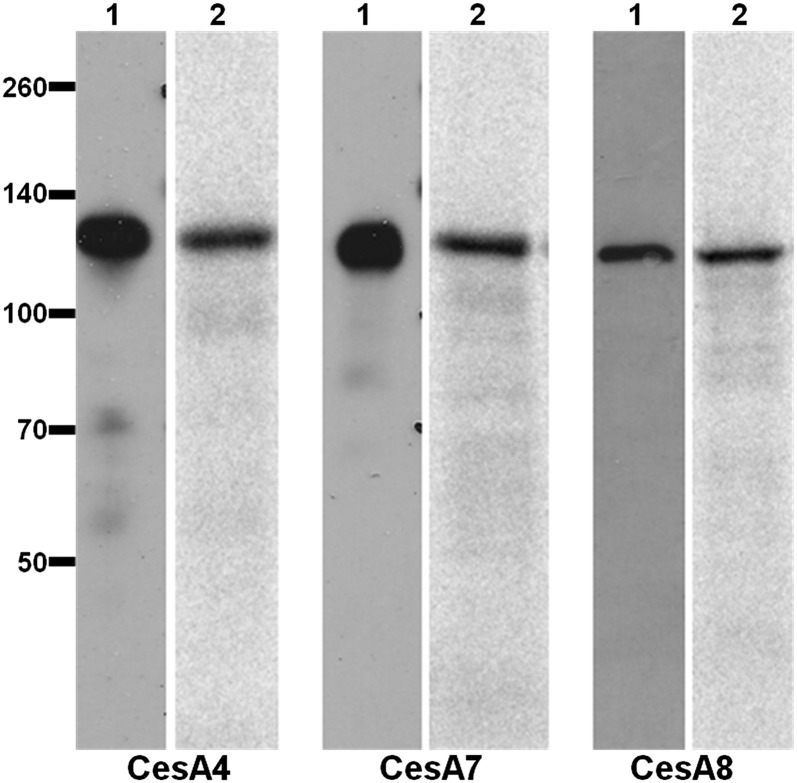
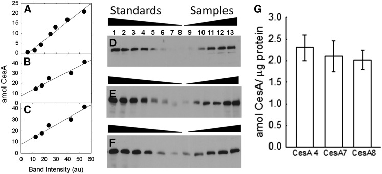
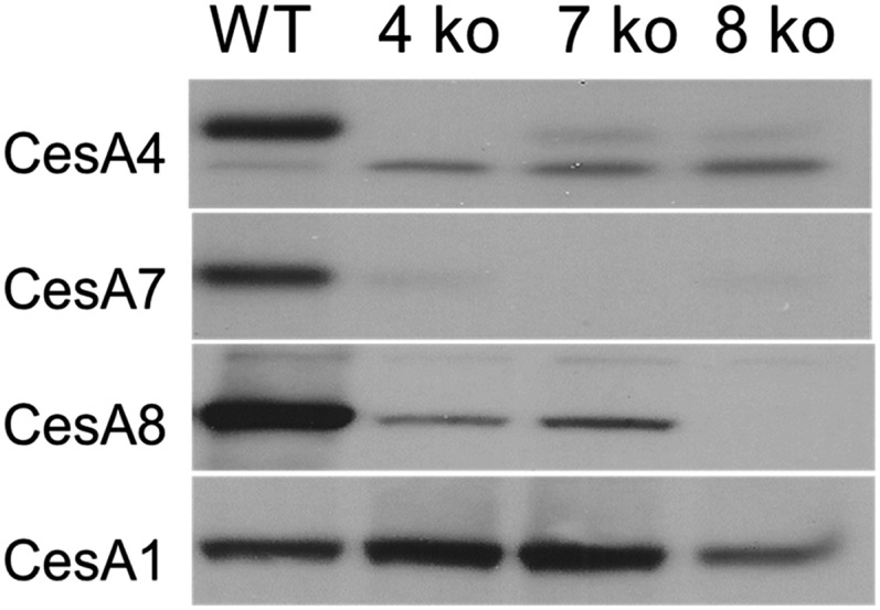
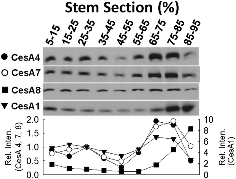
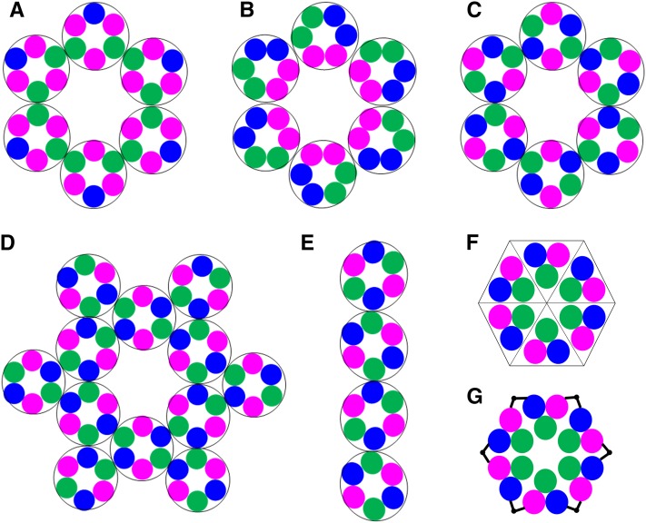
Cellulose is the most abundant renewable polymer on Earth and a major component of the plant cell wall. In vascular plants, cellulose synthesis is catalyzed by a large, plasma membrane-localized cellulose synthase complex (CSC), visualized as a hexameric rosette structure. Three unique cellulose synthase (CESA) isoforms are required for CSC assembly and function. However, elucidation of either the number or stoichiometry of CESAs within the CSC has remained elusive. In this study, we show a 1:1:1 stoichiometry between the three Arabidopsis thaliana secondary cell wall isozymes: CESA4, CESA7, and CESA8. This ratio was determined utilizing a simple but elegant method of quantitative immunoblotting using isoform-specific antibodies and (35)S-labeled protein standards for each CESA. Additionally, the observed equimolar stoichiometry was found to be fixed along the axis of the stem, which represents a developmental gradient. Our results complement recent spectroscopic analyses pointing toward an 18-chain cellulose microfibril. Taken together, we propose that the CSC is composed of a hexamer of catalytically active CESA trimers, with each CESA in equimolar amounts. This finding is a crucial advance in understanding how CESAs integrate to form higher order complexes, which is a key determinate of cellulose microfibril and cell wall properties.


Figures

Figure 1.
Figure 1.
Specificity of Antibody Populations. Equal amounts of protein from wild-type and cesa knockout stems were analyzed by immunoblot with affinity-purified populations of antibodies. Arrows indicate bands corresponding to CESA. Signals corresponding to other bands are cross-reactions with non-CESA proteins. Anti-CESA4.3, anti-CESA7.3, and anti-CESA8.2 were chosen for further use. (A) Antibodies to CESA4. Lane 1, the wild type; lane 2, cesa4. (B) Antibodies to CESA7. Lane 1, the wild type; lane 2, cesa7. (C) Antibodies to CESA8. Lane 1, the wild type; lane 2, cesa8.
Figure 2.
Figure 2.
Antibody Specificity by Analysis of Heterologously Expressed CESAs. The most abundant Arabidopsis CESAs (CESA1, CESA3, CESA6, CESA4, CESA7, and CESA8) were heterologously expressed in a cell-free wheat germ coupled transcription/translation system. Four identical SDS-PAGE gels were prepared and probed with different CESA antibodies. Immunodetection occurred only with the intended CESA isoform (i.e., anti-CESA1 detected CESA1 and failed to react with CESA3, CESA6, CESA4, CESA7, and CESA8).
Figure 3.
Figure 3.
Heterologous Expression of CESAs in Vitro. CESA4, CESA7, and CESA8 were heterologously expressed in a wheat germ cell-free coupled transcription/translation system in vitro and labeled with [35S]methionine. The major product was full-length CESA, which was detectable by immunoblot (lane 1) and autoradiogram (lane 2).
Figure 4.
Figure 4.
Quantitative Immunoblotting of CESA4, CESA7, and CESA8 Displays a 1:1:1 Stoichiometry. (A) to (C) Representative standard curves for CESA4, CESA7, and CESA8. au, arbitrary units. (D) to (F) Immunoblots corresponding to the standard curves shown in (A) to (C). Lanes 1 to 7, 35S-labeled, heterologously expressed CESA, decreasing amounts; lanes 8 to 13, increasing amounts of total protein from Arabidopsis stem (3, 4.5, 6, 9, 12, and 15 μg). (G) Calculated average concentration of each CESA with sd (n = 4 blots and n = 18 [CESA4], 20 [CESA7], and 22 [CESA8] points). There is no significant difference in CESA amount, signifying a 1:1:1 stoichiometry.
Figure 5.
Figure 5.
Immunoblot Analysis of cesa Knockout Lines. Equal amounts (20 µg) of protein from the wild type and three cesa knockout (ko) lines were separated by SDS-PAGE and immunoblotted against CESA4, CESA7, CESA8, and CESA1 (as specified). Each knockout line shows elimination of that CESA as well as a severe reduction in the interacting partner CESAs. By contrast, levels of CESA1 remain relatively constant.
Figure 6.
Figure 6.
Immunoblot Analysis of Arabidopsis Stem Sections. Equal amounts (30 μg*) of protein from sections of wild-type stem were immunoblotted against CESA4, CESA7, CESA8, and CESA1. The stem section designation is shown above the blots, while the CESA antibody used is shown to the left of the blots. Immunoblot intensity values were normalized to the 35 to 45% section and are plotted at bottom. The levels of CESA4 (closed circles), CESA7 (open circles), and CESA8 (closed triangles) show a close correlation along the entire stem, with maximal CESA levels in the sections representing 65 to 85% of the stem length as measured from the stem base. While CESA1 (closed squares) follows SCW CESA levels from 15 to 75%, indicating a consistent ratio at those points, there is a significant deviation at both the distal and apical regions of the stem. *Fifteen micrograms of sample was used for the 85 to 95% section probed for CESA1, so measured intensity was doubled.
Figure 7.
Figure 7.
Models of the CSC Rosette. Each colored circle represents one of three CESA isoforms. (A) The hexamer of hexamers model in a 1:2:3 stoichiometry. (B) to (G) Possible complexes formed with the 1:1:1 stoichiometry. (B) Pairs of CESAs in homodimers as the fundamental unit, resulting in inconsistent protein-protein interactions. (C) and (D) A hexamer of hexamers is shown where protein-protein contacts are consistent throughout (C). However, as shown in (D), this model can yield higher ordered CSCs wherein additional lobes are added to the rosette shown in (C). (E) A linear complex also can be formed with the green-to-blue contacts. (F) The hexamer of trimers rosette model. Protein contacts are consistent and the rosette is self-contained. (G) A possible mechanism for rosette assembly wherein adjacent trimers oligomerize through their N-terminal domains (black bars).

References

    1. Alonso J.M., et al. (2003). Genome-wide insertional mutagenesis of Arabidopsis thaliana. Science 301: 653–657. - PubMed
    1. Atanassov I.I., Pittman J.K., Turner S.R. (2009). Elucidating the mechanisms of assembly and subunit interaction of the cellulose synthase complex of Arabidopsis secondary cell walls. J. Biol. Chem. 284: 3833–3841. - PubMed
    1. Bashline L., Li S., Anderson C.T., Lei L., Gu Y. (2013). The endocytosis of cellulose synthase in Arabidopsis is dependent on μ2, a clathrin-mediated endocytosis adaptin. Plant Physiol. 163: 150–160. - PMC - PubMed
    1. Brown D.M., Zeef L.A.H., Ellis J., Goodacre R., Turner S.R. (2005). Identification of novel genes in Arabidopsis involved in secondary cell wall formation using expression profiling and reverse genetics. Plant Cell 17: 2281–2295. - PMC - PubMed
    1. Brown R.M. Jr., Willison J.H., Richardson C.L. (1976). Cellulose biosynthesis in Acetobacter xylinum: Visualization of the site of synthesis and direct measurement of the in vivo process. Proc. Natl. Acad. Sci. USA 73: 4565–4569. - PMC - PubMed

Publication types