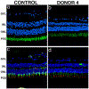
Histopathological comparison of eyes from patients with autosomal recessive retinitis pigmentosa caused by novel EYS mutations
- PMID: 25491159
- PMCID: PMC10846590
- DOI: 10.1007/s00417-014-2868-z
Histopathological comparison of eyes from patients with autosomal recessive retinitis pigmentosa caused by novel EYS mutations
Abstract
To evaluate the retinal histopathology in donor eyes from patients with autosomal recessive retinitis pigmentosa (arRP) caused by EYS mutations. Eyes from a 72-year-old female (donor 1, family 1), a 91-year-old female (donor 2, family 2), and her 97-year-old sister (donor 3, family 2) were evaluated with macroscopic, scanning laser ophthalmoscopy (SLO) and optical coherence tomography (OCT) imaging. Age-similar normal eyes and an eye donated by donor 1's asymptomatic mother (donor 4, family 1) were used as controls. The perifovea and peripheral retina were processed for microscopy and immunocytochemistry with markers for cone and rod photoreceptor cells. DNA analysis revealed EYS mutations c.2259 + 1G > A and c.2620C > T (p.Q874X) in family 1, and c.4350_4356del (p.I1451Pfs*3) and c.2739-?_3244 + ?del in family 2. Imaging studies revealed the presence of bone spicule pigment in arRP donor retinas. Histology of all three affected donor eyes showed very thin retinas with little evidence of stratified nuclear layers in the periphery. In contrast, the perifovea displayed a prominent inner nuclear layer. Immunocytochemistry analysis demonstrated advanced retinal degenerative changes in all eyes, with near-total absence of rod photoreceptors. In addition, we found that the perifoveal cones were more preserved in retinas from the donor with the midsize genomic rearrangement (c.4350_4356del (p.I1451Pfs*3) and c.2739-?_3244 + ?del) than in retinas from the donors with the truncating (c.2259 + 1G > A and c.2620C > T (p.Q874X) mutations. Advanced retinal degenerative changes with near-total absence of rods and preservation of some perifoveal cones are observed in arRP donor retinas with EYS mutations.
Figures


Similar articles
-
Retinal histopathology in eyes from patients with autosomal dominant retinitis pigmentosa caused by rhodopsin mutations.Graefes Arch Clin Exp Ophthalmol. 2015 Dec;253(12):2161-9. doi: 10.1007/s00417-015-3099-7. Epub 2015 Jul 23. Graefes Arch Clin Exp Ophthalmol. 2015. PMID: 26202387 Free PMC article.
-
Autosomal recessive cone-rod dystrophy associated with compound heterozygous mutations in the EYS gene.Doc Ophthalmol. 2014 Jun;128(3):211-7. doi: 10.1007/s10633-014-9435-0. Epub 2014 Mar 21. Doc Ophthalmol. 2014. PMID: 24652164
-
Disease expression in X-linked retinitis pigmentosa caused by a putative null mutation in the RPGR gene.Invest Ophthalmol Vis Sci. 1997 Sep;38(10):1983-97. Invest Ophthalmol Vis Sci. 1997. PMID: 9331262
-
Identification of a novel homozygous nonsense mutation in EYS in a Chinese family with autosomal recessive retinitis pigmentosa.BMC Med Genet. 2010 Aug 10;11:121. doi: 10.1186/1471-2350-11-121. BMC Med Genet. 2010. PMID: 20696082 Free PMC article.
-
Mutations in the EYS gene account for approximately 5% of autosomal recessive retinitis pigmentosa and cause a fairly homogeneous phenotype.Ophthalmology. 2010 Oct;117(10):2026-33, 2033.e1-7. doi: 10.1016/j.ophtha.2010.01.040. Ophthalmology. 2010. PMID: 20537394
Cited by
-
Eyes shut homolog (EYS) interacts with matriglycan of O-mannosyl glycans whose deficiency results in EYS mislocalization and degeneration of photoreceptors.Sci Rep. 2020 May 8;10(1):7795. doi: 10.1038/s41598-020-64752-4. Sci Rep. 2020. PMID: 32385361 Free PMC article.
-
Deletion of POMT2 in Zebrafish Causes Degeneration of Photoreceptors.Int J Mol Sci. 2022 Nov 26;23(23):14809. doi: 10.3390/ijms232314809. Int J Mol Sci. 2022. PMID: 36499139 Free PMC article.
-
Digenic heterozygous mutations in EYS/LRP5 in a Chinese family with retinitis pigmentosa.Int J Ophthalmol. 2017 Feb 18;10(2):325-328. doi: 10.18240/ijo.2017.02.25. eCollection 2017. Int J Ophthalmol. 2017. PMID: 28251098 Free PMC article. No abstract available.
-
A hypomorphic variant in EYS detected by genome-wide association study contributes toward retinitis pigmentosa.Commun Biol. 2021 Jan 29;4(1):140. doi: 10.1038/s42003-021-01662-9. Commun Biol. 2021. PMID: 33514863 Free PMC article.
-
Eyes shut homolog is required for maintaining the ciliary pocket and survival of photoreceptors in zebrafish.Biol Open. 2016 Nov 15;5(11):1662-1673. doi: 10.1242/bio.021584. Biol Open. 2016. PMID: 27737822 Free PMC article.
References
-
- Bunker CH, Berson EL, Bromley WC, Hayes RP, Roderick TH (1984) Prevalence of retinitis pigmentosa in Maine. Am J Ophthalmol 97:357–365. - PubMed
-
- Dryja TP, McGee TL, Hahn LB, Cowley GS, Olsson JE, Reichel E, Sandberg MA, Berson EL (1990) Mutations within the rhodopsin gene in patients with autosomal dominant retinitis pigmentosa. N Engl J Med 323:1302–1307. - PubMed
-
- Tsang SH, Wolpert K (2010) The Genetics of Retinitis Pigmentosa. Retinal Physician.
-
- Collin RW, Littink KW, Klevering BJ, van den Born LI, Koenekoop RK, Zonneveld MN, Blokland EA, Strom TM, Hoyng CB, den Hollander AI, Cremers FP (2008) Identification of a 2 Mb human ortholog of Drosophila eyes shut/spacemaker that is mutated in patients with retinitis pigmentosa. Am J Hum Genet 83:594–603. - PMC - PubMed
Publication types
MeSH terms
Substances
Supplementary concepts
Grants and funding
LinkOut - more resources
Full Text Sources
Other Literature Sources

