. 2014 Dec 10:9:190.
doi: 10.1186/s13023-014-0190-9.

Expanding the phenotype of PRPS1 syndromes in females: neuropathy, hearing loss and retinopathy

Affiliations

Expanding the phenotype of PRPS1 syndromes in females: neuropathy, hearing loss and retinopathy

Berta Almoguera et al. Orphanet J Rare Dis. .

Abstract

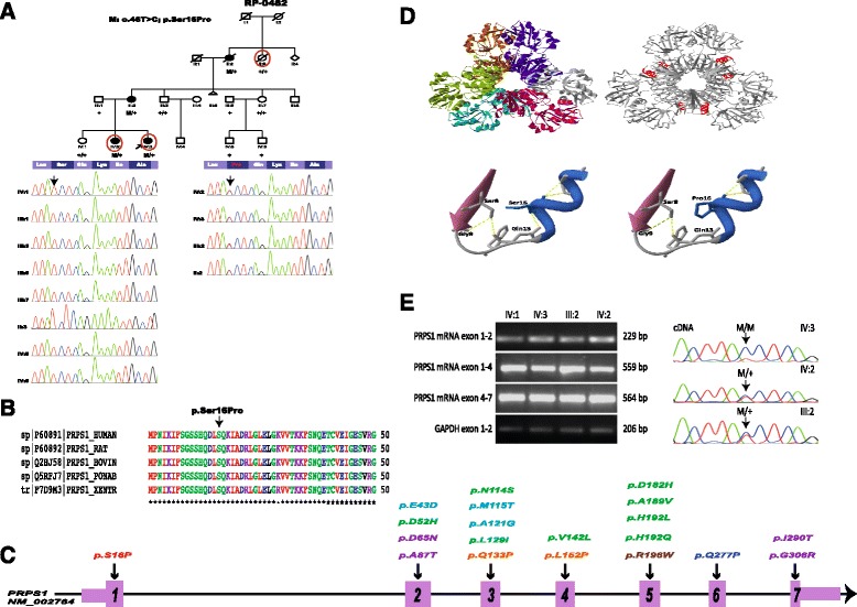
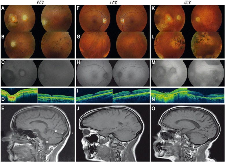
Background: Phosphoribosyl pyrophosphate synthetase (PRS) I deficiency is a rare medical condition caused by missense mutations in PRPS1 that lead to three different phenotypes: Arts Syndrome (MIM 301835), X-linked Charcot-Marie-Tooth (CMTX5, MIM 311070) or X-linked non-syndromic sensorineural deafness (DFN2, MIM 304500). All three are X-linked recessively inherited and males affected display variable degree of central and peripheral neuropathy. We applied whole exome sequencing to a three-generation family with optic atrophy followed by retinitis pigmentosa (RP) in all three cases, and ataxia, progressive peripheral neuropathy and hearing loss with variable presentation.

Methods: Whole exome sequencing was performed in two affecteds and one unaffected member of the family. Sanger sequencing was used to validate and segregate the 12 candidate mutations in the family and to confirm the absence of the novel variant in PRPS1 in 191 controls. The pathogenic role of the novel mutation in PRPS1 was assessed in silico and confirmed by enzymatic determination of PRS activity, mRNA expression and sequencing, and X-chromosome inactivation.

Results: A novel missense mutation was identified in PRPS1 in the affected females. Age of onset, presentation and severity of the phenotype are highly variable in the family: both the proband and her mother have neurological and ophthalmological symptoms, whereas the phenotype of the affected sister is milder and currently confined to the eye. Moreover, only the proband displayed a complete lack of expression of the wild type allele in leukocytes that seems to correlate with the degree of PRS deficiency and the severity of the phenotype. Interestingly, optic atrophy and RP are the only common manifestations to all three females and the only phenotype correlating with the degree of enzyme deficiency.

Conclusions: These results are in line with recent evidence of the existence of intermediate phenotypes in PRS-I deficiency syndromes and demonstrate that females can exhibit a disease phenotype as severe and complex as their male counterparts.


Figures

Figure 1
Figure 1
PSPS1 is mutated in females with syndromic retinitis pigmentosa from a three-generation family. A. Pedigree of family RP-0482 and validation by Sanger sequencing of p.Ser16Pro demonstrating the correct segregation in the family. All affected individuals (II:2, III:2; IV:2 and IV:3) are heterozygous for the variant. Red circles indicate the individuals analysed by WES. Although II:2 and II:3 died during the course of the study, DNA samples and informed consents were obtained before deceasing, allowing us to include those subjects in the segregation analysis. B. Multiple sequence alignment of PRPS1 across species using ClustalW [14] confirms that p.Ser16 is identical from human to zebrafish. C. Schematic representation of PRPS1 with the location of the novel heterozygous mutation p.Ser16Pro in exon 1 (in red) and previously known mutations in Arts syndrome (in orange), PRS-I Superactivity (in green), Charcot-Marie-Tooth disease-5 (in blue), X-linked nonsyndromic sensorineural deafness (in purple), and also the recently reported mutation associated with retinal dystrophy (in brown). Exons are indicated by rectangles that are wider for the coding regions. Nucleotide numbering reflects cDNA in the reference sequence NM_002764. D. Model of PRS-I with p.Ser16Pro based on the crystal structure of human PRS-I (PDB: 2H06) and close-up of the mutation showing the loss of a hydrogen bound with residue Gln13. E. RT-PCR and sequencing analysis of p.Ser16Pro in mRNA. RNA was derived from peripheral blood lymphocytes of the three patients (IV:2, IV:3 and III:2) and an unaffected control (IV:1). Normally spliced amplicons of exons 1–2, 1–4 and 4–7 of PRPS1 comprising 229, 559 and 564 nucleotides, respectively, were found in all cases. Amplification of GAPDH mRNA analysis was used as positive control. Sanger sequencing of RT-PCR products evidences the absence of the wild-type allele in the cDNA of the proband (IV:3).
Figure 2
Figure 2
Retinal and head MRI imaging of patients carrying the p.Ser16Pro variant in PRPS1 . A-E. Retinal imaging of both eyes (BE) of the proband IV:3 at age 35. (A-B) Fundus photographies show pale and atrophic papilla, narrowed vessels, diffuse hypopigmentation, bone spicules, and retinal pigment epithelium (RPE) atrophy in the mid-periphery of BE, and a well-defined atrophic macular lesion in the right eye (RE). (C) Autofluorescence reveals hypofluorescence areas corresponding with pigmented spicules and atrophic lesions. (D) Optical coherence tomography (OCT) confirms the generalized atrophy in all retinal layers in both perifoveal and foveal regions in RE. Well-conserved retinal architecture was found in the left eye (LE) and the atrophy is restricted to RPE and external layers. (E) Head MRI scan of the proband IV:3 demonstrates a moderate cerebellar atrophy (white arrows). F-J. Retinal imaging of BE of IV:2 at age 37. (F-G) Fundus photographies show a slight pallor in the papilla, slight arteriolar attenuation mainly around the optic nerve, a normal macula and an incipient RPE atrophy. (H) Normal autofluorescence. (I) OCTs display atrophy of photoreceptors layer in the perifoveal region and normal foveal thickness. (J) Head MRI scan of individual IV:2 does not evidence any sign of cerebellar atrophy. K-O. Retinal imaging of BE of III:2 at age 70. (K-L) Fundus photographies evidence pale and atrophic papilla, substantially narrowed vessels, sparse pigmentation, patches of chorioretinal atrophy in the mid-periphery of BE, and a well-defined atrophic macular lesion in RE and RPE alteration in LE. (M) Autofluorescence imaging shows alternating areas of hypo and hyperfluorescence. (N) OCT shows subfoveal atrophy lesion with loss of retinal architecture, an epiretinal membrane in RE and defects in external layer in LE. (O) Head MRI scan of individual III:2 exhibits a mild cerebellar atrophy (white arrow).

References

    1. Roessler BJ, Bell G, Heidler S, Seino S, Becker M, Palella TD. Cloning of two distinct copies of human phosphoribosylpyrophosphate synthetase cDNA. Nucleic Acids Res. 1990;18(1):193. doi: 10.1093/nar/18.1.193. - DOI - PMC - PubMed
    1. Taira M, Ishijima S, Kita K, Yamada K, Iizasa T, Tatibana M. Nucleotide and deduced amino acid sequences of two distinct cDNAs for rat phosphoribosylpyrophosphate synthetase. J Biol Chem. 1987;262(31):14867–14870. - PubMed
    1. de Brouwer AP, Williams KL, Duley JA, van Kuilenburg AB, Nabuurs SB, Egmont-Petersen M, Lugtenberg D, Zoetekouw L, Banning MJ, Roeffen M, Hamel BC, Weaving L, Ouvrier RA, Donald JA, Wevers RA, Christodoulou J, van Bokhoven H. Arts syndrome is caused by loss-of-function mutations in PRPS1. Am J Hum Genet. 2007;81(3):507–518. doi: 10.1086/520706. - DOI - PMC - PubMed
    1. Kim HJ, Sohn KM, Shy ME, Krajewski KM, Hwang M, Park JH, Jang SY, Won HH, Choi BO, Hong SH, Kim BJ, Suh YL, Ki CS, Lee SY, Kim SH, Kim JW. Mutations in PRPS1, which encodes the phosphoribosyl pyrophosphate synthetase enzyme critical for nucleotide biosynthesis, cause hereditary peripheral neuropathy with hearing loss and optic neuropathy (cmtx5) Am J Hum Genet. 2007;81(3):552–558. doi: 10.1086/519529. - DOI - PMC - PubMed
    1. Liu X, Han D, Li J, Han B, Ouyang X, Cheng J, Li X, Jin Z, Wang Y, Bitner-Glindzicz M, Kong X, Xu H, Kantardzhieva A, Eavey RD, Seidman CE, Seidman JG, Du LL, Chen ZY, Dai P, Teng M, Yan D, Yuan H. Loss-of-function mutations in the PRPS1 gene cause a type of nonsyndromic X-linked sensorineural deafness, DFN2. Am J Hum Genet. 2010;86(1):65–71. doi: 10.1016/j.ajhg.2009.11.015. - DOI - PMC - PubMed

Publication types

MeSH terms

Substances

Supplementary concepts

LinkOut - more resources