Review
. 2015 Mar;467(3):465-74.
doi: 10.1007/s00424-014-1662-4. Epub 2014 Dec 10.

Sodium sensing in the brain

Affiliations
Review

Sodium sensing in the brain

Masaharu Noda et al. Pflugers Arch. 2015 Mar.

Abstract

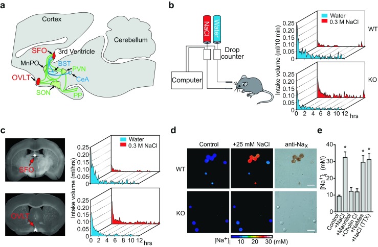
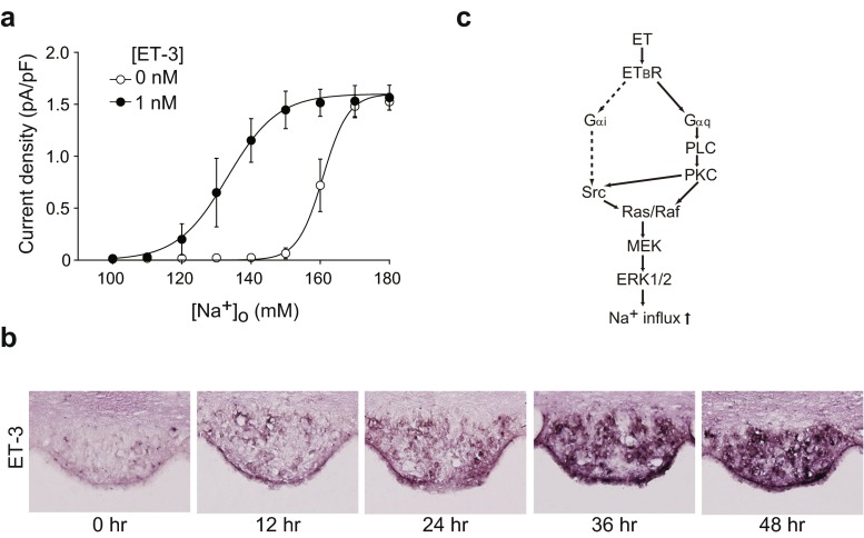
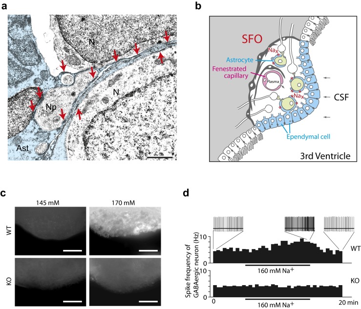
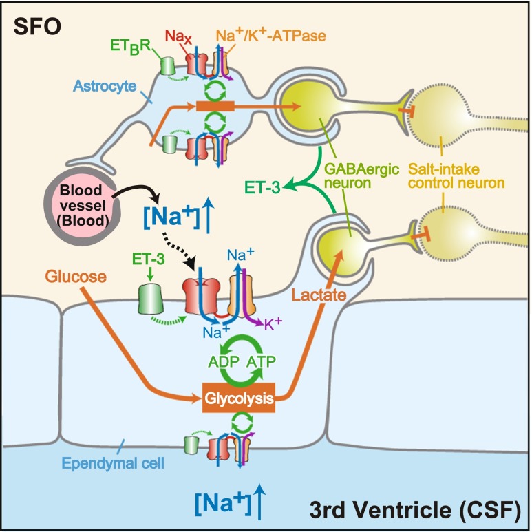
Sodium (Na) homeostasis is crucial for life, and the Na(+) level ([Na(+)]) of body fluids is strictly maintained at a range of 135-145 mM. However, the existence of a [Na(+)] sensor in the brain has long been controversial until Nax was identified as the molecular entity of the sensor. This review provides an overview of the [Na(+)]-sensing mechanism in the brain for the regulation of salt intake by summarizing a series of our studies on Nax. Nax is a Na channel expressed in the circumventricular organs (CVOs) in the brain. Among the CVOs, the subfornical organ (SFO) is the principal site for the control of salt intake behavior, where Nax populates the cellular processes of astrocytes and ependymal cells enveloping neurons. A local expression of endothelin-3 in the SFO modulates the [Na(+)] sensitivity for Nax activation, and thereby Nax is likely to be activated in the physiological [Na(+)] range. Nax stably interacts with Na(+)/K(+)-ATPase whereby Na(+) influx via Nax is coupled with activation of Na(+)/K(+)-ATPase associated with the consumption of ATP. The consequent activation of anaerobic glucose metabolism of Nax-positive glial cells upregulates the cellular release of lactate, and this lactate functions as a gliotransmitter to activate GABAergic neurons in the SFO. The GABAergic neurons presumably regulate hypothetic neurons involved in the control of salt intake behavior. Recently, a patient with essential hypernatremia caused by autoimmunity to Nax was found. In this case, the hypernatremia was considered to be induced by the complement-mediated cell death in the CVOs, where Nax specifically populates.


Figures

Fig. 1
Fig. 1
SFO is the primary locus of [Na+] sensing by Nax channel for the control of salt intake behavior. a Neural connections for body fluid control in the brain. Nax-positive sensory circumventricular organs in the midsagittal section are schematically represented in red. Of note, Nax is not expressed in the MnPO. Green lines indicate the neural connections involved in regulating the release of vasopressin; blue lines indicate putative neural connections involved in the control of water or salt intake. SFO subfornical organ, MnPO median preoptic area, OVLT organum vasculosum of the lamina terminalis, BST bed nucleus of the stria terminalis, SON supraoptic nucleus, PVN paraventricular nucleus, CeA central nucleus of the amygdala, PP posterior pituitary. b Averaged time course of water and saline (0.3 M NaCl) intake in wild-type (WT) and Na x-KO (KO) mice during the dark phase immediately after 48-h dehydration. Each point shows the averaged quantity per 10-min period of ten mice. c Coronal sections of mouse brains obtained from Na x-KO mice showing the infected loci by the expression of EGFP (left column). Time course of water and saline (0.3 M NaCl) intake by the infected mice after 48-h dehydration (right column). Behavioral data are the average of six mice that were successfully infected at a specific site in the brain by vectors encoding Na x and egfp. All of the Na x-KO mice that were conferred with the Na aversion behavior showed a common infection in the SFO (upper). On the other hand, transduction of the Nax gene into the OVLT could not rescue the abnormal salt intake behavior of the Na x-KO mice (lower). d Pseudocolor images showing the [Na+]i of the cells in the control ([Na+]o = 145 mM) and high-Na+ ([Na+]o = 170 mM) solutions (left and middle columns) and immunocytochemical images using anti-Nax antibody (right column). Scale bars, 50 μm. e The [Na+]i response to various stimulations. The response was dependent on [Na+]o, but not on extracellular [Cl]o or osmotic pressure. Instead of 25 mM NaCl, 50 mM mannitol, 25 mM choline chloride (Cholin Cl), or 25 mM sodium methanesulfonate (NaMes) was added to the control solution. The response was not affected by 1 μM tetrodotoxin (TTX). *P < 0.001 by one-tailed Mann-Whitney tests. Reproduced with permission from [55] (a), [33] (b, c), and [34] (d, e)
Fig. 2
Fig. 2
Endothelin-3 (ET-3) signaling shifted the [Na+]o dependency of Nax activation to the lower side. a Relationships between the current density and [Na+]o in the presence or absence of 1 nM ET-3; n = 6 for each. b In situ hybridization for detection of ET-3 expression in the SFO of WT mice. Brains were obtained from mice provided freely with food and water (0 h) or from those provided only with food during the indicated period (12, 24, 36, and 48 h). Sections on the same slide are shown. c Activation cascades of Nax by ETBR signaling. The pathway indicated by dotted lines was suggested not to work for the Nax activation. Reproduced with permission from [35]
Fig. 3
Fig. 3
Nax channels control lactate signaling from glial cells to neurons for [Na+] sensing in the SFO. a Immunoelectron microscopy of the SFO using anti-Nax antibody. Neurons (N) and their processes (Np) are enveloped with the immunopositive thin processes of an astrocyte (Ast; blue). Red arrows point to immunopositive signals. Neurons and their processes, including synapses, are surrounded by immunopositive thin processes of astrocytes. Scale bars, 1 μm. b Schematic drawing of Nax-positive ependymal cells and astrocytes in the SFO. The SFO is characterized by the presence of neuronal cell bodies and extensive networks of fenestrated capillaries that allow components of the plasma to leak into the intercellular space. The SFO has contact with the CSF through a single layer of Nax-positive ependymal cells. c Imaging analyses of the uptake of glucose in the SFO using a fluorescent glucose derivative. The SFO tissues isolated from wild-type (WT) and Na x-KO (KO) mice were incubated with the fluorescent glucose analog in 145 mM (left column) or 170 mM (right column) Na+ solution. Scale bars, 50 μm. d Control of spike frequency of GABAergic neurons in the SFO by Na+. The SFO tissues from WT and Na x-KO mice were treated with the high-Na+ solution. Nax is indispensable for [Na+]-dependent potentiation of the GABAergic firing in the SFO. Reproduced with permission from [67] (a), [52] (b), and [60] (c, d)
Fig. 4
Fig. 4
Overview of the [Na+]-sensing mechanism and Nax-dependent regulation of neuronal activity in the SFO. Reproduced with permission from [55]

References

    1. Adams JM, Bardgett ME, Stocker SD. Ventral lamina terminalis mediates enhanced cardiovascular responses of rostral ventrolateral medulla neurons during increased dietary salt. Hypertension. 2009;54:308–314. doi: 10.1161/HYPERTENSIONAHA.108.127803. - DOI - PMC - PubMed
    1. Akopian AN, Souslova V, Sivilotti L, Wood JN. Structure and distribution of a broadly expressed atypical sodium channel. FEBS Lett. 1997;400:183–187. doi: 10.1016/S0014-5793(96)01389-0. - DOI - PubMed
    1. Andersson B. Thirst—and brain control of water balance. Am Sci. 1971;59:408–415. - PubMed
    1. Andersson B. Regulation of water intake. Physiol Rev. 1978;58:582–603. - PubMed
    1. Arieff AI. Management of hyponatraemia. BMJ. 1993;307:305–308. doi: 10.1136/bmj.307.6899.305. - DOI - PMC - PubMed

MeSH terms