. 2014 Dec 10:4:7395.
doi: 10.1038/srep07395.

The dengue virus conceals double-stranded RNA in the intracellular membrane to escape from an interferon response

Affiliations

The dengue virus conceals double-stranded RNA in the intracellular membrane to escape from an interferon response

Leo Uchida et al. Sci Rep. .

Abstract

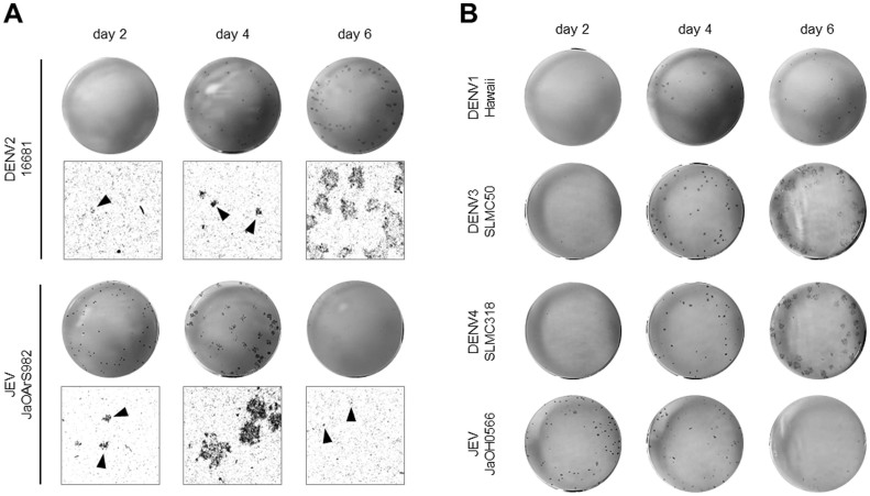
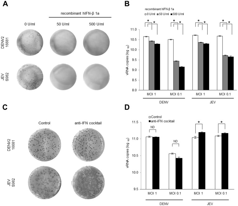
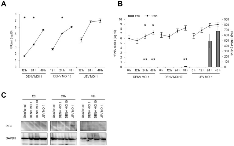
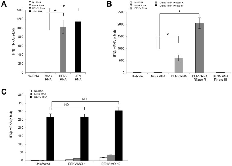
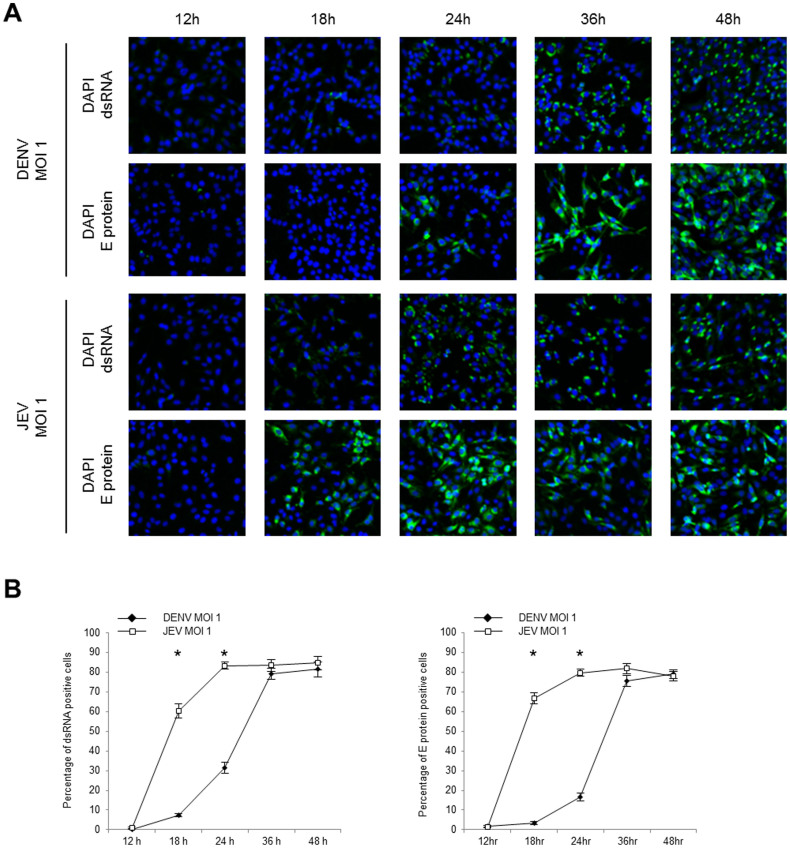
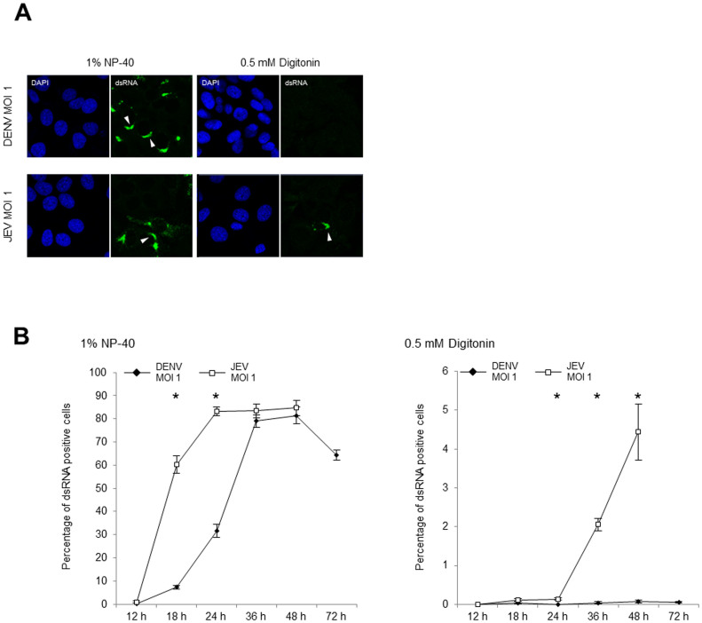
The dengue virus (DENV) circulates between humans and mosquitoes and requires no other mammals or birds for its maintenance in nature. The virus is well-adapted to humans, as reflected by high-level viraemia in patients. To investigate its high adaptability, the DENV induction of host type-I interferon (IFN) was assessed in vitro in human-derived HeLa cells and compared with that induced by the Japanese encephalitis virus (JEV), a closely related arbovirus that generally exhibits low viraemia in humans. A sustained viral spread with a poor IFN induction was observed in the DENV-infected cells, whereas the JEV infection resulted in a self-limiting and abortive infection with a high IFN induction. There was no difference between DENV and JEV double-stranded RNA (dsRNA) as IFN inducers. Instead, the dsRNA was poorly exposed in the cytosol as late as 48 h post-infection (p.i.), despite the high level of DENV replication in the infected cells. In contrast, the JEV-derived dsRNA appeared in the cytosol as early as 24 h p.i. Our results provided evidence for the first time in DENV, that concealing dsRNA in the intracellular membrane diminishes the effect of the host defence mechanism, a strategy that differs from an active suppression of IFN activity.


Figures

Figure 1
Figure 1. DENV and JEV focus formation in HeLa cells.
(A) HeLa cells were infected with DENV2 strain 16681 or JEV strain JaOArS982 at 40 focus forming units (FFU)/well. At the indicated days p.i., the foci were detected by immunostaining. The upper figures show macroscopic images, and the lower figures show 100× magnified images. The black arrowheads indicate the foci. (B) HeLa cells were infected with DENV1 strain Hawaii, DENV3 strain SLMC-50, DENV4 strain SLMC-318, or JEV strain JaOH0566 at 40 FFU/well. At the indicated days p.i., foci were detected by immunostaining.
Figure 2
Figure 2. Effect of IFN-β on DENV and JEV focus formation in HeLa cells.
HeLa cells were infected with DENV2 strain 16681 or JEV strain JaOArS982. At 12 h p.i., the culture medium was replaced with 0 to 500 U/ml of the human recombinant-IFN-β1a in MEM. At 4 days p.i., foci were detected by immunostaining (A) and the vRNA in the culture fluid was quantified by RT-qPCR (B). The HeLa cells were treated with anti-IFN cocktail consisting of 2,000 neutralization U/ml of the anti-human IFN-β Ab and 20 μg/ml of the anti-human IFN-α/βR2 (CD118) 1 h before the viral infection. The concentration of the anti-IFN cocktail was maintained after the viral infection. At 4 days p.i. the foci were detected by immunostaining (C), and the vRNA in the culture fluid was quantified by RT-qPCR (D). The mean ± SE of the vRNA copy number was obtained from two independent duplicate experiments. The values between two groups were tested by Welch test analysis. The asterisks indicate a statistical significance at p < 0.01, and ND indicates no significant difference.
Figure 3
Figure 3. Virus growth and innate immune response against DENV and JEV infection.
HeLa cells were infected with DENV2 strain 16681 at MOI 1 or 10 or JEV strain JaOArS982 at MOI 1. At the indicated time points, the culture fluid and the cells were harvested. (A) The virus titers in the culture fluid were determined by FFA. (B) The vRNA and the IFN-β gene expression in the DENV- and the JEV-infected cells that were quantified by RT-qPCR. The absolute vRNA copy numbers and the relative-fold increase in the IFN-β mRNA levels were normalized with GAPDH mRNA levels. The mean ± SE of the vRNA copy number was obtained from three independent duplicate experiments. The values between the DENV- (MOI 1) and JEV- (MOI 1) infected cells or the DENV- (MOI 10) and the JEV- (MOI 1) infected cells were tested by Welch test analysis. The single asterisks indicate a significant difference of vRNA (* p < 0.01) and double asterisks indicate a significant difference of IFN-β mRNA (** p < 0.01). (C) Representative cropped blots of RIG-I and GAPDH in DENV- and JEV-infected cells are shown. The infected cells were harvested and the target proteins were detected by the immunoblotting assay. GAPDH was used as an internal control. All the samples were derived from the same experiment and blotting was processed in parallel. The full-length blotting images are presented in Supplemental Fig. S4.
Figure 4
Figure 4. Effect of viral dsRNA on the IFN activation pathway.
(A) One microgram/well of the mock RNA, the DENV RNA, or the JEV RNA was transfected into uninfected cells. (B) Five hundred nanogram/well of the non-treated, RNase R-treated or the RNase III-treated DENV RNA was transfected into uninfected cells. (C) The cells were infected with DENV2 strain 16681 at MOI 1 or 10 prior to the IFN-β stimulating assay. After 48 h p.i., 500 ng/well of the mock RNA or the DENV RNA were transfected into the cells with the uninfected control. Six hours after the RNA transfection, the cells were harvested, and the IFN-β and the GAPDH mRNA levels were quantified by RT-qPCR. The relative-fold increase of the IFN-β mRNA levels was normalized with the GAPDH mRNA levels. The mean ± SE of the IFN-β mRNA levels were obtained from two independent duplicate experiments. The values between two groups were tested by Welch test analysis. The asterisks indicate statistical significance at p < 0.01, and ND indicate no significant difference.
Figure 5
Figure 5. dsRNA and E protein expression in DENV- and JEV-infected cells.
HeLa cells were infected with DENV2 strain 16681 or JEV strain JaOArS982 at MOI 1 in chamber slides. At the indicated time points, the infected cells were fixed and permeabilized by 1% NP-40. (A) The viral dsRNA and E protein were visualized by using the immunofluorescence assay. The nuclei were stained with DAPI. (B) The percentages of the dsRNA and the E protein expression were obtained by calculating the mean percentage of positively stained cells in 3 different fields (241.1 ± 10.8 total cells/field). The percentages of positively stained cells were arcsine transformed prior to the Welch test analysis. The statistically significant differences between DENV- and JEV-infected cells are indicated (* p < 0.01). The error bars indicate standard error of the means.
Figure 6
Figure 6. Immunofluorescence detection of the cytosolic DENV and JEV dsRNA.
(A) The localization of the dsRNA in DENV2 strain 16681 (MOI 1) and JEV strain JaOArS982 (MOI 1) infected cells were observed at 24 h p.i. The infected cells were permeabilized with 1% NP-40 or 0.5 mM of digitonin and stained for dsRNA using the immunofluorescence assay. The white arrowheads indicate the viral dsRNA. (B) The percentage of cells positively stained for dsRNA with 1% NP-40 or 0.5 mM digitonin permeabilization was calculated in 3 different fields (241.1 ± 10.8 total cells/field for NP-40, and 1241.4 ± 37.4 total cells/field for digitonin permeabilization). The percentages of positively stained cells were arcsine transformed prior to the Welch test analysis. The statistically significant differences between the DENV- and JEV-infected cells are indicated (* p < 0.01). The error bars indicate standard error of the means. The experiment was continued until the cells started dying (72 h p.i. for DENV and 48 h p.i. for JEV).

References

    1. Bhatt S. et al. The global distribution and burden of dengue. Nature 496, 504–507 (2013). - PMC - PubMed
    1. Bhamarapravati N. et al. World Health Organization technical report series. Viral Haemorrhagic Fevers. Ch. 2, 13–24 (World Health Organization, Geneva, 1985). - PubMed
    1. Guabiraba R. & Ryffel B. Dengue virus infection: current concepts in immune mechanisms and lessons from murine models. Immunology 141, 143–156 (2014). - PMC - PubMed
    1. Clyde K., Kyle J. L. & Harris E. Recent advances in deciphering viral and host determinants of dengue virus replication and pathogenesis. J. Virol. 80, 11418–11431 (2006). - PMC - PubMed
    1. Jessie K., Fong M. Y., Devi S., Lam S. K. & Wong K. T. Localization of dengue virus in naturally infected human tissues, by immunohistochemistry and in situ hybridization. J. Infect. Dis. 189, 1411–1418 (2004). - PubMed

Publication types

MeSH terms

LinkOut - more resources