. 2015 Dec;54(12):1710-21.
doi: 10.1002/mc.22242. Epub 2014 Dec 9.

Honokiol inhibits melanoma stem cells by targeting notch signaling

Affiliations

Honokiol inhibits melanoma stem cells by targeting notch signaling

Gaurav Kaushik et al. Mol Carcinog. 2015 Dec.

Abstract

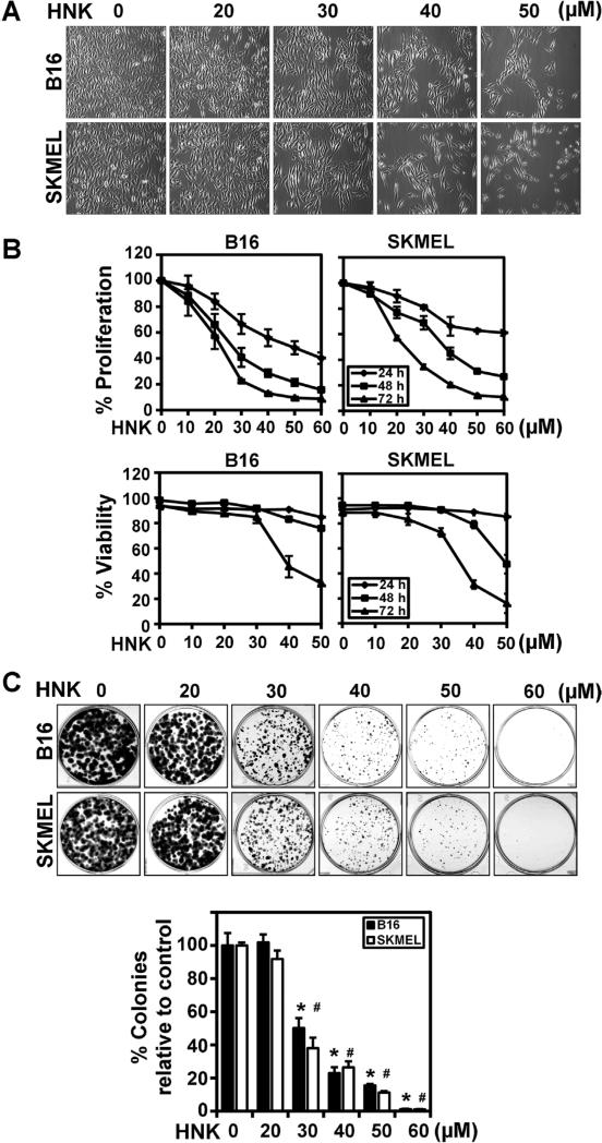
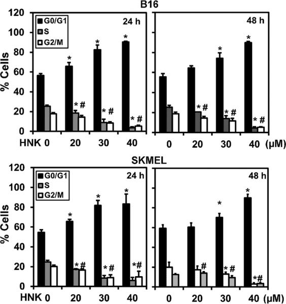
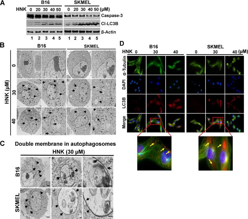
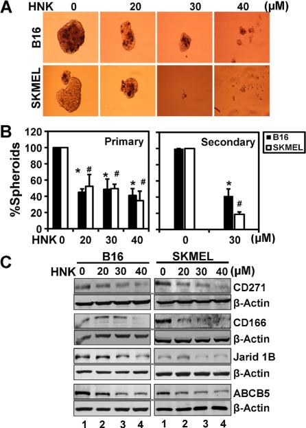
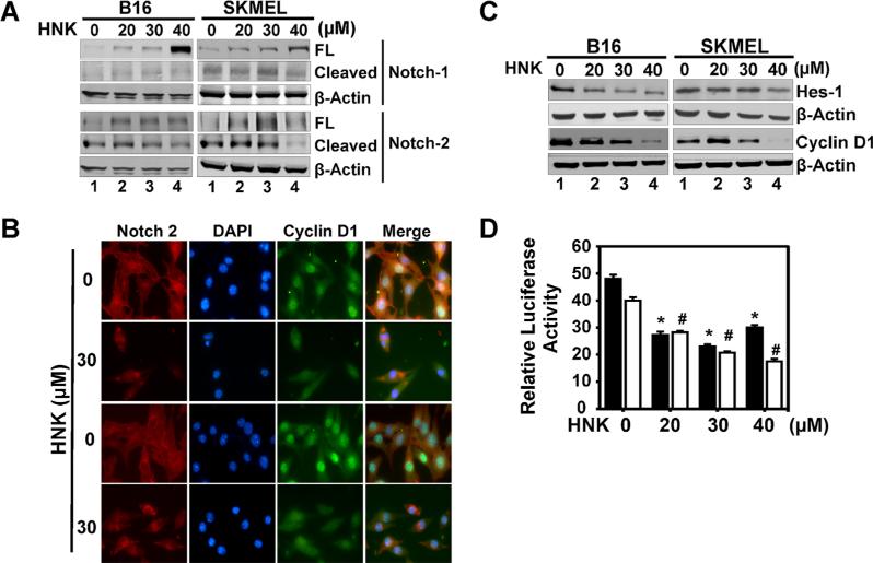
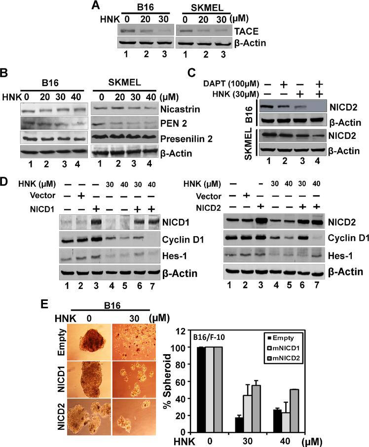
Melanoma is an aggressive disease with limited therapeutic options. Here, we determined the effects of honokiol (HNK), a biphenolic natural compound on melanoma cells and stemness. HNK significantly inhibited melanoma cell proliferation, viability, clonogenicity and induced autophagy. In addition, HNK significantly inhibited melanosphere formation in a dose dependent manner. Western blot analyses also demonstrated reduction in stem cell markers CD271, CD166, Jarid1b, and ABCB5. We next examined the effect of HNK on Notch signaling, a pathway involved in stem cell self-renewal. Four different Notch receptors exist in cells, which when cleaved by a series of enzymatic reactions catalyzed by Tumor Necrosis Factor-α-Converting Enzyme (TACE) and γ-secretase protein complex, results in the release of the Notch intracellular domain (NICD), which then translocates to the nucleus and induces target gene expression. Western blot analyses demonstrated that in HNK treated cells there is a significant reduction in the expression of cleaved Notch-2. In addition, there was a reduction in the expression of downstream target proteins, Hes-1 and cyclin D1. Moreover, HNK treatment suppressed the expression of TACE and γ-secretase complex proteins in melanoma cells. To confirm that suppression of Notch-2 activation is critical for HNK activity, we overexpressed NICD1, NICD2, and performed HNK treatment. NICD2, but not NICD1, partially restored the expression of Hes-1 and cyclin D1, and increased melanosphere formation. Taken together, these data suggest that HNK is a potent inhibitor of melanoma cells, in part, through the targeting of melanoma stem cells by suppressing Notch-2 signaling.

Keywords: Cancer stem cells; Notch-1; Notch-2; autophagy; cell cycle arrest.


Figures

Figure 1
Figure 1
HNK inhibit growth and clonogenic potential of melanoma cells. (A) Representative photomicrographs of B16/F-10 and SKMEL-28 cells after 24 h of HNK treatment (magnification 20×). Cells were seeded in 6 well plates, allowed to grow overnight and treated with increasing concentrations of HNK (0–50 μM) for 24 h. Significant changes in cell morphology were observed in both the cell lines. (B) Cell proliferation assay showed HNK induced growth inhibitory effects on melanoma cells. Cells were treated with increasing concentrations of HNK (0–60 μM) for 24, 48, and 72 h. Experiments were conducted at n = 6, and repeated at least three times. The data was analyzed as percent of control, where the control wells were treated with equivalent amounts of DMSO alone, and the analyzed data was presented as average ± SD. The differences among mean values were deemed significant at P < 0.05. HNK decrease cell proliferation in dose and time-dependent manner (P < 0.05). Cell proliferation assay showed HNK mediated decrease in viability of melanoma cells. Cells were treated with increasing concentrations of HNK (0–50 μM) for 24, 48, and 72 h. The data was analyzed as percent of control, where the control wells were treated with equivalent amounts of DMSO alone, and the analyzed data was presented as average ± SD. The differences among mean values were deemed significant at P < 0.05. (C) HNK inhibited the clonogenic potential of melanoma cells. Cells were incubated with increasing concentrations of HNK for 72 h. HNK significantly inhibited colony formation in cells in a dose and time dependent manner (data not shown). Results are representative of three independent experiments. Colonies were counted, analyzed and data was presented as an average ± SD. The differences among mean values were deemed significant at P < 0.05. (* #, P < 0.05).
Figure 2
Figure 2
HNK induce G0/G1 cell cycle arrest in melanoma cells. After 24 and 48 h of HNK (0–40 μM) treatment, cells were stained by PI/RNase staining buffer and analyzed by flow cytometry to quantify the cellular DNA content in cells. Data is percentage change normalized to control and the average of three replicate assays. HNK treatment significantly induced G0/G1 cell cycle arrest with 24 h of HNK treatment in both the cell types (*P < 0.05).
Figure 3
Figure 3
HNK induce autophagy in melanoma cells. (A) Effect of HNK on cleavage of LC3B and caspase-3 in melanoma cells. Cells after 48 h of HNK (0–40 μM) treatment were lysed and used for western blot to study the cell death markers in melanoma cells. HNK induced autophagic cell death in melanoma cells that were evident from cleavage of LC3B in cells. However, cleavage of apoptotic marker, like caspase-3 and PAPR-γ (data not shown) was not detected after HNK treatment in any of the cell lines. (B) Transmission electron micrographs (TEM) of melanoma cells treated with HNK. After HNK (0–40 μM) treatment for 48 h, cells showed formation of numerous pre-autophagosomal, and autophagosomal vacuoles in cells. All images were taken at 2000 and 4000 magnifications. (C) Transmission electron micrographs [at higher magnification (≤20,000×)] of cells after HNK treatment showed double wall in autophagosomes. (D) Cells treated with 30 μM HNK for 48 h were subjected to immuno-fluorescent staining using specific antibodies for α-tubulin and cleaved LC3B proteins in cells. HNK treatment showed cytoplasmic accumulation of cleaved LC3B in both the cell types. Yellow arrows in lower panel showing LC3 staining in both the cells after HNK treatment (30 μM).
Figure 4
Figure 4
HNK affects melanoma stem cells. (A) to perform melanosphere formation assay, cells were grown in ultralow attachment plates and treated with increasing concentrations of HNK (0–50 μM). After 6–7d, the spheroids were photographed and counted. (B) HNK (30 μM) treatment significantly inhibited primary and secondary melanospheres in 3D culture (* #, P < 0.05). Primary melanospheres were counted and performed bar diagram. For secondary spheroids, primary melanospheres were collected after 6–7 d of HNK treatment and dissociated into single cell suspension and replated for secondary spheroid formation without HNK treatment. (C) HNK decreased the expression of melanoma stem cell markers in both the cell types. Western blot analyses of B16/F-10 and SKMEL-28 cells after HNK treatment showed decreased levels of CD271, CD166, Jarid1b, and ABCB5.
Figure 5
Figure 5
HNK affects Notch signaling in melanoma cells. (A) HNK inhibited Notch signaling by decreasing the levels of cleaved Notch-2 receptor at 48 h of HNK treatment in melanoma cells in a dose dependent manner. (B) Cells treated with 30 μM of HNK for 48 h were subjected to immuno-fluorescent staining using antibodies for Notch-2 and cyclin D1. HNK treatment resulted in reduced levels of Notch-2 and cyclin D1 expression in cells. (C) Western blot analyses further showed decreased levels of Hes-1 and cyclin D1 Notch target proteins in cells. (D) Melanoma cells, transfected with HES-1 responsive luciferase plasmid, showed a HNK dependent reduction of luciferase activity in melanoma cells at 48 h of HNK treatment (* #, P < 0.05).
Figure 6
Figure 6
Mechanism involved in HNK induced changes in melanoma cells. (A) and (B) Western blot analyses showed levels of proteins involved in S2 and S3 cleavage of Notch receptors in cells. HNK showed reduced levels of TACE and γ-secretase complex proteins (presenilin-1 and 2, nicastrin, and PEN2) involved in S2 and S3 cleavage of Notch receptors in melanoma cells. (C) Western blot analyses showed effect of DAPT and/or HNK on cleavage of Notch2 receptor in both cell lines. DAPT showed reduced levels of NICD2 in both the cells line with more robust change in B16/F-10 cells. HNK alone showed more promising effect in inhibiting Notch2 receptor cleavage. However, both DAPT and HNK in combination further enhanced inhibition of cleavage of Notch2 receptor in melanoma cells. (D) ectopic overexpression of NICD1 and 2 in B16/F-10 melanoma cells. NICD1 and 2 overexpression overcomes HNK-mediated suppression of Hes-1 and cyclin D1 expression in cells. Cells transiently expressing NICD were treated with honokiol for 48 h. Lysates were analyzed by Western blotting. Hes-1 and cyclin D1 levels were increased in the NICD-expressing cells when compared with vector-transfected controls. (E) NICD overexpression recapitulates HNK-inhibited melanosphere formation. Melanoma cells transfected with NICD overexpressing plasmid were grown in melanosphere culture media in Ultra Low attachment plates for 6–7 d in the presence and absence of HNK. Ectopic expression of NICD rescued HNK-mediated inhibition of melanosphere formation of B16/F-10 cells (*, P < 0.05).

References

    1. Linos E, Swetter SM, Cockburn MG, Colditz GA, Clarke CA. Increasing burden of melanoma in the United States. J Invest Dermatol. 2009;129:1666–1674. - PMC - PubMed
    1. Perlis C, Herlyn M. Recent advances in melanoma biology. Oncologist. 2004;9:182–187. - PubMed
    1. Hendrix MJ, Seftor EA, Seftor RE, Kasemeier-Kulesa J, Kulesa PM, Postovit LM. Reprogramming metastatic tumour cells with embryonic microenvironments. Nat Rev Cancer. 2007;7:246–255. - PubMed
    1. Koch U, Radtke F. Notch signaling in solid tumors. Cur Top Develop Biol. 2010;92:411–455. - PubMed
    1. Koch U, Lehal R, Radtke F. Stem cells living with a Notch. Development. 2013;140:689–704. - PubMed

Publication types

MeSH terms