. 2015 Jan 26;54(5):1587-90.
doi: 10.1002/anie.201410647. Epub 2014 Dec 9.

High-resolution N(6) -methyladenosine (m(6) A) map using photo-crosslinking-assisted m(6) A sequencing

Affiliations

High-resolution N(6) -methyladenosine (m(6) A) map using photo-crosslinking-assisted m(6) A sequencing

Kai Chen et al. Angew Chem Int Ed Engl. .

Abstract

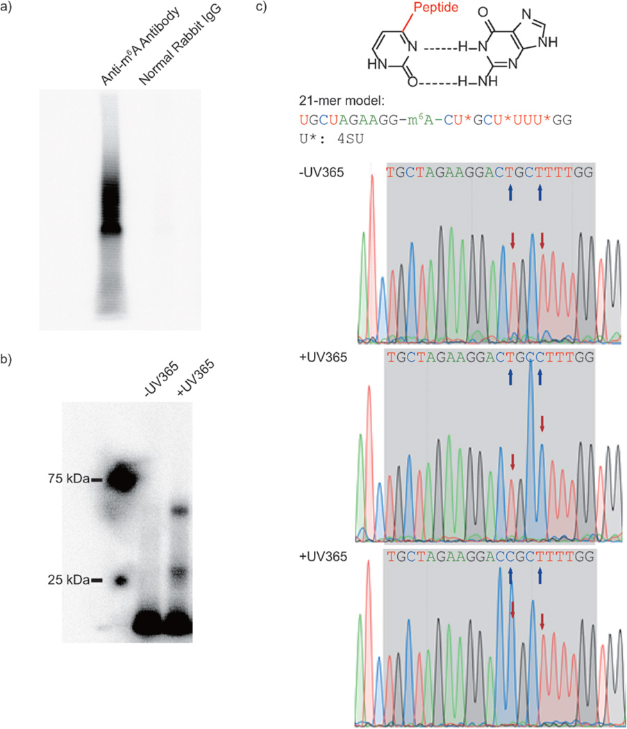
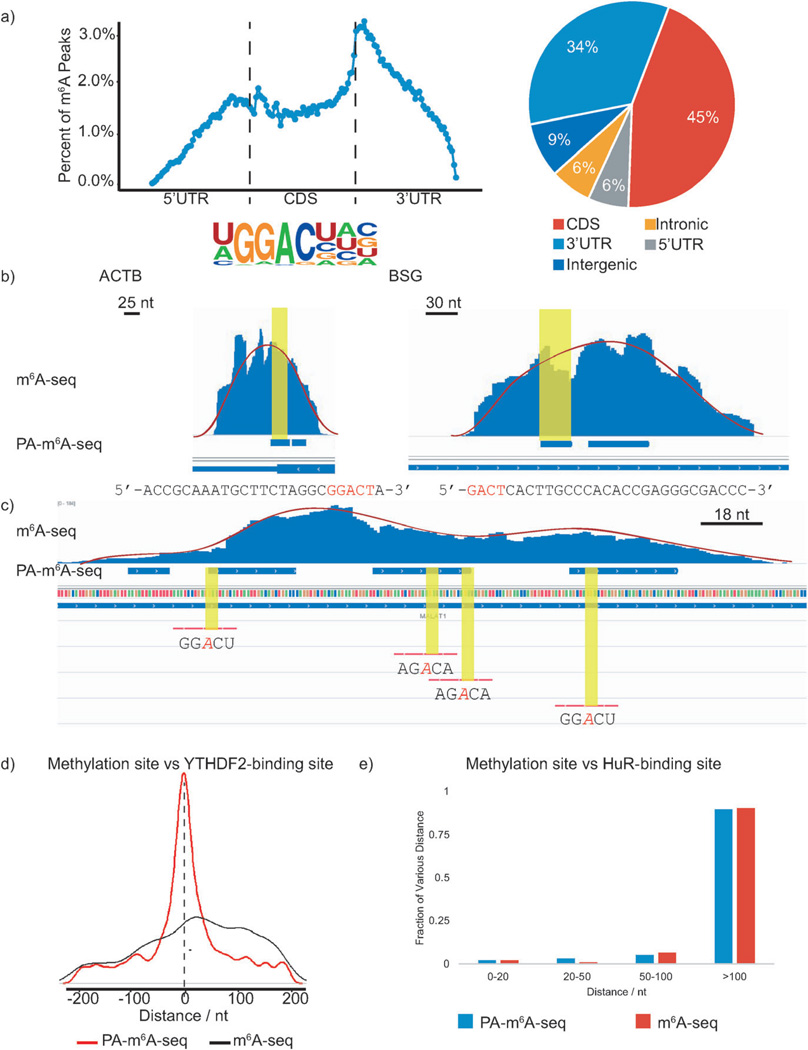
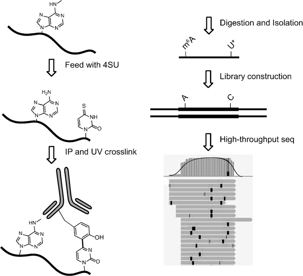
N(6) -methyladenosine (m(6) A) is an abundant internal modification in eukaryotic mRNA and plays regulatory roles in mRNA metabolism. However, methods to precisely locate the m(6) A modification remain limited. We present here a photo-crosslinking-assisted m(6) A sequencing strategy (PA-m(6) A-seq) to more accurately define sites with m(6) A modification. Using this strategy, we obtained a high-resolution map of m(6) A in a human transcriptome. The map resembles the general distribution pattern observed previously, and reveals new m(6) A sites at base resolution. Our results provide insight into the relationship between the methylation regions and the binding sites of RNA-binding proteins.

Keywords: N6-methyladenosine; RNA modification; photo-crosslinking; transcriptome sequencing.


Figures

Figure 1
Figure 1
Model study using PA-m6A-seq. a) Control study to confirm crosslinking is based on recognition of antibody against m6A-containing RNA. b) Control study to prove UV365 is the trigger of covalent crosslinking by using a synthesized 21-mer RNA oligonucleotide. c) T-to-C transition introduced by 4SU and UV irradiation. Proposed mechanism on how the 4SU crosslinked with protein changes the Watson–Crick base pair (top). The sequence of 21-mer model is shown in the middle. Sanger sequencing results of 21-mer model with and without UV365 proved the crosslinked 4SU near m6A was read as C. The blue arrows indicate the 4SU sites on the model, whereas red arrows point out the chromatogram reads of corresponding 4SU with or without crosslinking.
Figure 2
Figure 2
PA-m6A-seq applied to poly(A)-tailed RNA purified from HeLa cells. In following figures, blue bars represent methylation sites identified by PA-m6A-seq, whereas blue ‘peaks’ above those bars are from normal m6A-seq. a) Validation of PA-m6A-seq strategy. Metagene profile and pie-chart of the enrichment of RNA segments are consistent with previous reported distribution of m6A, and the motif search yielded GGACU as the predominant one, which was the same as the result from normal m6A-seq. b) Comparison of predicted methylation sites in β-actin (ACTB) and homo sapien basigin (BSG) from PA-m6A-seq with peaks from normal m6A-seq and single sites by SCARLET. Sequences of predicted sites are shown below, consensus motif containing m6A in red. All input background of normal m6A-seq has been subtracted. Both sites were verified by SCARLET. c) Multiple methylation sites in MALAT1 transcript. The methylation sites confirmed by SCARLET, which were covered by peaks from normal m6A-seq, were also identified by PA-m6A-seq with higher resolution. Yellow regions indicate the RRACH motif. Red lines are the probes used to verify these sites with SCARLET. d) Distance of predicted methylation sites using PA-m6A-seq versus peaks obtained from normal m6A-seq to YTHDF2-binding sites. e) Distance of predicted methylation sites obtained from PA-m6A-seq versus peaks obtained from normal m6A-seq to HuR-binding sites.
Scheme 1
Scheme 1
The strategy of photo-crosslinking-assisted m6A-seq (PA-m6A-seq). Covalently crosslinked 4SU is labeled as U*, which is read as C in RT-PCR. The example of the high-throughput sequencing result is shown on the bottom right. Black bold vertical bars represent T-to-C transition induced by 4SU and covalent crosslinking, compared to reference genome hg19.

References

    1. He C. Nat. Chem. Biol. 2010;6:863–865. - PubMed
    1. Desrosiers RC, Friderici KH, Rottman FM. Proc. Natl. Acad. Sci. USA. 1974;71:3971–3975. - PMC - PubMed
    2. Desrosiers RC, Friderici KH, Rottman FM. Biochemistry. 1975;14:4367–4374. - PubMed
    3. Jia G, Fu Y, He C. Trends Genet. 2013;29:108–115. - PMC - PubMed
    4. Fu Y, Dominissini D, Rechavi G, He C. Nat. Rev. Genet. 2014;15:293–306. - PubMed
    1. Bokar JA, Rath-Shambaugh ME, Ludwiczak R, Narayan P, Rottman F. J. Biol. Chem. 1994;269:17697–17704. - PubMed
    2. Bokar JA, Shambaugh ME, Polayes D, Matera AG, Rottman FM. RNA. 1997;3:1233–1247. - PMC - PubMed
    3. Liu J, et al. Nat. Chem. Biol. 2014;10:93–95. - PMC - PubMed
    4. Ping XL, et al. Cell Res. 2014;24:177–189. - PMC - PubMed
    5. Wang Y, Li Y, Toth JI, Petroski MD, Zhang Z, Zhao JC. Nat. Cell Biol. 2014;16:191–198. - PMC - PubMed
    1. Jia G, et al. Nat. Chem. Biol. 2011;7:885–887. - PMC - PubMed
    2. Zheng G, et al. Mol. Cell. 2013;49:18–29. - PMC - PubMed
    1. Wang X, et al. Nature. 2014;505:117–120. - PMC - PubMed

Publication types