. 2015 Jul;8(4):896-905.
doi: 10.1038/mi.2014.120. Epub 2014 Dec 10.

Thrombospondin-1 restrains neutrophil granule serine protease function and regulates the innate immune response during Klebsiella pneumoniae infection

Affiliations

Thrombospondin-1 restrains neutrophil granule serine protease function and regulates the innate immune response during Klebsiella pneumoniae infection

Y Zhao et al. Mucosal Immunol. 2015 Jul.

Abstract

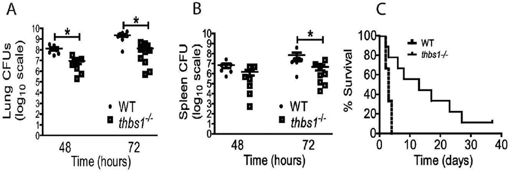
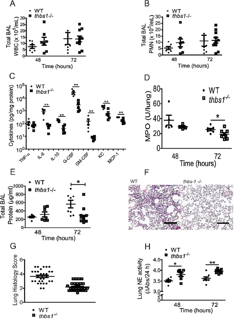
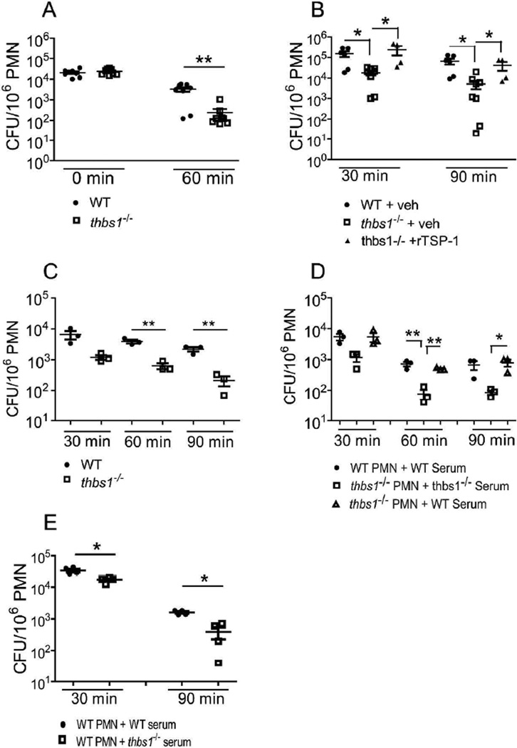
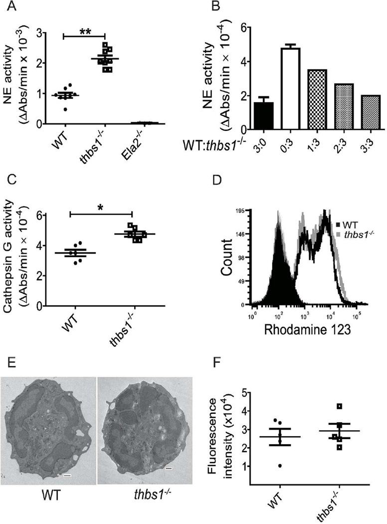
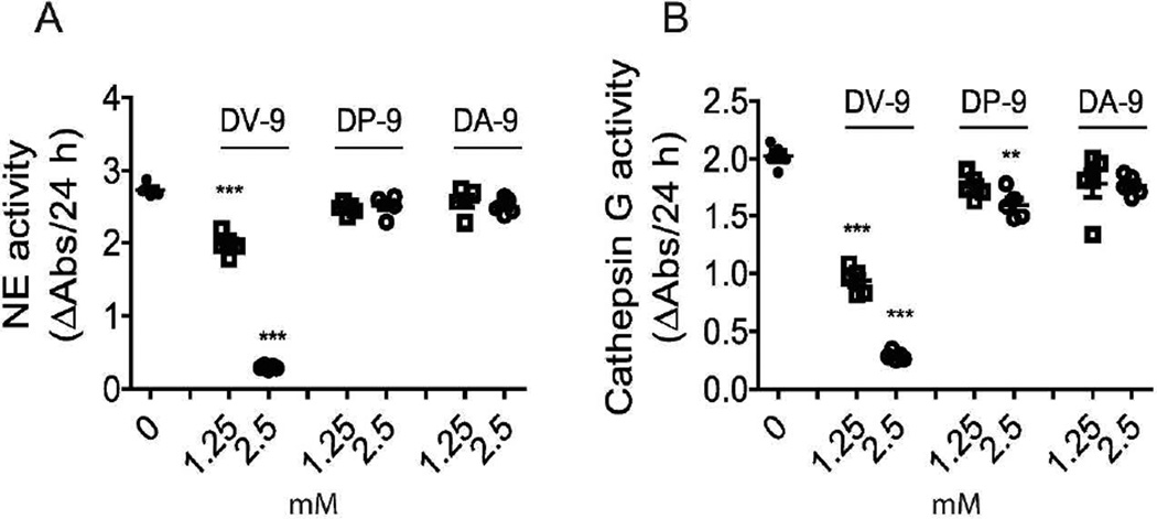
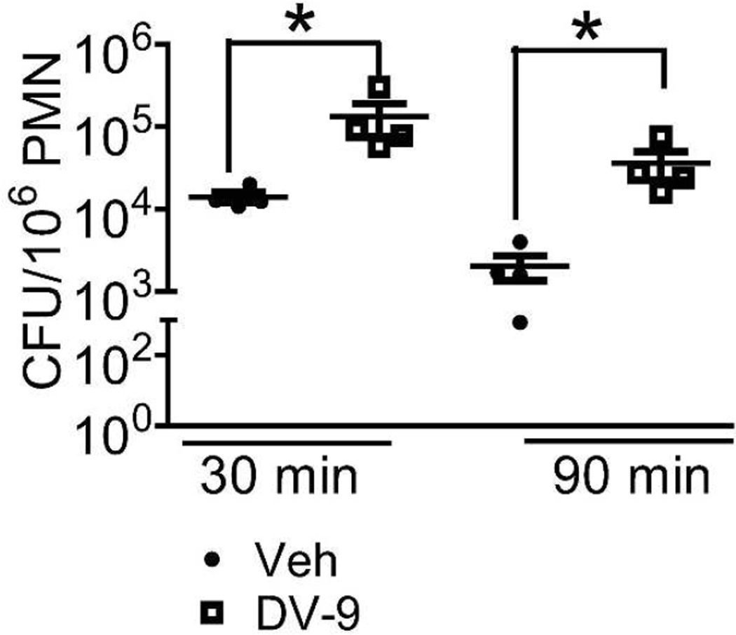
Neutrophil elastase (NE) and cathepsin G (CG) contribute to intracellular microbial killing but, if left unchecked and released extracellularly, promote tissue damage. Conversely, mechanisms that constrain neutrophil serine protease activity protect against tissue damage but may have the untoward effect of disabling the microbial killing arsenal. The host elaborates thrombospondin-1 (TSP-1), a matricellular protein released during inflammation, but its role during neutrophil activation following microbial pathogen challenge remains uncertain. Mice deficient in TSP-1 (thbs1(-/-)) showed enhanced lung bacterial clearance, reduced splenic dissemination, and increased survival compared with wild-type (WT) controls during intrapulmonary Klebsiella pneumoniae infection. More effective pathogen containment was associated with reduced burden of inflammation in thbs1(-/-) mouse lungs compared with WT controls. Lung NE activity was increased in thbs1(-/-) mice following K. pneumoniae challenge, and thbs1(-/-) neutrophils showed enhanced intracellular microbial killing that was abrogated with recombinant TSP-1 administration or WT serum. Thbs1(-/-) neutrophils exhibited enhanced NE and CG enzymatic activity, and a peptide corresponding to amino-acid residues 793-801 within the type-III repeat domain of TSP-1 bridled neutrophil proteolytic function and microbial killing in vitro. Thus, TSP-1 restrains proteolytic action during neutrophilic inflammation elicited by K. pneumoniae, providing a mechanism that may regulate the microbial killing arsenal.


Conflict of interest statement

Conflict of Interest: The authors declare no conflict of interest.

Figures

Figure 1
Figure 1. thbs1−/− mice show enhanced lung bacterial clearance, reduced splenic dissemination, and enhanced survival following intratracheal K. pneumoniae inoculation
Colony forming units (CFU) obtained from (A) lung tissue homogenate and (B) splenic homogenate cultures of WT and thbs1−/− mice 48 and 72 hours after i.t. inoculation with K. pneumoniae, n=7–10 mice/group. Kruskal-Wallis test with Dunn’s multiple comparisons, *p<0.05. (C) A Kaplan-Meier survival curve of WT and thbs1−/− mice following i.t. infection with K. pneumoniae (n=20 mice/genotype, p=0.02). The median survival for WT mice was 72 h (3 days) and 312 h (13 days) for thbs1−/− mice.
Figure 2
Figure 2. Reduced inflammatory response in the lung parenchyma of thbs1−/− mice is associated with enhanced NE activity following bacterial pneumonia with K. pneumoniae
(A) Total BAL leukocyte counts and (B) Total BAL PMN counts obtained from WT and thbs1−/− mice 48 and 72 hours following i.t. inoculation with K. pneumoniae. (C) Lung cytokine measurements in pg/mL from 100 µg protein of tissue homogenates obtained from the left lung of mice 48 hours following i.t. K. pneumoniae. Cytokines were measured individually by ELISA, and data is presented in one graph for simplicity. (D) MPO activity in left lung tissue homogenates measured as U/lung from WT and thbs1−/− mice 48 and 72 hours after i.t. inoculation with K. pneumoniae. (E) Total BAL protein concentrations in µg/mL from WT and thbs1−/− mice 48 and 72 hours after i.t. inoculation with K. pneumoniae. (F) Representative H&E sections of R lung tissue obtained from WT and thbs1−/− mice at 72 hours post-K. pneumoniae instillation. Experimental mice underwent bronchoalveolar lavage of the lung prior to tissue fixation. Scale bar = 100 µm. (G) Lung histology shows higher inflammation score in WT compared to thbs1−/− mouse lung tissue sections from 72 h post-K. pneumoniae instillation, n=60 random high powered images from 3 sections/group scored by 3 blinded reviewers. The averaged score for each image is shown as individual point. (H) NE activity measured in lung tissue homogenates at 48 and 72 hours following i.t. K. pneumoniae. (A–E, H) n=7–10 mice/group, Mann-Whitney U rank sum test, ***p<0.0001, **p <0.01, *p<0.05.
Figure 3
Figure 3. thbs1−/− neutrophils show enhanced intracellular microbial killing that is reversed by administration of recombinant TSP-1 or WT serum
(A) Intracellular killing by neutrophils from WT and thbs1−/− mice exposed to K. pneumoniae intraperitoneally. Neutrophils were immediately harvested and subsequently plated to quantify CFU or incubated further ex vivo for an additional 60 min prior to plating. CFU/106 PMN was obtained for each sample. Data points indicate 8 samples/group using harvested neutrophils from 4 thbs1−/− and 4 WT mice. (B) Mice were pre-treated with recombinant TSP-1 (10 µg/mouse) or PBS vehicle 30 min prior to intraperitoneal instillation of K. pneumoniae. Neutrophils were harvested and subsequently plated to quantify CFU at 30 min or incubated further ex vivo for an additional 60 min prior to plating. CFU/106 PMN was obtained for each sample. Data points indicate 4–8 samples/group using harvested neutrophils from 6 thbs1−/− and 3 WT mice.(C) In vitro microbial killing assay with WT and thbs1−/− neutrophils. Neutrophils were harvested from the peritoneum 6 h following 3% thioglycollate as detailed in the methods. K. pneumoniae was opsonized with 20% homologous serum on ice for 15 minutes. Neutrophils at 106 per well were infected with K. pneumoniae at a multiplicity of infection of 50 bacteria: 1 neutrophil in vitro. Neutrophils were washed with HBSS + gentamicin to remove extracellular or membrane-attached bacteria, lysed with 0.1% Triton-X, and CFU determined at the time points indicated. Data points indicate neutrophils harvested from individual mouse, n=3 mice/group. (D) In vitro neutrophil microbial killing assay with cross transfer of WT serum to thbs1−/− neutrophils. Conditions are the same as indicated in (C) except thbs1−/− neutrophils received bacteria opsonized with either 20% homologous or WT serum. Data points indicate neutrophils harvested from individual mouse, n=3 mice/group. (E) In vitro neutrophil microbial killing assay with WT neutrophils receiving either bacteria in WT or thbs1−/− serum. Data points indicate neutrophils harvested from individual mouse, n=4 mice/group. Mann-Whitney U rank sum test for 2 group comparisons, Kruskal-Wallis test followed by a Dunn’s Multiple Comparisons test for 3 group comparisons, *p<0.05, **p<0.01.
Figure 4
Figure 4. thbs1−/− neutrophils show increased NE and CG activity but normal neutrophil oxidative burst, in vitro phagocytosis, and morphology
Neutrophils were harvested from the peritoneum 6 h following 3% thioglycollate injection. (A) NE activity measured as the rate of enzymatic hydrolysis of synthetic NE substrate N-Methoxysuccinyl-Ala-Ala-Pro-Val p-nitroanilide reflected by the increase in Absorbance (Abs) at 405 nm over time utilizing lysates obtained from WT, thbs1−/−, and Ela2−/− neutrophils. Data points indicate neutrophils obtained from 4 mice/group performed in duplicates. Student’s t-test, **p< 0.001. (B) NE activity measured in WT, thbs1−/− and mixtures of WT: thbs1−/− neutrophil lysates at differing ratios normalized to protein concentrations. The mixture of neutrophil lysates was generated and pooled from WT and thbs1−/− mice at a ratio of 1:3, 2:3, 3:3 where 3 indicates 30 µL lysate containing 10 µg protein. Data obtained from WT (3:0, black bar graph) and thbs1−/− (0:3, white bar graph) neutrophil lysates. (C) Cathepsin G activity measured as the rate of enzymatic hydrolysis of synthetic CG substrate N-succinyl-Ala-Ala-Pro-Phe-P-nitroanilide reflected by the increase in Absorbance (Abs) at 405 nm over time utilizing lysates obtained from WT, thbs1−/− neutrophils. Data points indicate neutrophils obtained from 3 mice/group performed in duplicates. Student’s t-test, *p<0.01. (D) Respiratory burst of WT and thbs1−/− peritoneal neutrophils as indicated by the shift in fluorescence following oxidation of dihydro-rhodamine [0.4 µg/mL] to rhodamine. Gray filled histogram: thbs1−/− neutrophils at baseline; Black filled histogram: WT neutrophils at baseline; Gray unfilled histogram: thbs1−/− neutrophils stimulated with PMA 2 µg/ml; Black unfilled histogram: WT neutrophils stimulated with PMA 2 µg/ml. Data obtained from neutrophils pooled from 5 mice/group. (E) Transmission electron microscopy of WT and thbs1−/− neutrophils pooled from 5 mice/group. Images are representative of 15 neutrophils examined in each group. Scale bars = 500 nm. (F) In vitro phagocytosis of fluorescence labeled E. Coli bioparticles by WT and thbs1−/− neutrophils, as indicated by fluorescence intensity. Data points indicate neutrophils obtained from 5 mice/group performed in quadruplicates.
Figure 5
Figure 5. Peptides generated from the thrombospondin-1 type III repeats domain inhibit neutrophil proteolytic function
(A) NE activity measured in WT neutrophil lysates with 1.25 mM or 2.5 mM peptides DV-9 (DNCQYVYNV), DP-9 (DNCPFHYNP), and DA-9 (DNCPYVPNA). Data points indicate neutrophils obtained from 5 mice/group. (B) Cathepsin G activity measured in WT neutrophil lysates 1.25 mM or 2.5 mM peptides DV-9 (DNCQYVYNV), DP-9 (DNCPFHYNP), and DA-9 (DNCPYVPNA). Data points indicate neutrophils obtained from 5 mice/group. ANOVA with Bonferroni multiple comparisons test, **p <0.01, ***p <0.001.
Figure 6
Figure 6. DV-9 peptide impairs neutrophil microbial killing of K. pneumoniae
Intracellular killing by peritoneal neutrophils from WT mice treated with peptide DV-9 or vehicle DMSO just prior to instillation of K. pneumoniae in vivo. 30 min represents the initial time point sampled, allowing for neutrophils to engulf bacteria in vivo. Neutrophils were harvested and subsequently plated to quantify CFU at 30 min or incubated further ex vivo for an additional 60 min prior to plating. CFU/106 PMN was obtained for each sample. Data points indicate neutrophils harvested from individual mouse, n=4 mice/group. *p<0.05. Mean-fold reduction in CFU/106 PMN ± SEM over time: Vehicle treatment, 8.3 ± 2.0 compared to DV-9 treatment, 3.5 ± 0.6, Mann-Whitney U test, *p < 0.05.

References

    1. Frazier WA. Thrombospondin: a modular adhesive glycoprotein of platelets and nucleated cells. The Journal of cell biology. 1987;105:625–632. - PMC - PubMed
    1. Bornstein P. Diversity of function is inherent in matricellular proteins: an appraisal of thrombospondin 1. The Journal of cell biology. 1995;130:503–506. - PMC - PubMed
    1. Hogg PJ, Owensby DA, Chesterman CN. Thrombospondin 1 is a tight-binding competitive inhibitor of neutrophil cathepsin G. Determination of the kinetic mechanism of inhibition and localization of cathepsin G binding to the thrombospondin 1 type 3 repeats. J Biol Chem. 1993;268:21811–21818. - PubMed
    1. Hogg PJ, Owensby DA, Mosher DF, Misenheimer TM, Chesterman CN. Thrombospondin is a tight-binding competitive inhibitor of neutrophil elastase. J Biol Chem. 1993;268:7139–7146. - PubMed
    1. Lawler J. The structural and functional properties of thrombospondin. Blood. 1986;67:1197–1209. - PubMed

Publication types

MeSH terms

LinkOut - more resources