. 2015 Mar;23(3):533-48.
doi: 10.1038/mt.2014.236. Epub 2014 Dec 10.

Transplantation of glial progenitors that overexpress glutamate transporter GLT1 preserves diaphragm function following cervical SCI

Affiliations

Transplantation of glial progenitors that overexpress glutamate transporter GLT1 preserves diaphragm function following cervical SCI

Ke Li et al. Mol Ther. 2015 Mar.

Abstract

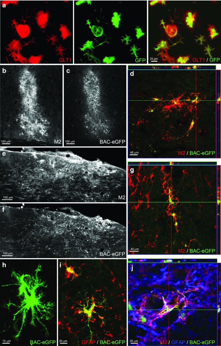
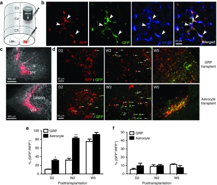
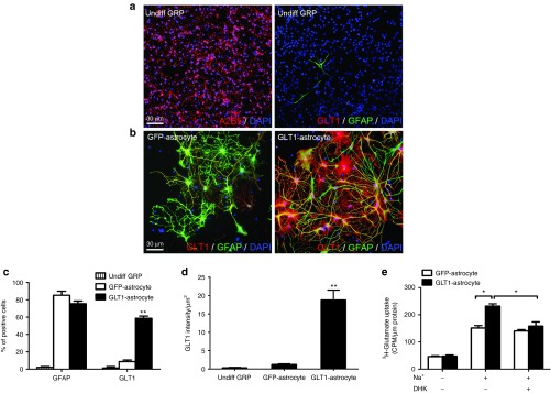
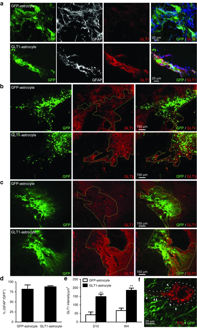
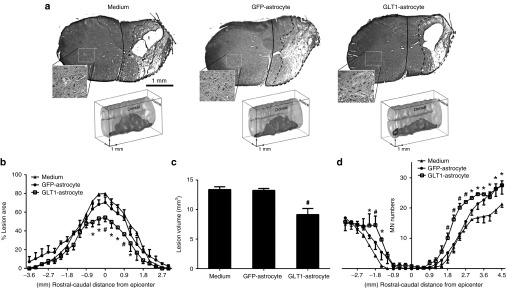
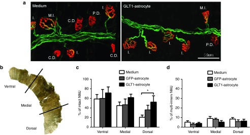
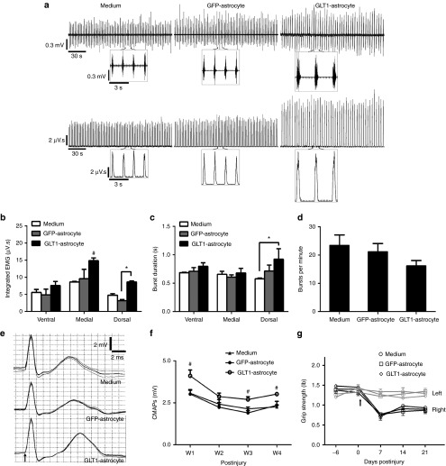
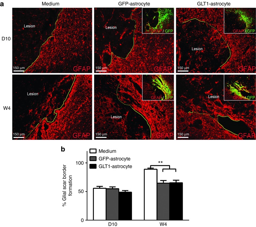
Approximately half of traumatic spinal cord injury (SCI) cases affect cervical regions, resulting in chronic respiratory compromise. The majority of these injuries affect midcervical levels, the location of phrenic motor neurons (PMNs) that innervate the diaphragm. A valuable opportunity exists following SCI for preventing PMN loss that occurs during secondary degeneration. One of the primary causes of secondary injury is excitotoxicity due to dysregulation of extracellular glutamate homeostasis. Astrocytes express glutamate transporter 1 (GLT1), which is responsible for the majority of CNS glutamate clearance. Given our observations of GLT1 dysfunction post-SCI, we evaluated intraspinal transplantation of Glial-Restricted Precursors (GRPs)--a class of lineage-restricted astrocyte progenitors--into ventral horn following cervical hemicontusion as a novel strategy for reconstituting GLT1 function, preventing excitotoxicity and protecting PMNs in the acutely injured spinal cord. We find that unmodified transplants express low levels of GLT1 in the injured spinal cord. To enhance their therapeutic properties, we engineered GRPs with AAV8 to overexpress GLT1 only in astrocytes using the GFA2 promoter, resulting in significantly increased GLT1 protein expression and functional glutamate uptake following astrocyte differentiation in vitro and after transplantation into C4 hemicontusion. Compared to medium-only control and unmodified GRPs, GLT1-overexpressing transplants reduced lesion size, diaphragm denervation and diaphragm dysfunction. Our findings demonstrate transplantation-based replacement of astrocyte GLT1 is a promising approach for SCI.


Figures

Figure 1
Figure 1
BAC-GLT1-eGFP reporter cells can be used to spatiotemporally track GLT1 expression by transplants in the spinal cord. (a) BAC-GLT1-eGFP GRP-derived astrocytes expressed both GLT1 protein and BAC-GLT1-eGFP reporter when cultured with neuronal-conditioned medium. At 5 weeks after transplantation of undifferentiated BAC-GLT1-eGFP GRPs into the uninjured spinal cord, we detected surviving cells in host rat spinal cord with the mouse-specific marker, M2 (b: ventral horn gray matter; e: surrounding white matter). M2+ transplant-derived cells expressed BAC-GLT1-eGFP reporter in (c) gray matter ((d) confocal image) and (f) white matter ((g) confocal image). BAC-GLT1-eGFP+ cells (h) elaborated astrocyte-like morphology and (i,j) expressed GFAP.
Figure 2
Figure 2
Transplanted glial progenitor-derived astrocytes do not express GLT1 in the injured cervical spinal cord. (a) Undifferentiated BAC-GLT1-eGFP GRPs and GRP-derived astrocytes were transplanted into the injured spinal cord, (c) where they survived in both the C4 lesion site (lower panel) and surrounding C5 tissue where tissue damage was not observed (upper panel). (b) RFP reporter labeling allowed us to track the expression of the BAC-GLT1-eGFP reporter in transplant-derived GFAP+ astrocytes. (d) At day 2 (D2), week 2 (W2), and W5 after transplantation, transplant survival (RFP+) and GLT1 expression (BAC-GLT1-eGFP+) were detected in both caudal C5 and intralesion (data not shown) locations. Percentage of GLT1 expressing transplant-derived cells (% of RFP+ cells that are BAC-GLT1-eGFP+) was quantified for transplants of both BAC-GLT1-eGFP undifferentiated GRPs and GRP-derived astrocytes ((e) surrounding intact tissue; (f) lesion site). Results were expressed as means ± SEM. *P < 0.05, **P < 0.01, n = 6 per group.
Figure 3
Figure 3
AAV8-GLT1 transduction in vitro increases GLT1 protein expression and GLT1-mediated functional glutamate uptake. (a) Undifferentiated rat GRPs expressed the glial progenitor marker A2B5 (left panel), but not the astrocyte marker GFAP (right panel) in vitro. Cultured undifferentiated rat GRPs (right panel) expressed little-to-no GLT1 protein. (b) During GRP differentiation into GFAP+ astrocytes, cells transduced with AAV8-Gfa2-GLT1 significantly increased GLT1 protein overexpression (right panel), while cells transduced with AAV8-Gfa2 control vector did not express GLT1 (left panel). (c) Percentage of DAPI+ cells in vitro expressing GFAP and GLT1 protein. (d) Quantification of GLT1 protein expression level in vitro. (e) In vitro 3H-glutamate uptake assay was performed to detect functional Na+-dependent GLT1-mediated glutamate uptake in GRP-derived astrocytes using the GLT1 inhibitor DHK. Results were expressed as means ± SEM. *P < 0.05, **P < 0.01, n = 6 per group for GLT1 and GFAP expression analyses, n = 3 per group for 3H-glutamate uptake assay.
Figure 4
Figure 4
GLT1 overexpression significantly enhances GLT1 protein expression by transplanted astrocytes in the injured cervical spinal cord. We transplanted (i) rat GRP-derived astrocytes transduced prior to transplantation with AAV8-GFA2 control vector (GFP-astrocyte) or (ii) rat GRP-derived astrocytes transduced prior to transplantation with AAV8-GFA2-GLT1 vector (GLT1-astrocyte) into the injured spinal cord immediately following C4 hemicontusion SCI. Both cell groups were also transduced with lentiviral vector for labeling of all cells (regardless of their phenotype) with a GFP reporter. (a) At day 10 after injection, the vast majority of transplant-derived GFP+ cells in both groups expressed GFAP (quantification in d). At both (b) D10 and (c) W5 after transplantation into the injured spinal cord, the control transplants expressed very low levels of GLT1 protein, while the overexpressing transplants expressed greatly increased levels of GLT1 protein (area of transplant-derived GFP+ cells marked by dotted line). GLT1 protein signal intensity was quantified within the regions of surviving GFP+ transplant-derived cells at both (e) D10 and W4 after transplantation/injury. (f) Transplant-derived cells spatially localized around PMNs, which were retrogradely labeled from the ipsilateral hemidiaphragm with the tracer, CTβ. Results were expressed as means ± SEM. *P < 0.05, **P < 0.01, n = 5–7 per group.
Figure 5
Figure 5
GLT1 overexpressing astrocyte transplants reduce lesion size following cervical contusion SCI. GLT1-overexpressing astrocytes (GLT1-astrocyte), control astrocytes (GFP-astrocytes) or medium-only control were transplanted immediately following C4 hemicontusion SCI. (a) Representative images of Cresyl-violet staining and three dimensional reconstruction illustrate the lesion epicenter and entire lesion extent, respectively, in the groups at 10 days after injury. (b) Lesion size and (c) total lesion volume were reduced in the GLT1-astrocyte group compared to the two control groups. (d) Large motor neuron populations in the ventral horn ipsilateral to the contusion site were identified (inset in a shows contralateral ventral horn as an example of motor neuron labeling), quantified, and plotted at multiple distances from the epicenter. Results were expressed as means ± SEM. *P < 0.05, GLT1-astrocyte group versus medium control group. #P < 0.05, GLT1-astrocyte group versus both control groups. n = 6 rats per group.
Figure 6
Figure 6
GLT1 overexpressing astrocyte transplants preserve diaphragm innervation by phrenic motor neurons following cervical contusion SCI. Diaphragm NMJs ipsilateral to the hemicontusion were assessed via labeling with Alexa-647 conjugated α-bungarotoxin (red), SMI-312R (green) and SV2-s (green). Areas of muscle denervation were observed in all three groups. (a) Representative confocal images are shown for the medium control and GLT1-astrocyte groups. Reduced denervation can be observed in the GLT1-astrocyte muscle compared to the medium control group. Individual NMJs were characterized as: intact (I.); completely denervated (C.D.); partially denervated (P.D.); multiply innervated (M.I.). (b) For analysis, the hemidiaphragm was divided into three anatomical subregions (ventral, medial, and dorsal). (c) GLT1 overexpressing transplants significantly increased the percentage of intact junctions in the dorsal diaphragm compared to medium-only control by two- to threefold. (d) No differences amongst groups were noted in the percentage of multiply-innervated NMJs at any subregion of the hemidiaphragm. Results were expressed as means ± SEM. *P < 0.05, GLT1-astrocyte group versus medium control group. n = 5–7 rats per group.
Figure 7
Figure 7
GLT1 overexpressing astrocyte transplants preserve diaphragm function following cervical contusion SCI. (a) At D10 after injury/transplantation, spontaneous diaphragm electromyography (EMG) recordings were performed and are shown as raw voltage output (upper panels) and integrated signals (lower panels), and the individual breaths are shown in a (inset). (b) Integrated EMG amplitude, (c) inspiratory burst duration, and (d) inspiratory bursts per minute in the ipsilateral hemidiaphragm were averaged over a 2 minute period. (e) Diaphragm compound muscle action potentials (CMAPs) obtained following supramaximal stimulation (arrow in e denotes time of stimulation) were recorded in the ipsilateral hemidiaphragm. (f) Compared to the two control groups, peak CMAP amplitudes were significantly larger in the GLT1-astrocyte group at W1, W3, and W4 after injury/transplantation. No differences were observed amongst groups in forelimb motor performance using grip strength testing (g). Results were expressed as means ± SEM. *P < 0.05, GLT1-astrocyte group versus ether medium control group or GFP-astrocyte control group only. #P < 0.05, GLT1-astrocyte group versus both control groups. n = 6 rats per group for CMAP recordings; n = 3 rats per group for EMG recordings.
Figure 8
Figure 8
Glial scar reduction is not responsible for the therapeutic effects of GLT1 overexpressing transplants. (a) At 10 days (D10) and 4 weeks (W4) after injury, immunostaining with anti-GFAP antibody was performed. In cell transplantation groups, the border containing GFP+ transplant-derived cells was analyzed (a, inset). Astroglial scar border is highlighted as yellow solid line. At W4 after injury (but not at D10 after injury), we observed (b) reduced glial scar formation with both control and GLT1 overexpressing transplant groups compared to medium-only control. However, we found no differences in the degree of reduced scar formation between the two transplant groups at this W4 time point (b). Results were expressed as means ± SEM. **P < 0.01, n = 6 rats per group for analysis of scar formation.

Similar articles

Cited by

References

    1. Lane MA, Fuller DD, White TE, Reier PJ. Respiratory neuroplasticity and cervical spinal cord injury: translational perspectives. Trends Neurosci. 2008;31:538–547. - PMC - PubMed
    1. Lane MA, Lee KZ, Fuller DD, Reier PJ. Spinal circuitry and respiratory recovery following spinal cord injury. Respir Physiol Neurobiol. 2009;169:123–132. - PMC - PubMed
    1. Zimmer MB, Nantwi K, Goshgarian HG. Effect of spinal cord injury on the respiratory system: basic research and current clinical treatment options. J Spinal Cord Med. 2007;30:319–330. - PMC - PubMed
    1. Shanmuganathan K, Gullapalli RP, Zhuo J, Mirvis SE. Diffusion tensor MR imaging in cervical spine trauma. AJNR Am J Neuroradiol. 2008;29:655–659. - PMC - PubMed
    1. Strakowski JA, Pease WS, Johnson EW. Phrenic nerve stimulation in the evaluation of ventilator-dependent individuals with C4- and C5-level spinal cord injury. Am J Phys Med Rehabil. 2007;86:153–157. - PubMed

Publication types

MeSH terms

Substances