. 2015 Aug 1;32(15):1158-67.
doi: 10.1089/neu.2014.3703. Epub 2015 Mar 9.

Fibronectin Matrix Assembly after Spinal Cord Injury

Affiliations

Fibronectin Matrix Assembly after Spinal Cord Injury

Yunjiao Zhu et al. J Neurotrauma. .

Abstract

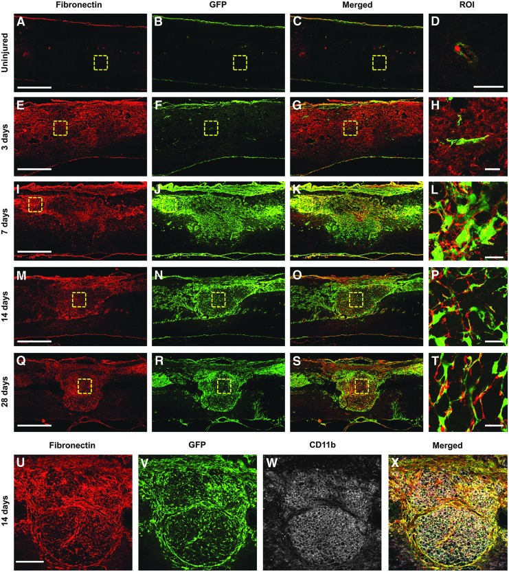
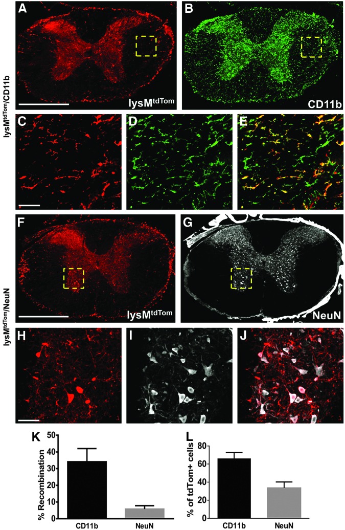
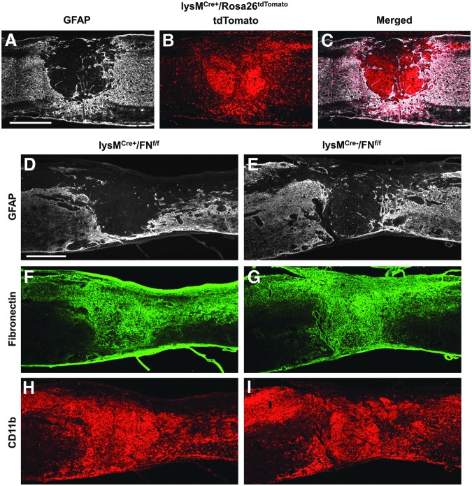
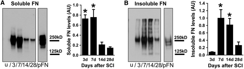
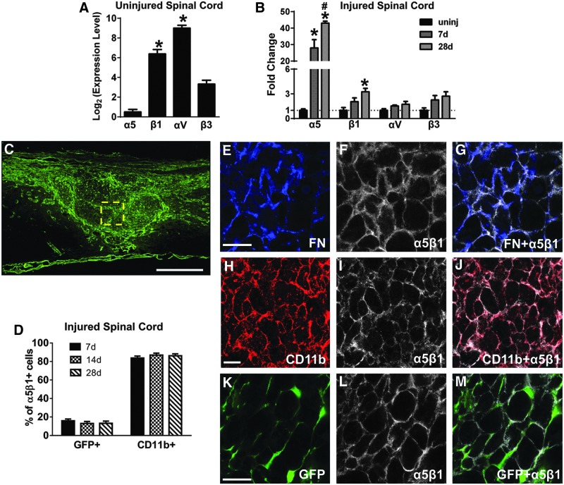
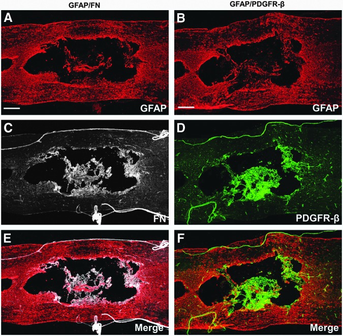
After spinal cord injury (SCI), a fibrotic scar forms at the injury site that is best characterized by the accumulation of perivascular fibroblasts and deposition of the extracellular matrix protein fibronectin. While fibronectin is a growth-permissive substrate for axons, the fibrotic scar is inhibitory to axon regeneration. The mechanism behind how fibronectin contributes to the inhibitory environment and how the fibronectin matrix is assembled in the fibrotic scar is unknown. By deleting fibronectin in myeloid cells, we demonstrate that fibroblasts are most likely the major source of fibronectin in the fibrotic scar. In addition, we demonstrate that fibronectin is initially present in a soluble form and is assembled into a matrix at 7 d post-SCI. Assembly of the fibronectin matrix may be mediated by the canonical fibronectin receptor, integrin α5β1, which is primarily expressed by activated macrophages/microglia in the fibrotic scar. Despite the pronounced cavitation after rat SCI, fibrotic scar also is observed in a rat SCI model, which is considered to be more similar to human pathology. Taken together, our study provides insight into the mechanism of fibrotic scar formation after spinal cord injury.

Keywords: fibronectin; fibrotic scar; macrophages; microglia; spinal cord injury.


Figures

<b>FIG. 1.</b>
FIG. 1.
Fibrotic scar formation after contusive spinal cord injury. In the uninjured spinal cord (A-D), fibronectin (red) expression and fibroblasts (green) are found only around blood vessels (D) and in the dura. At 3 d after injury (E-H), there is diffuse fibronectin expression throughout the injury site (E) while fibroblasts are not present in significant numbers (F). At 7 d (I-L), when fibroblasts densely populate the injury site (J), the area of fibronectin expression closely matches that of fibroblasts (K) and appears more punctate (L). At 14 d (M-P, U-X), when the fibrotic scar has matured, it is characterized by a dense population of mostly fibroblasts and CD11b+ leukocytes (W) and a fibronectin expression network that closely follows the distribution of fibroblasts throughout the injury site. This pattern continues at 28 d (Q-T). n=5 per group. Scale bar in A, E, I, M, Q=500 μm; in D, H, L, P, T=50 μm; in U=200 μm.
<b>FIG. 2.</b>
FIG. 2.
Recombination rate in uninjured spinal cord of lysM-Cre mice. lysM-Cre/Rosa26-tdTomato mice demonstrate that approximately 34% of all microglia (CD11b) have undergone recombination (tdTomato) (A-E, K). A small percent of neurons (NeuN, 6%) also were labeled with tdTomato (F-J, K). Of all the cells that underwent recombination (tdTomato), approximately 66% were microglia and 33% were neurons (L). n=3. Scale bar in A, F=500 μm; in C, H=50 μm. Error bars are standard error of the mean.
<b>FIG. 3.</b>
FIG. 3.
Activated macrophages/microglia are not a major source of fibronectin after spinal cord injury. Breeding lysM-Cre mice to Rosa26-tdTomato reporter mice demonstrates the distribution of myeloid cells at the injury site that have undergone recombination (A-C, 14 d after injury, n=3). Genetic deletion of fibronectin (FN) in myeloid cells (lysM-Cre bred to floxed FN mouse; D, F, H; n=5) does not show visible differences in CD11b+ leukocyte infiltration and FN expression at the injury site, compared with Cre controls (E, G, I; 14 d after injury; n=5). Scale bar in A, D=500 μm.
<b>FIG. 4.</b>
FIG. 4.
Fibronectin matrix assembly after spinal cord injury (SCI). Soluble fibronectin is significantly increased at 3 and 7 d after SCI and then decreases over time, albeit still at higher levels than uninjured spinal cord (A; n=5 per group). Insoluble fibronectin (indicated by multiple bands above the 250 kD monomer band) is highest at 7 and 14 d after injury (B, n=5 per group). Purified plasma fibronectin (pFN) was used as a positive control and loaded in equal amounts in both blots. Note the absence of>250 kD bands in the soluble fraction (A) even after a longer film exposure time (as indicated by darker pFN band in A compared with B). The entire blot is shown in Supplementary Figure 1. *p<0.05 compared with 14 d, 28 d in A; 3 d, 28 d in B using one way analysis of variance with Tukey's post-test. Error bars are standard error of the mean.
<b>FIG. 5.</b>
FIG. 5.
Integrin receptor α5β1 is expressed by macrophages/microglia and fibroblasts after spinal cord injury (SCI). In the uninjured spinal cord, αV and β1 integrin subunit messenger RNA (mRNA) levels are higher than α5 and β3 (A, n=3 per group). After SCI, both α5 and β1 mRNA are significantly increased, while αV and β3 mRNA levels are not significantly altered (B, n=3 per group). Within the fibrotic scar at 14 d after injury (C), fibronectin is present mostly in regions of α5β1 expression (E-G), which is present on CD11b+ macrophages/microglia (H-J, D; n=3 per group) and fibroblasts (K-M, D; n=3 per group). C, E, H, K are from different animals. Scale bar in C=500 μm; in E, H, K=50 μm. A: *p<0.05 compared with α5, β3 using one way analysis of variance (ANOVA) with Tukey's post-test. B: *p<0.05 compared with uninjured (uninj), #p<0.05 compared with 7 d, two-way ANOVA with Bonferonni post-test. Error bars are standard error of the mean.
<b>FIG. 6.</b>
FIG. 6.
Presence of fibrotic scar in rat injury site. Images (A-F) are taken from horizontal sections of rat spinal cord 56 d after a moderate contusion (n=4). GFAP represents the astroglial scar and is expressed throughout the entire injury site (A, B) after spinal cord injury. Expression of fibronectin (FN) and PDGFR-β (a marker for fibroblasts) is found on blood vessels, meninges, and lining the cavity (C-F). Scale bar in A, B=500 μm.

References

    1. Cregg J.M., Depaul M.A., Filous A.R., Lang B.T., Tran A. and Silver J. (2014). Functional regeneration beyond the glial scar. Exp. Neurol. 253C, 197–207 - PMC - PubMed
    1. Burda J.E. and Sofroniew M.V. (2014). Reactive gliosis and the multicellular response to CNS damage and disease. Neuron 81, 229–248 - PMC - PubMed
    1. Shearer M.C. and Fawcett J.W. (2001). The astrocyte/meningeal cell interface–a barrier to successful nerve regeneration? Cell Tissue Res. 305, 267–273 - PubMed
    1. Kimura-Kuroda J., Teng X., Komuta Y., Yoshioka N., Sango K., Kawamura K., Raisman G., and Kawano H. (2010). An in vitro model of the inhibition of axon growth in the lesion scar formed after central nervous system injury. Mol. Cell. Neurosci. 43, 177–187 - PubMed
    1. Wanner I.B., Deik A., Torres M., Rosendahl A., Neary J.T., Lemmon V.P., and Bixby J.L. (2008). A new in vitro model of the glial scar inhibits axon growth. Glia 56, 1691–1709 - PMC - PubMed

Publication types

LinkOut - more resources