Review
. 2015 Feb;129(2):207-20.
doi: 10.1007/s00401-014-1371-2. Epub 2014 Dec 10.

Tau aggregation and its interplay with amyloid-β

Affiliations
Review

Tau aggregation and its interplay with amyloid-β

Rebecca M Nisbet et al. Acta Neuropathol. 2015 Feb.

Abstract

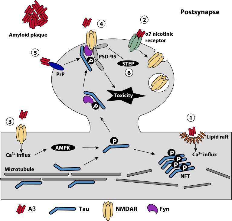
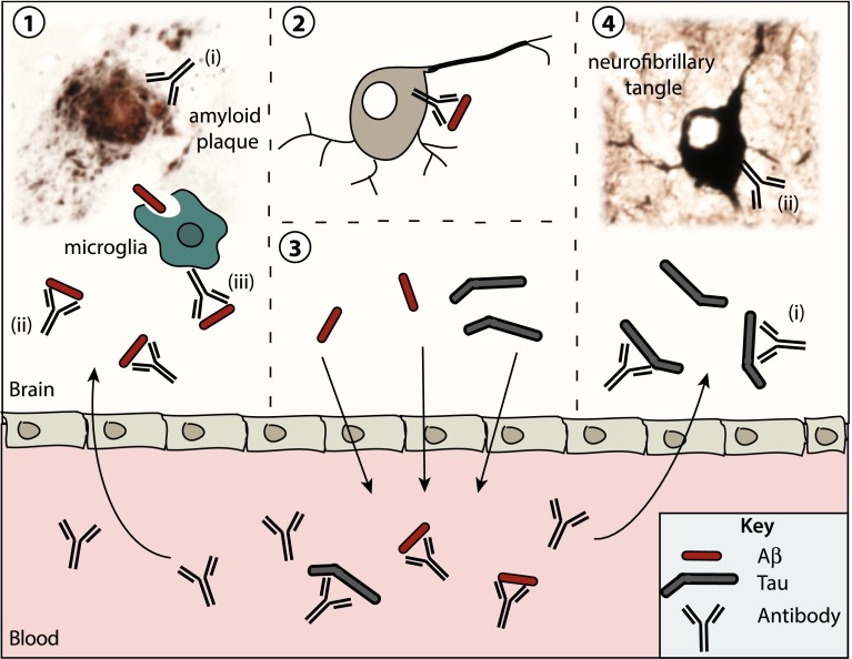
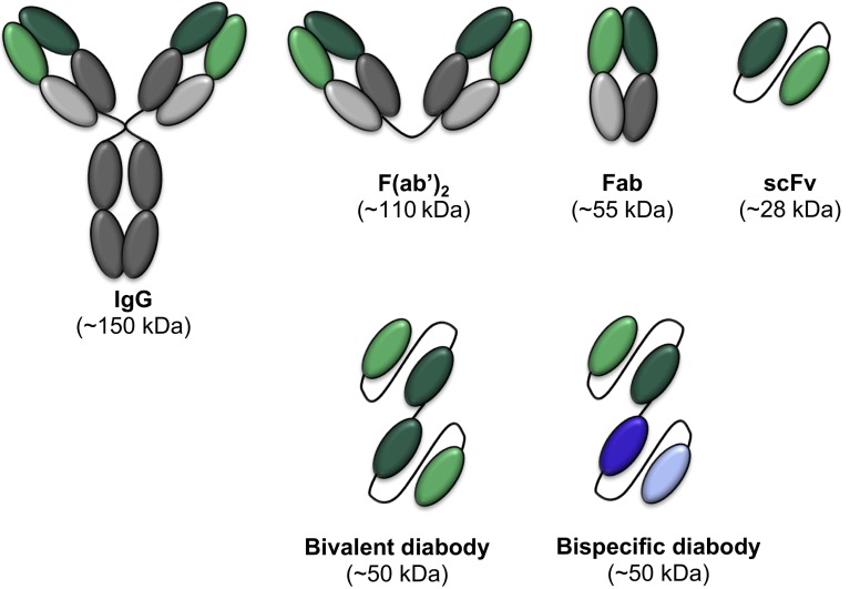
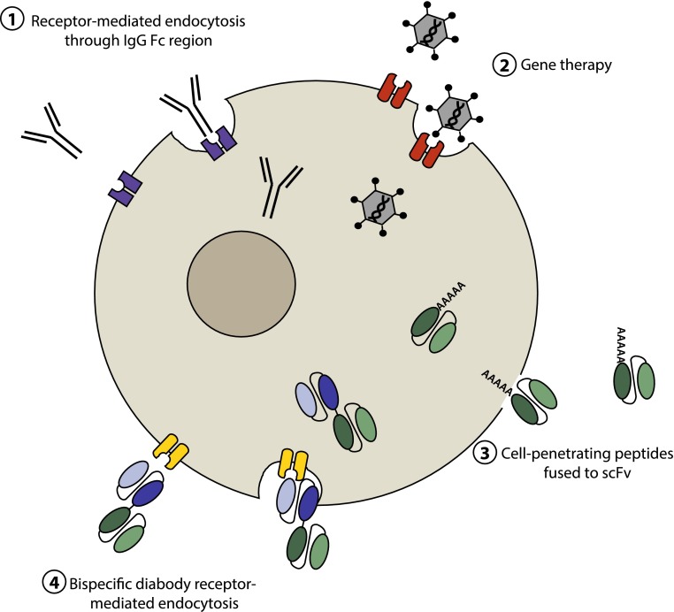
Neurofibrillary tangles and amyloid plaques constitute the hallmark brain lesions of Alzheimer's disease (AD) patients. Tangles are composed of fibrillar aggregates of the microtubule-associated protein tau, and plaques comprise fibrillar forms of a proteolytic cleavage product, amyloid-β (Aβ). Although plaques and tangles are the end-stage lesions in AD, small oligomers of Aβ and tau are now receiving increased attention as they are shown to correlate best with neurotoxicity. One key question of debate, however, is which of these pathologies appears first and hence is upstream in the pathocascade. Studies suggest that there is an intense crosstalk between the two molecules and, based on work in animal models, there is increasing evidence that Aβ, at least in part, exerts its toxicity via tau, with the Src kinase Fyn playing a crucial role in this process. In other experimental paradigms, Aβ and tau have been found to exert both separate and synergistic modes of toxicity. The challenge, however, is to integrate these different scenarios into a coherent picture. Furthermore, the ability of therapeutic interventions targeting just one of these molecules, to successfully neutralize the toxicity of the other, needs to be ascertained to improve current therapeutic strategies, such as immunotherapy, for the treatment of AD. Although this article is not intended to provide a comprehensive review of the currently pursued therapeutic strategies, we will discuss what has been achieved by immunotherapy and, in particular, how the inherent limitations of this approach can possibly be overcome by novel strategies that involve single-chain antibodies.


Figures

Fig. 1
Fig. 1
Proposed mechanisms underlying the toxic interplay between Aβ and tau at the synapse. Aβ oligomers have been demonstrated to exert their toxicity at the synapse through a number of mechanisms: (1) Binding of Aβ to the plasma membrane forms pores in the membrane, which may be facilitated by lipid rafts, resulting in calcium influx into the cell and the downstream activation of kinases implicated in tau phosphorylation. (2) Aβ can mediate the internalization of synaptic NMDARs indirectly through the binding of α7 nicotinic receptors. This results in a reduction of NMDARs at the synapse and causes synaptic spine shrinkage and retraction. (3) Aβ can mediate the activation of extrasynaptic NMDARs, which also induces a calcium influx into the neuron and in turn activates kinases such as AMPK. Activated kinases can phosphorylate dendritic tau which not only causes tau to detach from microtubules and aggregate into NFTs, but also enhances its binding to Fyn and results in the migration of tau and Fyn into the dendritic spine. (4) Within the dendritic spine, Fyn phosphorylates NMDARs and thereby mediates their interaction with PSD-95—an interaction required for Aβ toxicity. (5) Binding of Aβ to PrPc also can activate Fyn to phosphorylate the NMDARs. (6) As the disease progresses, Aβ can activate the Fyn phosphatase STEP, which inactivates Fyn, resulting in the loss of synapses and dendritic spine collapse
Fig. 2
Fig. 2
Proposed mechanisms of action of Alzheimer’s disease immunotherapies. AD is characterized pathologically by the aggregation of Aβ and tau and their deposition as extracellular amyloid plaques and intracellular NFTs, respectively. On their pathway to forming plaques and NFTs, Aβ and tau exist as small oligomers that aggregate to fibrils and are the best correlate of neurotoxicity. Preventing Aβ and tau aggregation and promoting their clearance from the brain are the main goals of current immunotherapeutics for the treatment of AD and are depicted in this model. (1) Anti-Aβ antibodies that cross the blood–brain barrier (i) bind to aggregated Aβ and aid in its disaggregation, (ii) bind to soluble Aβ and prevent further Aβ aggregation and/or binding to neurons, and iii activate resident microglia to phagocytose soluble Aβ and help dissolve Aβ plaques. (2) Endocytosed antibody–target complexes are cleared via the neuronal lysosome. (3) ‘Peripheral sink hypothesis’ whereby the antibody-mediated depletion of peripheral Aβ and tau causes an efflux of these proteins from the brain into the blood. (4) Anti-tau antibodies in the brain (i) bind to secreted tau and prevent its ability to ‘seed’ neighboring neurons or (ii) are endocytosed by neurons and bind to intracellular tau. Included in this model is a Campbell–Switzer stained amyloid plaque from a mutant APP transgenic mouse and a Gallyas silver-stained NFT within a neuron of a mutant tau transgenic mouse
Fig. 3
Fig. 3
Engineered antibody formats. In addition to full-length ‘classic’ immunoglobulin (IgG) molecules, which are comprised of an antigen-binding fragment (Fab) and a crystallisable fragment (Fc), a variety of antibody variants are currently being exploited for therapeutic intervention. These include: F(ab′)2, which is generated by papain digestion of the whole IgG and lacks the Fc region; Fab, which comprises only one antigen-binding region; scFv, which is composed of the variable heavy and variable light chains of an IgG, joined by a flexible linker; bivalent diabodies, which are composed of two scFvs which have the same target, joined together by a linker; and bispecific diabodies, which are composed of two scFvs with different targets, joined together by a linker
Fig. 4
Fig. 4
Mechanisms of antibody-mediated cell internalization. The intracellular localization of proteins implicated in disease, such as tau, makes them difficult to target using an immunotherapeutic approach as only very few full-length antibodies are able to penetrate the cell membrane via receptor-mediated endocytosis (1). Antibody engineering, however, has generated a number of different avenues one can now pursue to effectively localize functional antibody fragments intracellularly, such as viral-mediated delivery of an scFv gene (2); cell membrane penetration via a cell-penetrating peptide fused or conjugated to an scFv (3); and receptor-mediated endocytosis of a bispecific scFv with one arm specific for a cell surface receptor (4)

Similar articles

Cited by

References

    1. Adolfsson O, Pihlgren M, Toni N, Varisco Y, Buccarello AL, Antoniello K, Lohmann S, Piorkowska K, Gafner V, Atwal JK, Maloney J, Chen M, Gogineni A, Weimer RM, Mortensen DL, Friesenhahn M, Ho C, Paul R, Pfeifer A, Muhs A, Watts RJ. An effector-reduced anti-beta-amyloid (Abeta) antibody with unique abeta binding properties promotes neuroprotection and glial engulfment of Abeta. J Neurosci Off J Soc Neurosci. 2012;32(28):9677–9689. - PMC - PubMed
    1. Ahmad ZA, Yeap SK, Ali AM, Ho WY, Alitheen NB, Hamid M. scFv antibody: principles and clinical application. Clin Dev Immunol. 2012;2012:980250. - PMC - PubMed
    1. Asuni AA, Boutajangout A, Quartermain D, Sigurdsson EM. Immunotherapy targeting pathological tau conformers in a tangle mouse model reduces brain pathology with associated functional improvements. J Neurosci Off J Soc Neurosci. 2007;27(34):9115–9129. - PMC - PubMed
    1. Baleriola J, Walker CA, Jean YY, Crary JF, Troy CM, Nagy PL, Hengst U. Axonally synthesized ATF4 transmits a neurodegenerative signal across brain regions. Cell. 2014;158(5):1159–1172. - PMC - PubMed
    1. Ballard C, Gauthier S, Corbett A, Brayne C, Aarsland D, Jones E. Alzheimer’s disease. Lancet. 2011;377(9770):1019–1031. - PubMed

Publication types