. 2015 Jan 23;290(4):2225-34.
doi: 10.1074/jbc.M114.625483. Epub 2014 Dec 9.

First evidence for substrate channeling between proline catabolic enzymes: a validation of domain fusion analysis for predicting protein-protein interactions

Affiliations

First evidence for substrate channeling between proline catabolic enzymes: a validation of domain fusion analysis for predicting protein-protein interactions

Nikhilesh Sanyal et al. J Biol Chem. .

Abstract

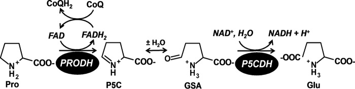
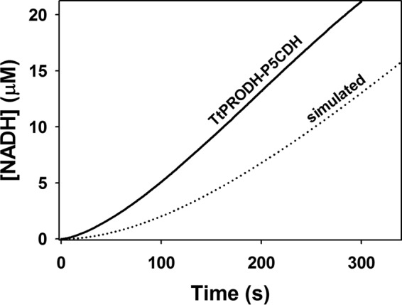
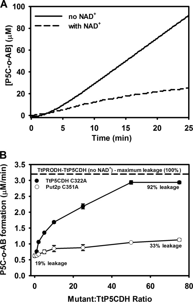
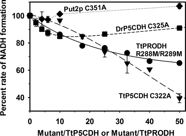
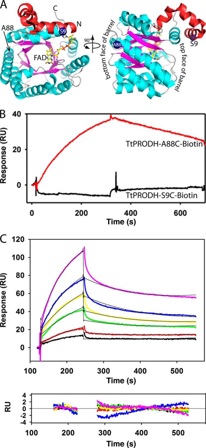
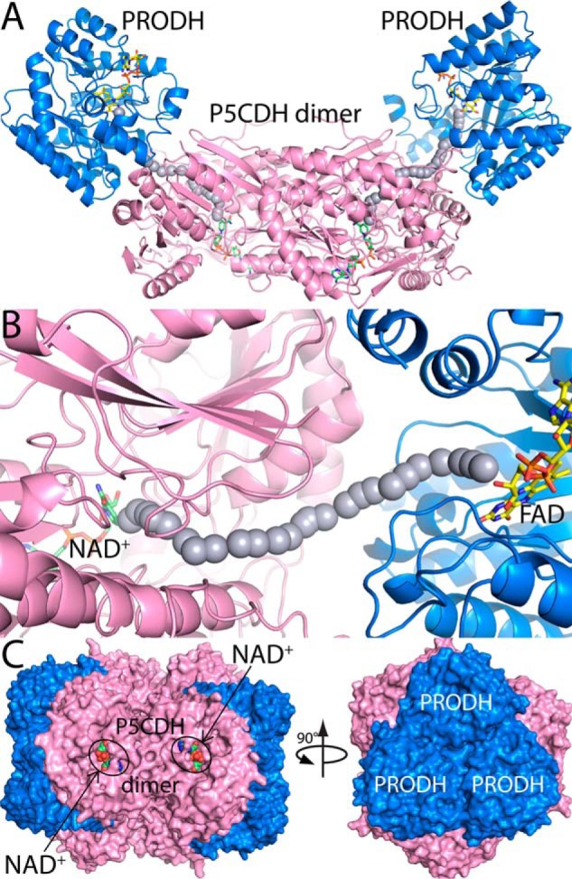
Proline dehydrogenase (PRODH) and Δ(1)-pyrroline-5-carboxylate (P5C) dehydrogenase (P5CDH) catalyze the four-electron oxidation of proline to glutamate via the intermediates P5C and l-glutamate-γ-semialdehyde (GSA). In Gram-negative bacteria, PRODH and P5CDH are fused together in the bifunctional enzyme proline utilization A (PutA) whereas in other organisms PRODH and P5CDH are expressed as separate monofunctional enzymes. Substrate channeling has previously been shown for bifunctional PutAs, but whether the monofunctional enzymes utilize an analogous channeling mechanism has not been examined. Here, we report the first evidence of substrate channeling in a PRODH-P5CDH two-enzyme pair. Kinetic data for the coupled reaction of PRODH and P5CDH from Thermus thermophilus are consistent with a substrate channeling mechanism, as the approach to steady-state formation of NADH does not fit a non-channeling two-enzyme model. Furthermore, inactive P5CDH and PRODH mutants inhibit NADH production and increase trapping of the P5C intermediate in coupled assays of wild-type PRODH-P5CDH enzyme pairs, indicating that the mutants disrupt PRODH-P5CDH channeling interactions. A dissociation constant of 3 μm was estimated for a putative PRODH-P5CDH complex by surface plasmon resonance (SPR). Interestingly, P5CDH binding to PRODH was only observed when PRODH was immobilized with the top face of its (βα)8 barrel exposed. Using the known x-ray crystal structures of PRODH and P5CDH from T. thermophilus, a model was built for a proposed PRODH-P5CDH enzyme channeling complex. The structural model predicts that the core channeling pathway of bifunctional PutA enzymes is conserved in monofunctional PRODH-P5CDH enzyme pairs.

Keywords: Amino Acid; Bacterial Metabolism; Dehydrogenase; Enzyme Kinetics; Enzyme Mechanism; Flavoprotein; Proline; Substrate Channeling.


Figures

FIGURE 1.
FIGURE 1.
Reactions catalyzed by PRODH and P5CDH.
FIGURE 2.
FIGURE 2.
Inhibition pattern of TtP5CDH with l-proline. A, non-linear least squares fit to the competitive inhibition model (Eq. 1) with 0 (●), 2 (○), 4 (▾), 6 (▵), 8 (■), 10 (□), and 15 mm (♦) proline and varied P5C. Best fit parameters were kcat = 0.43 ± 0.02 s−1, Km = 27.7 ± 4.3 μm, and KI (pro) = 3.9 ± 0.7 mm proline. B, double reciprocal plot of the same data in panel A showing competitive inhibition pattern.
FIGURE 3.
FIGURE 3.
Dependence of the NADH formation rate of P5CDH on TtPRODH concentration. The reaction mixture contained 0.5 μm TtP5CDH, 1 mm proline, 0.2 mm NAD+, and 0.1 mm CoQ1 with TtPRODH varied from 0.1 to 10 μm. Assays were performed in 50 mm potassium phosphate (pH 7.5, 25 mm NaCl), and NADH formation was monitored at 340 nm. Data points are the mean ± S.D. from three assays.
FIGURE 4.
FIGURE 4.
Experimental and simulated reaction progress curves of coupled TtPRODH and TtP5CDH activity. The solid line is the steady-state formation of NADH followed at 340 nm in an assay containing an equimolar mixture of TtPRODH (0.5 μm) and TtP5CDH (0.5 μm), 1 mm proline, 0.2 mm NAD+, and 0.1 mm CoQ1 (pH 7.5). The dotted line is simulated NADH formation from a non-channeling free diffusion model (Eq. 2) using experimentally determined values of v1 (0.08 μm/s), Km2 (43 μm), and v2 (0.26 μm/s).
FIGURE 5.
FIGURE 5.
Trapping of P5C intermediate shows evidence of substrate channeling. A, solid line is P5C-o-AB production from an equimolar mixture of TtPRODH (0.5 μm) and TtP5CDH (0.5 μm) with 1 mm proline and 0.2 mm CoQ1. The dotted line is the same reaction mixture but with 0.2 mm NAD+. Assays were performed in 50 mm potassium phosphate (pH 7.5) and 1 mm o-AB. Complex formation of P5C-o-AB is followed at 443 nm. B, steady-state P5C-o-AB formation rate from an equimolar mixture of TtPRODH (0.5 μm) and TtP5CDH (0.5 μm) as a function of increased ratio of inactive TtP5CDH C322A (filled circles) or Put2p C351A mutants (open circles) (mutant/wild-type TtP5CDH). Assays were performed in 50 mm potassium phosphate (pH 7.5) containing 1 mm proline, 0.2 mm NAD+, 0.2 mm CoQ1, and 1 mm o-AB. Percent leakage was estimated by dividing the rate of P5C-o-AB formation by the maximum rate of P5C-o-AB formation determined by assays without NAD+ (dotted line).
FIGURE 6.
FIGURE 6.
Test for substrate channeling. Percent rate of NADH formation using an equimolar mixture of TtPRODH (0.5 μm) and TtP5CDH (0.5 μm) is plotted as a function of increased ratio of inactive mutants of P5CDH (Mutant/wild-type TtP5CDH) or PRODH (mutant/wild-type TtPRODH). Shown are mutants TtP5CDH C322A (triangles), TtPRODH R288M/R289M (circles), DrP5CDH C325A (squares), and Put2p C351A (diamonds). Assays were performed in 50 mm potassium phosphate (pH 7.5) with 1 mm proline, 0.2 mm NAD+, and 0.1 mm CoQ1. Percent NADH formation rate is the rate of the initial velocity progress curve without mutant divided by the rate in the presence of mutant enzyme. The concentration of inactive mutants was increased from 0 to 25 μm. Data points are the mean ± S.D. from three assays.
FIGURE 7.
FIGURE 7.
TtP5CDH binds to immobilized TtPRODH. A, structure of TtPRODH (PDB ID 2G37) showing the sites of immobilization for SPR analysis (blue spheres, Ser-9 and Ala-88). The N-terminal domain is colored red and the (βα)8 barrel is in cyan/magenta. B, SPR analysis of TtP5CDH binding to TtPRODH immobilized in two different orientations via S9C and A88C. Sensorgrams show binding response of TtP5CDH (1 μm) injected onto surface with immobilized TtPRODH S9C (black curve) or A88C (red curve) at a flow rate of 30 μl/min in HEPES-EP buffer (pH 7.4) at 25 °C. The association and dissociation phases were 300 s each. C, SPR kinetic analysis of TtP5CDH binding to immobilized TtPRODH A88C in HEPES-EP buffer (pH 7.4) at 25 °C. Sensorgrams are of increasing TtP5CDH concentrations of 0.5 μm (black), 1 μm (red), 2 μm (green), 3 μm (yellow), 5 μm (blue), and 7.5 μm (pink). The association phase is the injection of TtP5CDH at 30 μl/min for 120 s and the dissociation phase is HEPES-EP buffer (pH 7.4) at 30 μl/min for 300 s. The data were fit by global analysis to a 1:1 Langmuir binding isotherm using BIAevaluation 4.1 software to yield a KD of 3 μm. Bottom panel shows the residuals from the fitting analysis (chi-square value 0.58). Signals from the control surface have been subtracted.
FIGURE 8.
FIGURE 8.
Models of TtPRODH-TtP5CDH complexes. A, model of a tetramer containing a P5CDH dimer (pink) and two copies of TtPRODH (blue). FAD and NAD+ are drawn as yellow and green sticks, respectively. The gray spheres represent the path between the PRODH and P5CDH active sites. This model was built by superimposing a TtP5CDH dimer (PDB 2BHQ) and two copies of TtPRODH (PDB 2G37) onto a dimer of BjPutA (PDB 3HAZ). B, close-up view of the modeled PRODH-P5CDH interface. C, model of a dodecamer generated by applying 3-fold symmetry to the tetramer in panel A. Each tetramer has a different color. NAD+ bound to P5CDH is shown in green spheres.

Similar articles

Cited by

References

    1. Phang J. M., Pandhare J., Liu Y. (2008) The metabolism of proline as microenvironmental stress substrate. J. Nutr. 138, 2008S–2015S - PMC - PubMed
    1. Takagi H. (2008) Proline as a stress protectant in yeast: physiological functions, metabolic regulations, and biotechnological applications. Appl. Microbiol. Biotechnol. 81, 211–223 - PubMed
    1. Krishnan N., Doster A. R., Duhamel G. E., Becker D. F. (2008) Characterization of a Helicobacter hepaticus putA mutant strain in host colonization and oxidative stress. Infect. Immun. 76, 3037–3044 - PMC - PubMed
    1. Curtis J., Shearer G., Kohl D. H. (2004) Bacteriod proline catabolism affects N2 fixation rate of drought-stressed soybeans. Plant Physiol. 136, 3313–3318 - PMC - PubMed
    1. Kohl D. H., Schubert K. R., Carter M. B., Hagedorn C. H., Shearer G. (1988) Proline metabolism in N2-fixing root nodules: Energy transfer and regulation of purine synthesis. Proc. Natl. Acad. Sci. U.S.A. 85, 2036–2040 - PMC - PubMed

Publication types

LinkOut - more resources