. 2014 Oct;1(10):822-32.
doi: 10.1002/acn3.120. Epub 2014 Oct 18.

High prevalence of NMDA receptor IgA/IgM antibodies in different dementia types

Affiliations

High prevalence of NMDA receptor IgA/IgM antibodies in different dementia types

Sarah Doss et al. Ann Clin Transl Neurol. 2014 Oct.

Abstract

Objective: To retrospectively determine the frequency of N-Methyl-D-Aspartate (NMDA) receptor (NMDAR) autoantibodies in patients with different forms of dementia.

Methods: Clinical characterization of 660 patients with dementia, neurodegenerative disease without dementia, other neurological disorders and age-matched healthy controls combined with retrospective analysis of serum or cerebrospinal fluid (CSF) for the presence of NMDAR antibodies. Antibody binding to receptor mutants and the effect of immunotherapy were determined in a subgroup of patients.

Results: Serum NMDAR antibodies of IgM, IgA, or IgG subtypes were detected in 16.1% of 286 dementia patients (9.5% IgM, 4.9% IgA, and 1.7% IgG) and in 2.8% of 217 cognitively healthy controls (1.9% IgM and 0.9% IgA). Antibodies were rarely found in CSF. The highest prevalence of serum antibodies was detected in patients with "unclassified dementia" followed by progressive supranuclear palsy, corticobasal syndrome, Parkinson's disease-related dementia, and primary progressive aphasia. Among the unclassified dementia group, 60% of 20 patients had NMDAR antibodies, accompanied by higher frequency of CSF abnormalities, and subacute or fluctuating disease progression. Immunotherapy in selected prospective cases resulted in clinical stabilization, loss of antibodies, and improvement of functional imaging parameters. Epitope mapping showed varied determinants in patients with NMDAR IgA-associated cognitive decline.

Interpretation: Serum IgA/IgM NMDAR antibodies occur in a significant number of patients with dementia. Whether these antibodies result from or contribute to the neurodegenerative disorder remains unknown, but our findings reveal a subgroup of patients with high antibody levels who can potentially benefit from immunotherapy.


Figures

Figure 1
Figure 1
High frequency of N-Methyl-D-Aspartate receptor (NMDAR) antibodies in dementia. Percentage of NMDAR antibody-positive patients (serum, A) and detection of NMDAR antibodies using transfected HEK cells and primary hippocampal neuronal cultures (B).
Figure 2
Figure 2
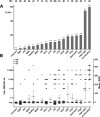
Frequency distribution, isotypes, and titers of serum N-Methyl-D-Aspartate receptor (NMDAR) antibodies in study subjects. Number of subjects per group, percentage of dementia patients and controls with positive NMDAR antibodies (IgM, IgA, or IgG) in serum (A). Antibody isotypes and serum titers across dementia groups and controls (B).
Figure 3
Figure 3
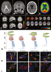
Imaging findings in dementia patients with NMDAR antibodies and epitope mapping with IgA-positive serum. Several patients with unclassified dementia and IgA/IgM NMDAR antibodies showed MRI and PET abnormalities that were not typical of primary neurodegenerative disorders. These included patients with otherwise unexplained marked bilateral leukoencephalopathy (A), global atrophy associated with very rapidly developing dementia (B and C), or patchy FDG uptake with reduction in paraventricular and cortical areas (D). NMDAR antibody levels in these patients were IgA 1:100 in serum and 1:10 in CSF (A), IgM 1:3200 in serum (B), IgA 1:1000 in serum (C and D). Imaging demonstrates treatment effects following immunotherapy with plasma exchange using fMRI (E) and PET (F). Functional connectivity of the default mode network (a set of brain regions with strongly correlated neural activity) was significantly decreased with the posterior cingulate cortex, the precuneus, and the superior parietal cortex in posttreatment scans in comparison with pretreatment scans (E). PET studies in a patient with unclassified dementia and IgA antibodies documented significant improvement of cerebral metabolism in cortical brain areas after plasma exchange (F). HEK cells were transfected with wild-type NR1a, or with NR1a mutants lacking the amino terminal domain (ATD-deleted), with amino acid 368 mutated (N368Q), or lacking the ligand-binding domain and first 3 transmembrane domains (ATD-TM4) (G). Subject A0 had IgA antibodies that strongly recognized NR1a. ATD deletion and N368Q mutation both only mildly reduced antibody binding while binding to the ATD-TM4 construct was increased (H; NR1a commercial antibody staining in red, human IgA antibodies in green; insert – corresponding grayscale images of human IgA). Subject A6 had IgA antibodies that recognized NR1a. For this subject, ATD deletion and N368Q mutation nearly eliminated staining of the NR1a construct and the antibodies had reduced binding to the ATD-TM4 construct (I). NMDAR, N-Methyl-D-Aspartate receptor; FDG, [F-18]-fluorodeoxyglucose; fMRI, functional magnetic resonance imaging; CSF, cerebrospinal fluid.

References

    1. Flanagan EP, McKeon A, Lennon VA, et al. Autoimmune dementia: clinical course and predictors of immunotherapy response. Mayo Clin Proc. 2010;85:881–897. - PMC - PubMed
    1. Flanagan EP, Caselli RJ. Autoimmune encephalopathy. Semin Neurol. 2011;31:144–157. - PubMed
    1. Lyons MK, Caselli RJ, Parisi JE. Nonvasculitic autoimmune inflammatory meningoencephalitis as a cause of potentially reversible dementia: report of 4 cases. J Neurosurg. 2008;108:1024–1027. - PubMed
    1. McKeon A, Marnane M, O’connell M, et al. Potassium channel antibody associated encephalopathy presenting with a frontotemporal dementia like syndrome. Arch Neurol. 2007;64:1528–1530. - PubMed
    1. Sabater L, Gaig C, Gelpi E, et al. A novel non-rapid-eye movement and rapid-eye-movement parasomnia with sleep breathing disorder associated with antibodies to IgLON5: a case series, characterisation of the antigen, and post-mortem study. Lancet Neurol. 2014;13:575–586. - PMC - PubMed