Review
. 2015:33:79-106.
doi: 10.1146/annurev-immunol-032414-112248. Epub 2014 Dec 10.

Programmed necrosis in the cross talk of cell death and inflammation

Affiliations
Review

Programmed necrosis in the cross talk of cell death and inflammation

Francis Ka-Ming Chan et al. Annu Rev Immunol. 2015.

Abstract

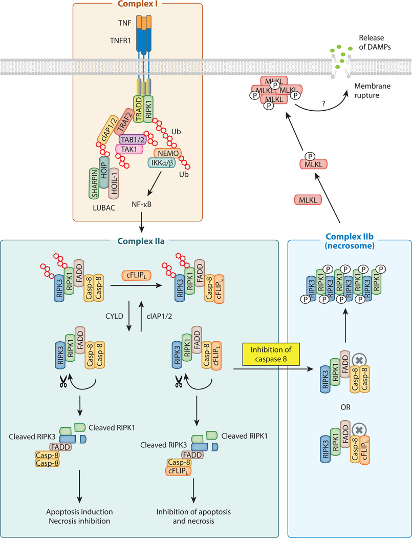
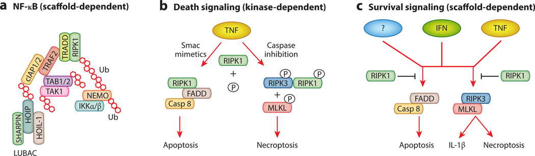
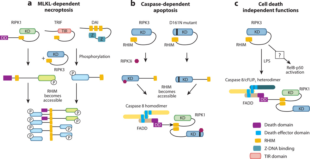
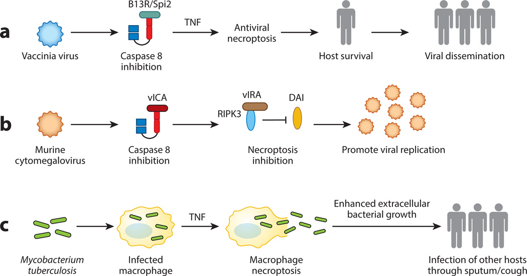
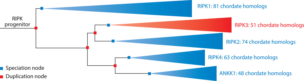
Cell proliferation and cell death are integral elements in maintaining homeostatic balance in metazoans. Disease pathologies ensue when these processes are disturbed. A plethora of evidence indicates that malfunction of cell death can lead to inflammation, autoimmunity, or immunodeficiency. Programmed necrosis or necroptosis is a form of nonapoptotic cell death driven by the receptor interacting protein kinase 3 (RIPK3) and its substrate, mixed lineage kinase domain-like (MLKL). RIPK3 partners with its upstream adaptors RIPK1, TRIF, or DAI to signal for necroptosis in response to death receptor or Toll-like receptor stimulation, pathogen infection, or sterile cell injury. Necroptosis promotes inflammation through leakage of cellular contents from damaged plasma membranes. Intriguingly, many of the signal adaptors of necroptosis have dual functions in innate immune signaling. This unique signature illustrates the cooperative nature of necroptosis and innate inflammatory signaling pathways in managing cell and organismal stresses from pathogen infection and sterile tissue injury.

Keywords: DAI; MCMV; MLKL; RIPK1; RIPK3; TNF; TRIF; inflammation; necroptosis; vaccinia virus.


Figures

Figure 1
Figure 1. Schematic diagram of TNF-induced signaling complexes
The membrane associated Complex I is chiefly responsible for NF-κB activation. The ubiquitin chains are represented by the red hexagons. Induction of cFLIPL expression by NF-κB inhibits apoptosis and necroptosis. Active caspase 8 in Complex IIa promotes apoptosis and inhibits necroptosis by cleavage of RIPK1, RIPK3 and CYLD. When caspase 8 is inactive, RIPK1 and RIPK3 initiate Complex IIb assembly, amyloid conversion, and recruitment of MLKL. Both Complex IIa and Complex IIb are also regulated by protein ubiquitination. CYLD acts as the de-ubiquitinase that promotes Complex II activity by removing ubiquitin chains on RIPK1 and RIPK3.
Figure 2
Figure 2. RIPK1 mediates cell survival and cell death through distinct mechanisms
(A) RIPK1 facilitates assembly of the ubiquitin scaffold that stimulates NF-κB activation. This function does not require the kinase activity of RIPK1. (B) The kinase activity of RIPK1 promotes apoptosis and necroptosis. (C) The scaffolding function of RIPK1 promotes survival and suppresses inflammation by inhibiting FADD-Caspase 8 and RIPK3-MLKL activation. This RIPK1 function is required to neutralize deleterious signals from interferon receptor, TNF receptor and other yet to be identified receptors. The kinase activity of RIPK1 is dispensable for this survival function.
Figure 3
Figure 3. The RHIM is a conserved signaling motif in innate immune and death signaling adaptors
Sequence alignment of mammalian (human), viral and Drosophila RHIM-containing adaptors. R45 and E45 are the M45 homologues in the Maastricht and English isolates of rat CMV, respectively. ICP6 and ICP10 are M45 homologues of herpes simplex virus 1 (HSV-1) and HSV-2 respectively. Although all the viral RHIM adaptors encode a ribonucleotide reductase domain, not all of them are active enzymes (146). The Drosophila “RHIM-like” adaptors and receptors are included for comparison, although there is no evidence currently to indicate that they function like the mammalian RHIMs. Other than the PGRPs, the Drosophila IMD and Relish also contain RHIM-like motifs (A. Kleino and N. Silverman, personal communication). The red box indicates the tetra-peptide core of the RHIM. The black and grey shades represent highly conserved and moderately conserved residues as defined by functional side chains. a.a. = amino acid position of the last residue shown in the sequence alignment.
Figure 4
Figure 4. RIPK3 signals for cell death and inflammation through diverse mechanisms
(A) RIPK3 mediates necroptosis by binding to RIPK1 or other RHIM-containing adaptors. This causes amyloid conversion of RIPK3, which serves as a platform for docking and recruitment of the RIPK3 substrate MLKL. (B) Binding of RIPK3 kinase inhibitor or introduction of the D161N mutation causes a conformational change that promotes a different form of RHIM-mediated interaction between RIPK1 and RIPK3 that leads to FADD and caspase 8 binding and apoptosis. (C) Although the mechanisms are yet to be defined, RIPK3 can also induce pro-IL-1β processing through caspase 1 and caspase 8. In over-expression studies, RIPK3 has also been shown to either enhance or inhibit NF-κB signaling.
Figure 5
Figure 5. Necroptosis in host-pathogen interaction
(A) Vaccinia virus inhibits caspase 8 via the viral inhibitor B13R/Spi2. This primes the cells towards necroptosis. Although necroptosis and the ensuing inflammation have anti-viral effects, it may in fact promote viral dissemination to another host by avoiding premature death of the infected host. (B) MCMV inhibits caspase 8 and necroptosis via the vICA and vIRA. Genetic experiments show that vIRA is essential to prevent premature death of the infected cells. Hence, vIRA-mediated necroptosis inhibition is important for the virus to complete its replication cycle and to generate more viral progenies. (C) Mycobacterium tuberculosis uses RIPK3-dependent necroptosis to release the bacteria into growth permissive environment, which in turn enhances spread of the pathogen to uninfected hosts via the sputum.
Figure 6
Figure 6. The RIPK gene family evolved through a series of gene duplication events
The reconstructed phylogeny was generated by the Ensembl genome browser (183). Internal nodes correspond to key speciation (purple) and gene duplication (red) events. Branch lengths correspond to rates of evolutionary change. ANKK1 (ankyrin repeat and protein kinase domain-containing protein 1) does not function as a RIP kinase but is closely related to RIPK4 and other RIPKs.

References

    1. Segawa K, Kurata S, Yanagihashi Y, Brummelkamp TR, Matsuda F, Nagata S. Caspase-mediated cleavage of phospholipid flippase for apoptotic phosphatidylserine exposure. Science. 2014;344:1164–1168. - PubMed
    1. Hochreiter-Hufford A, Ravichandran KS. Clearing the dead: apoptotic cell sensing, recognition, engulfment, and digestion. Cold Spring Harb Perspect Biol. 2013;5:a008748. - PMC - PubMed
    1. Kono H, Rock KL. How dying cells alert the immune system to danger. Nat Rev Immunol. 2008;8:279–289. - PMC - PubMed
    1. Sawai H, Domae N. Discrimination between primary necrosis and apoptosis by necrostatin-1 in Annexin V-positive/propidium iodide-negative cells. Biochem Biophys Res Commun. 2011;411:569–573. - PubMed
    1. Yamasaki S, Ishikawa E, Sakuma M, Hara H, Ogata K, Saito T. Mincle is an ITAM-coupled activating receptor that senses damaged cells. Nat Immunol. 2008;9:1179–1188. - PubMed

Publication types

MeSH terms