. 2014 Nov 8;15(6):4970.
doi: 10.1120/jacmp.v15i6.4970.

Evaluation of treatment plans using various treatment techniques for the radiotherapy of cutaneous Kaposi's sarcoma developed on the skin of feet

Affiliations

Evaluation of treatment plans using various treatment techniques for the radiotherapy of cutaneous Kaposi's sarcoma developed on the skin of feet

Jong Min Park et al. J Appl Clin Med Phys. .

Abstract

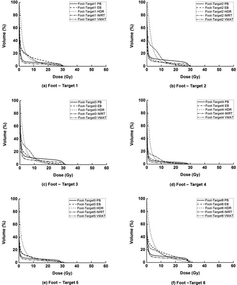
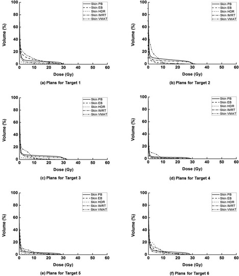
The purpose of this study was to investigate the plan qualities of various treatment modalities for the radiotherapy of cutaneous Kaposi's sarcoma developed on the skin of the foot. A total of six virtual targets were generated on the skin of the foot in CT images. Five types of treatment plans were generated using photon beams (PB), electron beams (EB), high-dose-rate (HDR) brachytherapy with a Freiburg flap applicator, intensity-modulated radiation therapy (IMRT), and volumetric- modulated arc therapy (VMAT) techniques. Plans for each of the six targets (single-target plans) and also for the combined target consisting of the six single targets combined (multitarget plans) were generated. Dose-volumetric analysis was performed for the targets and normal tissues. The averaged conformity index (CI) and homogeneity index (HI) values for each single target using PB, EB, HDR, IMRT, and VMAT techniques were 1.97, 2.39, 1.60, 4.60, and 0.80 and 1.05, 1.11, 1.52, 1.04, and 1.04, respectively. For the multitarget, the CI values were 3.99, 5.08, 1.38, 1.95, and 0.84, and the values of HI were 1.10, 1.36, 1.43, 1.06, and 1.04, respectively. The averaged mean doses to normal tissue were 2.5, 2.7, 3.6, 1.7, and 2.9 Gy for single-target plans, and 21.3, 14.6, 14.2, 14.3, and 13.0 Gy for the multitarget plans, respectively. The VMAT demonstrated dosimetric advantages and better treatment efficiency over other techniques for the radiotherapy of multifocal skin disease of the feet.


Figures

Figure 1
Figure 1
The custom‐made water reservoir for the skin treatment of the foot (a) and the patient setup using this water reservoir filled with water (b). The reservoir is made of acrylic and the level of water surface is marked. There is a slope on one side of the reservoir for the comfortable setup of patients when they put their foot inside the reservoir. The water reservoir could eliminate the buildup effect at cutaneous regions, thereby enabling the prescribed dose to be delivered to the target located at a superficial region.
Figure 2
Figure 2
The virtual clinical target volumes (CTVs) located at cutaneous regions are illustrated. A total of six targets on the skin of the foot are postulated for the treatment planning of cutaneous Kaposi's sarcoma. Targets 1 to 6 is defined at heel pad (a), inner ankle (b), inner arch (c), dorsal surface (d), inner big toe mound (e), and outer little toe mound (f) of foot. The thickness of target is 5 mm and the area of target 1 to 6 is 30.8, 20.6, 13.0, 9.4, 19.8, and 51.8cm2, respectively. Each single target is summed and postulated for the treatment planning of multifocal Kaposi's sarcoma. The total area of multifocal target is 145.4cm2.
Figure 3
Figure 3
The Freiburg flap applicator (a) and its application on the skin of foot (b) are shown. It is used for the radiation therapy of skin combined with high‐dose‐rate (HDR) brachytherapy. The Freiburg flap applicator consists of a flexible mesh style surface mold formed by 0.5 cm radius silicon spheres. The spheres are tunneled through the center with flexible catheters for the positioning of Ir‐192 sources. The design of the Freiburg flap applicator keeps the distance from source to skin constant.
Figure 4
Figure 4
Dose‐volume histograms (DVHs) of target 1 (a), target 2 (b), target 3 (c), target 4 (d), target 5 (e), and target 6 (f) are shown. For each target, DVHs from plans using tangential photon beam (PB), electron beam (EB), high‐dose‐rate (HDR) brachytherapy with surface applicator, intensity‐modulated radiation therapy (IMRT), and volumetric‐modulated arc therapy (VMAT) are shown. The VMAT shows better target coverage and homogeneity than the other techniques.
Figure 5
Figure 5
The dose‐volume histograms (DVHs) of the foot subtracted by the target (foot organ at risk, foot OAR) from the treatment plan for target 1 (a), target 2 (b), target 3 (c), target 4 (d), target 5 (e), and target 6 (f) are shown, respectively. For each target, DVHs from plans using tangential photon beam (PB), electron beam (EB), high‐dose‐rate (HDR) brachytherapy with surface applicator, intensity‐modulated radiation therapy (IMRT), and volumetric‐modulated arc therapy (VMAT) are shown. The doses delivered to the foot OAR in high‐dose regions are higher than the others when using the PB technique, and lower in the low‐dose regions than the other techniques. In the case of the HDR plans, the doses in the low‐dose region tend to be higher than the other techniques, while in high‐dose region, those are lower than the others.
Figure 6
Figure 6
The dose‐volume histograms (DVHs) of the skin from the treatment plan for target 1 (a), target 2 (b), target 3 (c), target 4 (d), target 5 (e), and target 6 (f) are shown, respectively. For each target, DVHs from plans using tangential photon beam (PB), electron beam (EB), high‐dose‐rate (HDR) brachytherapy with surface applicator, intensity‐modulated radiation therapy (IMRT), and volumetric‐modulated arc therapy (VMAT) are shown. The doses delivered to the skin in high‐dose regions are higher than the others when using the PB technique.
Figure 7
Figure 7
The dose‐volume histograms (DVHs) of the bone from the treatment plan for target 1 (a), target 2 (b), target 3 (c), target 4 (d), target 5 (e), and target 6 (f) are shown, respectively. For each target, DVHs from plans using tangential photon beam (PB), electron beam (EB), high‐dose‐rate (HDR) brachytherapy with surface applicator, intensity‐modulated radiation therapy (IMRT), and volumetric‐modulated arc therapy (VMAT) are shown. The doses delivered to the bone in high‐dose regions are higher than the others when using the PB technique. In the case of the HDR plans, the doses in the low‐dose region tend to be higher than the other techniques.
Figure 8
Figure 8
The dose‐volume histograms (DVHs) of multitarget (a), normal tissue (b), bone (c), and skin (d) from plans using 4‐box photon beam with multileaf collimator (PB), electron beam (EB), high‐dose‐rate (HDR) brachytherapy, intensity‐modulated radiation therapy (IMRT), and volumetric‐modulated arc therapy (VMAT) are shown. When using the EB technique, the maximum dose to normal tissue is higher than the other techniques. In the high‐dose region, the EB plan shows larger irradiated volume of normal tissue than others, while the HDR plan shows largest irradiated volume of normal tissue in the low‐dose region. When using the HDR technique, the doses to the skin in the intermediate and low‐dose regions were lowest.
Figure 9
Figure 9
The beam setup and the dose distributions of treatment plans for multitarget using 4‐box photon beam with multileaf collimator (PB), electron beam (EB), high‐dose‐rate (HDR) brachytherapy, intensity‐modulated radiation therapy (IMRT), and volumetric‐modulated arc therapy (VMAT) are shown. The beam setup of PB (a), EB (e), HDR (i), IMRT (m), and VMAT (q) are shown. The axial dose distribution of PB (b), EB (f), HDR (j), IMRT (n), and VMAT (r) are shown. The sagittal dose distribution of PB (d), EB (h), HDR (l), IMRT (p), and VMAT (t), as well as the coronal dose distribution of PB (c), EB (g), HDR (k), IMRT (o), and VMAT (s), are also shown.

References

    1. Weshler Z, Loewinger E, Loewenthal E, Levinson R, Fucks Z. Megavoltage radiotherapy using water bolus in the treatment of Kaposi's sarcoma. Int J Radiat Oncol Biol Phys. 1986;12(11):2029–32. - PubMed
    1. Aldenhoven M, Barlo NP, Sanders CJ. Therapeutic strategies for epidemic Kaposi's sarcoma. Int J STD AIDS. 2006;17(9):571–78. - PubMed
    1. Stelzer KJ and Griffin TW. A randomized prospective trial of radiation therapy for AIDS‐associated Kaposi's sarcoma. Int J Radiat Oncol Biol Phys. 1993;27(5):1057–61. - PubMed
    1. Cohen L, Palmer PE, Nickson JJ. Treatment of Kaposi's sarcoma by radiation. Acta Unio Int Contra Cancrum. 1961;18:502–09. - PubMed
    1. Chang JH and Kim IH. Role of radiotherapy in local control of non‐AIDS associated Kaposi's sarcoma patients in Korea: a single instution experience. Radiat Oncol J. 2012;30(4):153–57. - PMC - PubMed

MeSH terms

LinkOut - more resources