. 2014 Dec 10;9(12):e114628.
doi: 10.1371/journal.pone.0114628. eCollection 2014.

Triacylglycerol Storage in Lipid Droplets in Procyclic Trypanosoma brucei

Affiliations

Triacylglycerol Storage in Lipid Droplets in Procyclic Trypanosoma brucei

Stefan Allmann et al. PLoS One. .

Abstract

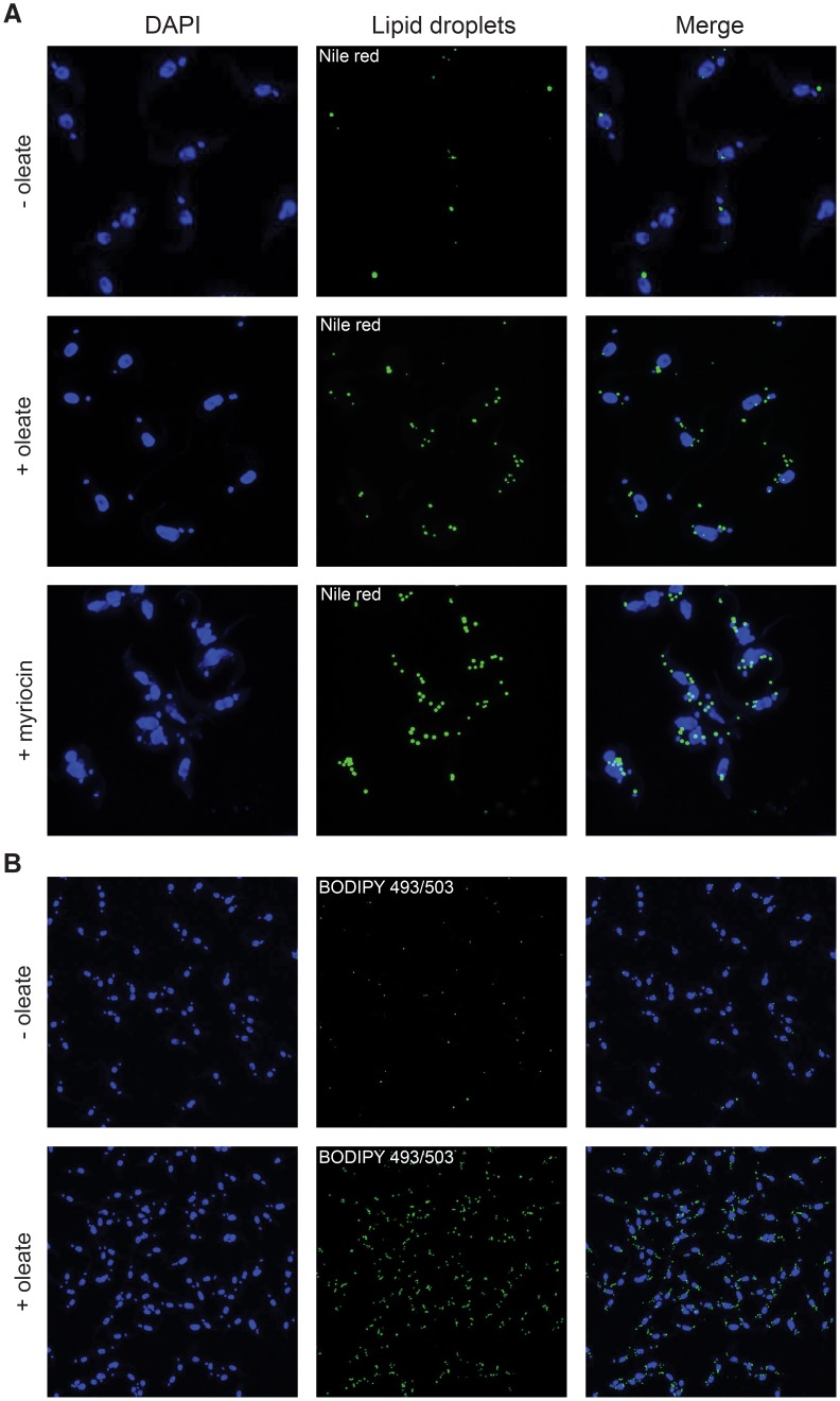
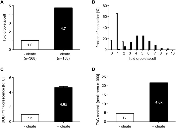
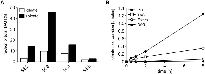
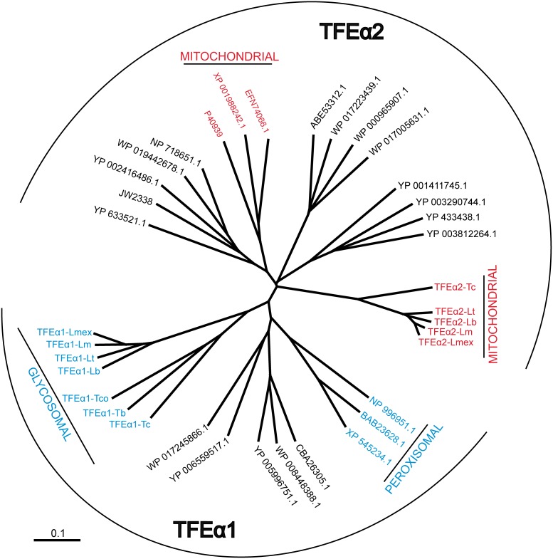
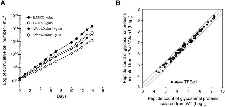
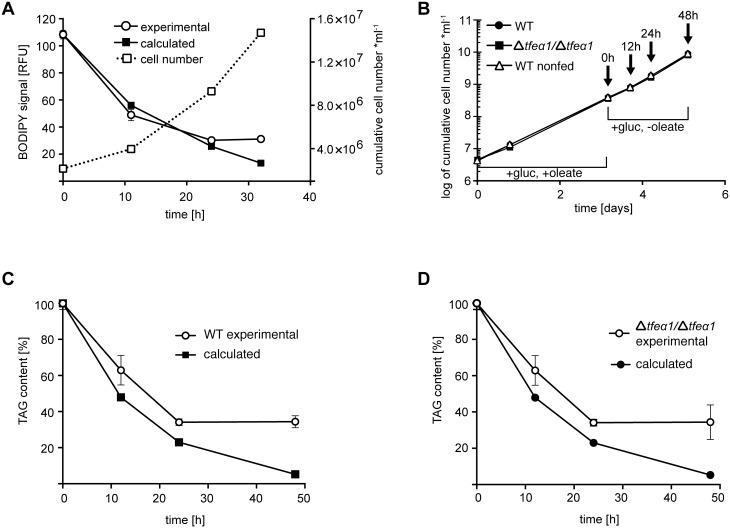
Carbon storage is likely to enable adaptation of trypanosomes to nutritional challenges or bottlenecks during their stage development and migration in the tsetse. Lipid droplets are candidates for this function. This report shows that feeding of T. brucei with oleate results in a 4-5 fold increase in the number of lipid droplets, as quantified by confocal fluorescence microscopy and by flow cytometry of BODIPY 493/503-stained cells. The triacylglycerol (TAG) content also increased 4-5 fold, and labeled oleate is incorporated into TAG. Fatty acid carbon can thus be stored as TAG in lipid droplets under physiological growth conditions in procyclic T. brucei. β-oxidation has been suggested as a possible catabolic pathway for lipids in T. brucei. A single candidate gene, TFEα1 with coding capacity for a subunit of the trifunctional enzyme complex was identified. TFEα1 is expressed in procyclic T. brucei and present in glycosomal proteomes, Unexpectedly, a TFEα1 gene knock-out mutant still expressed wild-type levels of previously reported NADP-dependent 3-hydroxyacyl-CoA dehydrogenase activity, and therefore, another gene encodes this enzymatic activity. Homozygous Δtfeα1/Δtfeα1 null mutant cells show a normal growth rate and an unchanged glycosomal proteome in procyclic T. brucei. The decay kinetics of accumulated lipid droplets upon oleate withdrawal can be fully accounted for by the dilution effect of cell division in wild-type and Δtfeα1/Δtfeα1 cells. The absence of net catabolism of stored TAG in procyclic T. brucei, even under strictly glucose-free conditions, does not formally exclude a flux through TAG, in which biosynthesis equals catabolism. Also, the possibility remains that TAG catabolism is completely repressed by other carbon sources in culture media or developmentally activated in post-procyclic stages in the tsetse.


Conflict of interest statement

Competing Interests: The authors have declared that no competing interests exist.

Figures

Figure 1
Figure 1. Oleate feeding stimulates lipid droplet formation in procyclic T. brucei cells.
Staining of lipid droplets with nile red (A) or BODIPY 493/503 (B) was as detailed in experimental procedures. Myriocin treatment (0.5 µM for 24 h) was included for comparison to a previous report . An example of several experiments is shown.
Figure 2
Figure 2. Quantification of the oleate-induced lipid droplet formation.
(A) BODIPY 493/503 stained LDs were counted in stacks of confocal laser scanning microscopy (CLSM) images; the average number of LDs per cell is given after oleate feeding (black column) or in the control (white column). (B) Distribution of LD numbers per cells in the population after oleate feeding (black columns) or in the control (white columns). (C) Quantification of BODIPY-stained LDs by flow cytometry after oleate feeding (black column) or in the control (white column). BODIPY 493/503 preferentially stains nonpolar lipids. Error bars give the SEM (n = 3) of values normalized to the control. (D) Quantification of TAG content by HPTLC and densitometry after oleate feeding (black columns) or in the control (white columns). Values are normalized to the control.
Figure 3
Figure 3. TAG species analysis and uptake of labeled oleate.
(A) Dominant TAG species in procyclic T. brucei cells identified by ESI/MS/MS after oleate feeding for three days (black columns) or in the control (white columns). For a complete list of TAG species detected see S1 Figure. The nomenclature 54:X indicates the total carbon number of all three acyl chains and the sum of all unsaturated double bonds within the acyl chains. (B) Uptake kinetics upon growth in the presence of radiolabeled oleate for up to 8 h. The incorporation of 14C oleate into lipid species was quantified by HPTLC and a Storm 860 phosphorimager. PPL, phospholipids; TAG, triacylglycerol; SE, Steryl-esters; DAG, diacylglycerol.
Figure 4
Figure 4. Dendrogram of trifunctional enzyme (TFE) isoforms.
Prokaryotic (black characters) and eukaryotic (colored characters) TFEα sequences are represented by their GenBank accession codes. Glycosomal/peroxisomal (TFEα1) or mitochondrial (TFEα2) proteins are highlighted in blue and red. Experimental evidence for glycosomal localization of trypanosomatid TFEα1 isoforms, which all contain a PTS2 motif, is limited to T. brucei TFEα1 (see and S4 Figure). Mitochondrial localization of the trypanosomatid TFEα2 isoforms is assumed due to an N-terminal mitochondrial targeting motif and the absence of a PTS motif. Abbreviations: Lb, Leishmania braziliensis; Lm, L. major; Lmex, L. mexicana; Lt, L. tarentolae; Tb, T. brucei; Tc, T. cruzi; Tco, T. congolense. The organisms corresponding to the accession numbers are: Canis lupus familiaris (XP_545234.1), Danio rerio (NP_996951.1), Mus musculus (BAB23628.1), Curvibacter putative symbiont of Hydra magnipapillata (CBA26305.1), Janthinobacterium sp. HH01 (WP_008448388.1), Marinobacter sp. BSs20148 (YP_006559517.1), Pseudomonas stutzeri (WP_017245866.1), Ralstonia solanacearum CMR15 (YP_005996751.1), Camponotus floridanus (EFN74066.1), Drosophila grimshawi (XP_001988242.1), γ-proteobacterium HdN1 (YP_003812264.1), Hahella chejuensis KCTC2396 (YP_433438.1), Homo sapiens (P40939), Moritella dasanensis (WP_017223439.1), Parvibaculum lavamentivorans DS-1 (YP_001411745.1), Rhodothermus marinus DSM4252 (YP_003290744.1), Shewanella denitrificans OS217 (ABE53312.1), Vibrio splendidus LGP32 (YP_002416486.1), Escherichia coli (JW2338), Enterovibrio norvegicus (WP_017005631.1), Moritella marina (WP_019442678.1), Myxococcus xanthus DK1622 (YP_633521.1), Shigella flexneri (WP_000965907.1), Shewanella oneidensis MR-1 (NP_718651.1).
Figure 5
Figure 5. Phenotypic analysis of Δtfeα1tfeα1 cell.
(A) growth curve of WT and Δtfeα1tfeα1 cell knock cells in glucose-rich (SDM79 with 10 mM glucose) or glucose-free (SDM79GluFree) conditions. (B) Global protein abundance in the partially purified glycosome fraction of WT (x-axis) and Δtfeα1tfeα1 cell knock cells (y-axis). Each protein identification is presented by a point at log10 of normalized peptide count values taken from the proteome data in S4 Figure. Proteins on the dashed grey line have identical normalized peptide counts in both samples; the grey lines represent a 2-fold abundance in one condition.
Figure 6
Figure 6. LD and TAG turnover in WT and Δtfeα1tfeα1 cells.
Cells were fed with oleate in glucose-rich SDM79 medium for three days, and after oleate withdrawal samples were taken at the time points indicated. (A) WT cells stained with BODIPY and analyzed by flow cytometry (left y-axis). Error bars represent the SEM of independent replicates (n = 3). The growth curve is given as dashed line (right y-axis). (B) Growth curve and sampling time points (arrows) for the experiments in panels (C) and (D). Total TAG content was determined in triplicate by HPTLC and densitometry in WT (C) and Δtfeα1tfeα1 (D) cells. Error bars represent the SEM of independent replicates (n = 3). The calculated values (filled symbols) account for dilution of LDs or TAG content by cell division, based on the matched growth data.

References

    1. Waltermann M, Steinbuchel A (2005) Neutral lipid bodies in prokaryotes: recent insights into structure, formation, and relationship to eukaryotic lipid depots. J Bacteriol 187:3607–3619. - PMC - PubMed
    1. Martin S, Parton RG (2006) Lipid droplets: a unified view of a dynamic organelle. Nat Rev Mol Cell Biol 7:373–378. - PubMed
    1. Igal RA, Coleman RA (1996) Acylglycerol recycling from triacylglycerol to phospholipid, not lipase activity, is defective in neutral lipid storage disease fibroblasts. J Biol Chem 271:16644–16651. - PubMed
    1. Igal RA, Coleman RA (1998) Neutral lipid storage disease: a genetic disorder with abnormalities in the regulation of phospholipid metabolism. J Lipid Res 39:31–43. - PubMed
    1. Leber R, Zinser E, Hrastnik C, Paltauf F, Daum G (1995) Export of steryl esters from lipid particles and release of free sterols in the yeast, Saccharomyces cerevisiae. Biochim Biophys Acta 1234:119–126. - PubMed

Publication types

LinkOut - more resources