. 2015 Mar 1;466(2):253-62.
doi: 10.1042/BJ20141211.

Catalytic surface radical in dye-decolorizing peroxidase: a computational, spectroscopic and site-directed mutagenesis study

Affiliations

Catalytic surface radical in dye-decolorizing peroxidase: a computational, spectroscopic and site-directed mutagenesis study

Dolores Linde et al. Biochem J. .

Abstract

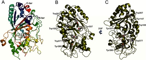
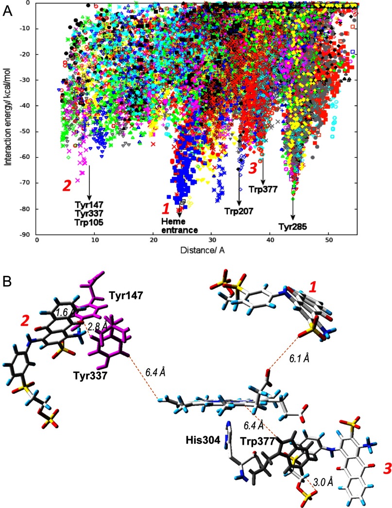
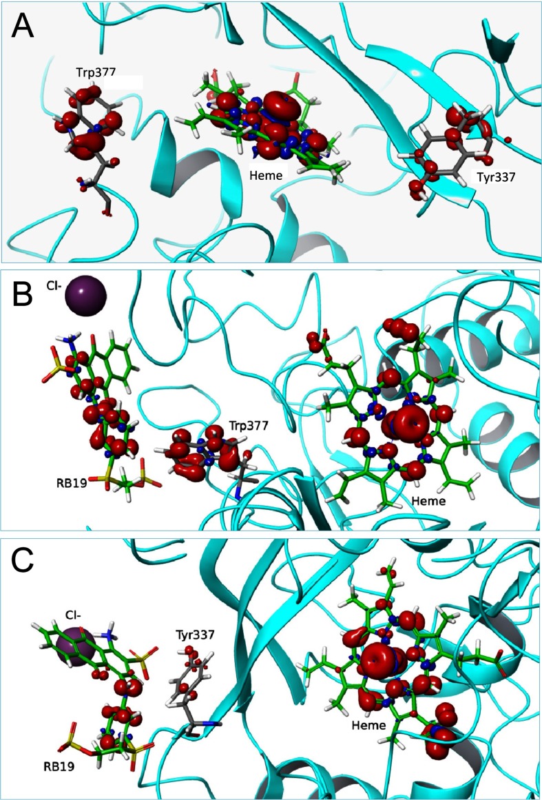
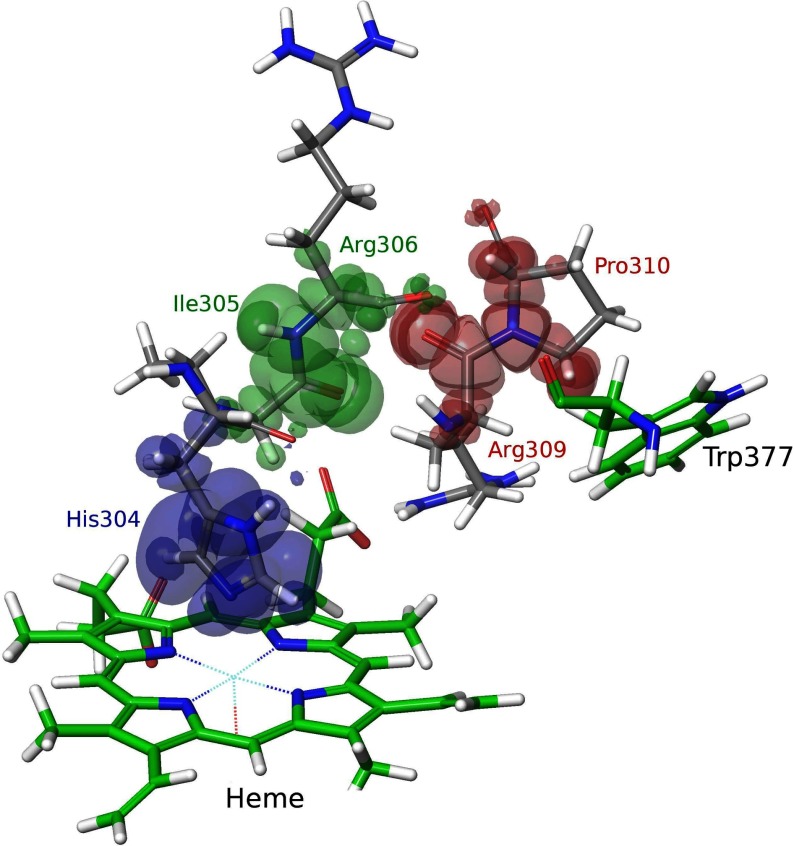
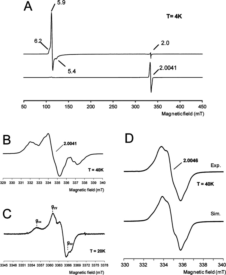
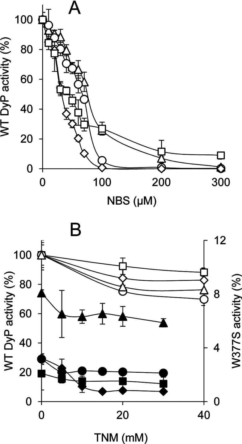
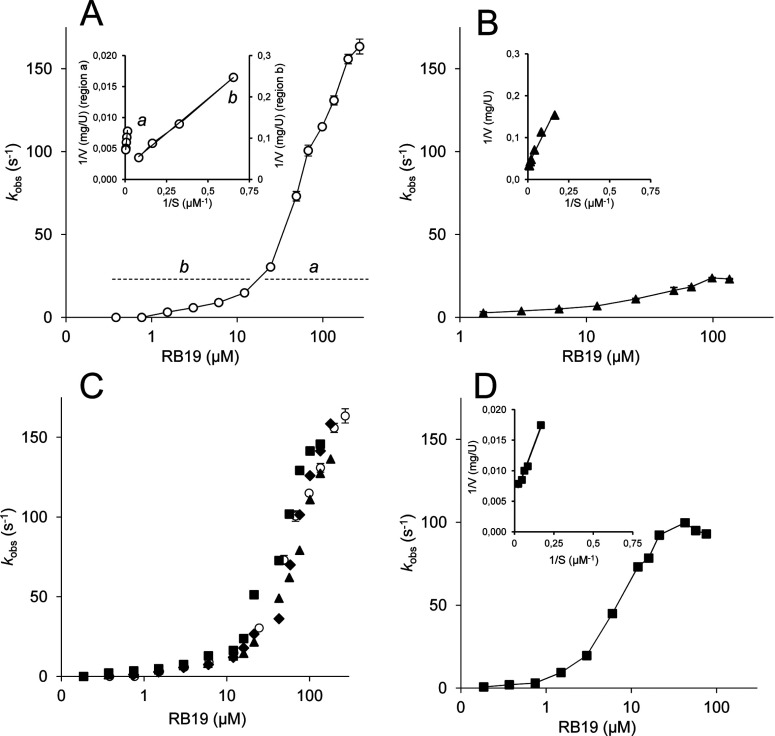
Dye-decolorizing peroxidase (DyP) of Auricularia auricula-judae has been expressed in Escherichia coli as a representative of a new DyP family, and subjected to mutagenic, spectroscopic, crystallographic and computational studies. The crystal structure of DyP shows a buried haem cofactor, and surface tryptophan and tyrosine residues potentially involved in long-range electron transfer from bulky dyes. Simulations using PELE (Protein Energy Landscape Exploration) software provided several binding-energy optima for the anthraquinone-type RB19 (Reactive Blue 19) near the above aromatic residues and the haem access-channel. Subsequent QM/MM (quantum mechanics/molecular mechanics) calculations showed a higher tendency of Trp-377 than other exposed haem-neighbouring residues to harbour a catalytic protein radical, and identified the electron-transfer pathway. The existence of such a radical in H₂O₂-activated DyP was shown by low-temperature EPR, being identified as a mixed tryptophanyl/tyrosyl radical in multifrequency experiments. The signal was dominated by the Trp-377 neutral radical contribution, which disappeared in the W377S variant, and included a tyrosyl contribution assigned to Tyr-337 after analysing the W377S spectra. Kinetics of substrate oxidation by DyP suggests the existence of high- and low-turnover sites. The high-turnover site for oxidation of RB19 (k(cat) > 200 s⁻¹) and other DyP substrates was assigned to Trp-377 since it was absent from the W377S variant. The low-turnover site/s (RB19 k(cat) ~20 s⁻¹) could correspond to the haem access-channel, since activity was decreased when the haem channel was occluded by the G169L mutation. If a tyrosine residue is also involved, it will be different from Tyr-337 since all activities are largely unaffected in the Y337S variant.


Figures

Figure 1
Figure 1. Folding of A. auricula-judae DyP and location of exposed aromatic residues
(A) General folding constituted by two domains, each of them including two large β-sheets and two or three helices, with the haem cofactor in the upper part of the lower domain [cartoon coloured from the N- to the C-terminus, with the haem shown as CPK (Corey–Pauling–Koltun) sticks]. (B and C) Location of exposed Trp-105, Tyr-147, Trp-207, Tyr-285, Tyr-337 and Trp-377 (as CPK spheres) in two different orientations of the DyP molecule (cartoon with the haem shown as CPK sticks). From PDB code 4W7J.
Figure 2
Figure 2. Substrate exploration on the DyP surface
(A) Local minima identified in the PELE [29] simulations of RB19 diffusion on the recombinant DyP crystal structure (PDB code 4W7J) showing interaction energy against distance to Tyr-147 (taken as a reference residue). The presence of RB19 in the vicinity of different surface residues and the haem-access channel is indicated. (B) Distances between the closest positions of RB19 (magenta sticks) with respect to haem (1), Tyr-147/Tyr-337 (2), and Trp-377 (3) shown by PELE (A), and between the above residues and the haem cofactor (the distances are measured including hydrogen atoms). RB19 is shown as CPK (Corey–Pauling–Koltun) sticks, Tyr-147/Tyr-337 as magenta sticks and Trp-377 as grey sticks.
Figure 3
Figure 3. QM/MM electron spin distribution on Trp-377 and Tyr-337 and RB19 substrate
(A) Total spin density when including Trp-377, Tyr-337 and haem compound I in the quantum region (in the absence of RB19). (B and C) Total spin density when, in addition to RB19 and compound I, the quantum region includes Trp-377 or Tyr-337 respectively. From PDB code 4W7J after 5 ns MD (A) and selected snapshots from two energy minima showing RB19 near Trp-377 and Tyr-337, during PELE [29] diffusion in Figure 2 (B and C respectively).
Figure 4
Figure 4. Electron transfer pathway from DyP Trp-377 to haem
The electron transfer pathway was obtained after three iterations of the QM/MM e-pathway approach [32] with a total of 15 residues (His-304–Arg-311, Leu-323–Ala-325, Leu-373–Gln-375 and Asp-395) included in the quantum region. Each iteration identifies the residue(s) with the highest affinity for the electron, and is shown in a different colour. The mapped route includes Pro-310 and Arg-309, followed by Arg-306, Ile-305 and His-304, as shown by the electron spin distribution.
Figure 5
Figure 5. EPR spectra of WT DyP and its W377S variant
(A) X-band EPR spectra of WT DyP at pH 3 before (top) and after (bottom) the addition of H2O2 (and rapid freezing). Experimental conditions: v=9.39 GHz, 0.2 mW microwave power, 0.4 mT modulation amplitude. (B and C) Narrow scan X-band (v=9.39 GHz) and W-band (v=94.29 GHz) respectively of the radical species. The positions of the three g-tensor components of the tyrosyl contribution are indicated. X-band experimental conditions: v=9.38 GHz, 1 mW microwave power and 0.05 mT modulation amplitude; W-band experimental conditions: v=94.29 GHz, 0.05 mW microwave power and 0.1 mT modulation amplitude. (D) X-band EPR spectrum of the radical intermediate formed in the W377S variant paired with its better simulation (Sim) (see magnetic parameters in Supplementary Table S3). Experimental conditions: v=9.39 GHz, 1 mW microwave power, 0.2 mT modulation amplitude.
Figure 6
Figure 6. Chemical modification of tryptophan and tyrosine residues in WT DyP and its W377S variant
(A) Residual activities of 3 μM WT DyP treated with increasing NBS concentrations (in 50 mM acetate, pH 4) for modification of tryptophan residues. (B) Residual activities of 3 μM WT DyP and W377S variant (white and black symbols respectively) treated with increasing TNM concentrations (in 50 mM Tris/HCl, pH 7, with 2.6% ethanol) for modification of tyrosine residues. The residual activities of the WT DyP and the W377S variant in (A) and (B) were monitored for oxidation of 180 μM RB19 (diamonds), 7.5 mM DMP (circles), 15 μM RB5 (squares) and 1.25 mM ABTS (triangles), and referred to activities of untreated WT DyP (taken as 100%).
Figure 7
Figure 7. Kinetics for RB19 oxidation by WT DyP and different variants
(A) WT DyP biphasic kinetics enabling calculation of two sets of constants in the 0.2–10 μM (b) and 50–270 μM (a) ranges (inset with Lineweaver–Burk inverse representation for the high, a, and low, b, turnover sites). (B) Simple kinetics yielding a single set of constants (inset, inverse representation) for the W377S variant. (C) Results from Y147S (squares), Y337S (diamonds) and Y147F/Y337F (triangles) variants yielding kinetic curves superimposable with that of WT DyP (circles). (D) Simple kinetics yielding a single set of constants (inset, inverse representation) for the G169L variant. A substrate concentration logarithmic scale is used in the main plots.

References

    1. Sugano Y. DyP-type peroxidases comprise a novel heme peroxidase family. Cell. Mol. Life Sci. 2009;66:1387–1403. doi: 10.1007/s00018-008-8651-8. - DOI - PMC - PubMed
    1. Hofrichter M., Ullrich R., Pecyna M.J., Liers C., Lundell T. New and classic families of secreted fungal heme peroxidases. Appl. Microbiol. Biotechnol. 2010;87:871–897. doi: 10.1007/s00253-010-2633-0. - DOI - PubMed
    1. Ruiz-Dueñas F.J., Martínez A.T. Structural and functional features of peroxidases with a potential as industrial biocatalysts. In: Torres E., Ayala M., editors. Biocatalysts Based on Heme Peroxidases. Berlin: Springer-Verlag; 2010. pp. 37–59. - DOI
    1. Colpa D.I., Fraaije M.W., van Bloois E. DyP-type peroxidases: a promising and versatile class of enzymes. J. Ind. Microbiol. Biotechnol. 2014;41:1–7. doi: 10.1007/s10295-013-1371-6. - DOI - PubMed
    1. Kim S.J., Shoda M. Purification and characterization of a novel peroxidase from Geotrichum candidum Dec 1 involved in decolorization of dyes. Appl. Environ. Microbiol. 1999;65:1029–1035. - PMC - PubMed

Publication types

MeSH terms

LinkOut - more resources