

Enhancing uterine fibroid research through utilization of biorepositories linked to electronic medical record data
- PMID: 25495367
- PMCID: PMC4267124
- DOI: 10.1089/jwh.2014.4978
Enhancing uterine fibroid research through utilization of biorepositories linked to electronic medical record data
Abstract
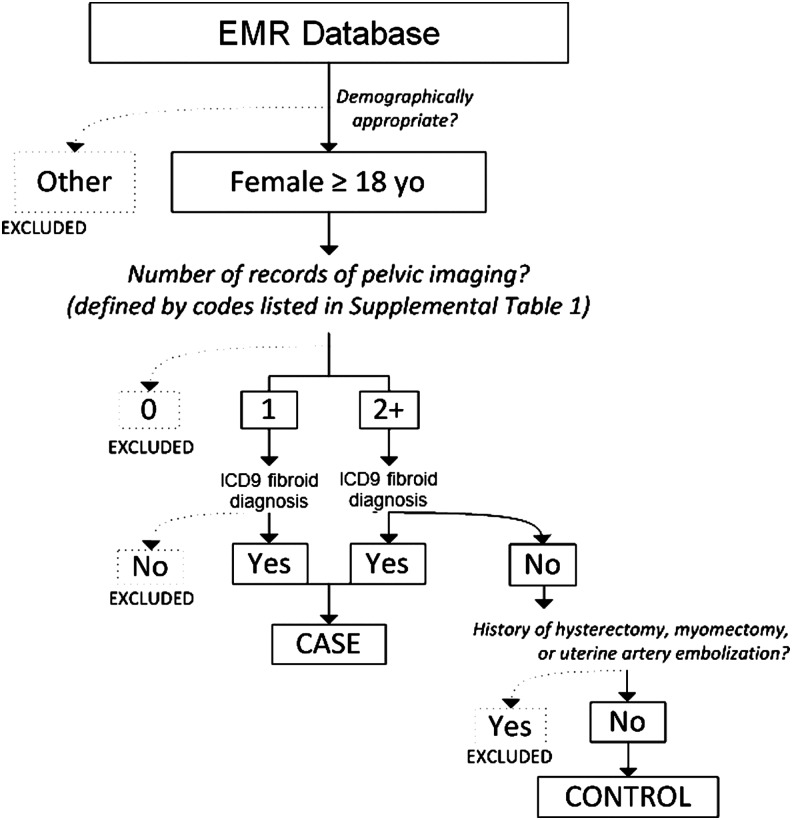
Background: Uterine leiomyomata (fibroids) affect up to 77% of women by menopause and account for $9.4 billion in yearly healthcare costs. Most studies rely on self-reported diagnosis, which may result in misclassification of controls since as many as 50% of cases are asymptomatic and thus undiagnosed. Our objective was to evaluate the performance and accuracy of a fibroid phenotyping algorithm constructed from electronic medical record (EMR) data, limiting to subjects with pelvic imaging.
Methods: Our study population includes women from a clinical population at Vanderbilt University Medical Center (2008-2012). Analyses were restricted to women 18 years and older with at least one fibroid diagnosis confirmed by imaging for cases or at least two separate pelvic imaging procedures without a diagnosis for controls. We randomly reviewed 218 records to evaluate the accuracy of our algorithm and assess the indications for pelvic imaging. Participant characteristics and indications for imaging were compared between cases and controls in unadjusted and adjusted logistic regression analyses.
Results: Our algorithm had a positive predictive value of 96% and negative predictive value of 98%. Increasing age (odds ratio=1.05, 95% confidence interval 1.03-1.08) and Black race (odds ratio=2.15, 95% confidence interval 1.18-3.94) were identified as risk factors for fibroids. The most common indications for imaging in both cases and controls were pain, bleeding, and reproductive factors, and the most common imaging modality was a pelvic ultrasound.
Conclusions: These data suggest that using biorepositories linked to EMR data is a feasible way to identify populations of imaged women that facilitate investigations of fibroid risk factors.
Figures
Comment in
-
Overcoming the challenges of studying uterine fibroids.J Womens Health (Larchmt). 2015 Feb;24(2):112-3. doi: 10.1089/jwh.2015.1520. J Womens Health (Larchmt). 2015. PMID: 25682815 No abstract available.
References
-
- Day BD, Dunson DB, Hill MC, Cousins D, Schectman JM. High cumulative incidence of uterine leiomyoma in black and white women: ultrasound evidence. Am J Obstet Gynecol 2003;188:100–107 - PubMed
-
- Hartmann KE, Birnbaum H, Ben-Hamadi R, et al. Annual costs associated with diagnosis of uterine leiomyomata. Obstet Gynecol 2006;108:930–937 - PubMed
-
- Cramer SF, Patel A. The frequency of uterine leiomyomas. Am J Clin Pathol 1990; 94:435–438 - PubMed
-
- Marshall LM, Spiegelman D, Barbieri RL, et al. Variation in the incidence of uterine leiomyoma among premenopausal women by age and race. Obstet Gynecol 1997;90:967–973 - PubMed
Publication types
MeSH terms
Grants and funding
LinkOut - more resources
Full Text Sources
Other Literature Sources
Medical
Molecular Biology Databases
