. 2015 Nov;25(6):701-11.
doi: 10.1111/bpa.12237. Epub 2015 Jan 27.

Huntington's Disease (HD): Neurodegeneration of Brodmann's Primary Visual Area 17 (BA17)

Affiliations

Huntington's Disease (HD): Neurodegeneration of Brodmann's Primary Visual Area 17 (BA17)

Udo Rüb et al. Brain Pathol. 2015 Nov.

Abstract

Huntington's disease (HD), an autosomal dominantly inherited polyglutamine or CAG repeat disease along with somatomotor, oculomotor, psychiatric and cognitive symptoms, presents clinically with impairments of elementary and complex visual functions as well as altered visual-evoked potentials (VEPs). Previous volumetric and pathoanatomical post-mortem investigations pointed to an involvement of Brodmann's primary visual area 17 (BA17) in HD. Because the involvement of BA17 could be interpreted as an early onset brain neurodegeneration, we further characterized this potential primary cortical site of HD-related neurodegeneration neuropathologically and performed an unbiased estimation of the absolute nerve cell number in thick gallocyanin-stained frontoparallel tissue sections through the striate area of seven control individuals and seven HD patients using Cavalieri's principle for volume and the optical disector for nerve and glial cell density estimations. This investigation showed a reduction of the estimated absolute nerve cell number of BA17 in the HD patients (71,044,037 ± 12,740,515 nerve cells) of 32% in comparison with the control individuals (104,075,067 ± 9,424,491 nerve cells) (Mann-Whitney U-test; P < 0.001). Additional pathoanatomical studies showed that nerve cell loss was most prominent in the outer pyramidal layer III, the inner granular layers IVa and IVc as well as in the multiform layer VI of BA17 of the HD patients. Our neuropathological results in BA17 confirm and extend previous post-mortem, biochemical and in vivo neuroradiological HD findings and offer suitable explanations for the elementary and complex visual dysfunctions, as well as for the altered VEP observed in HD patients.

Keywords: Huntington's disease; area 17; cerebral cortex; polyglutamine diseases; visual system; visual-evoked potentials (VEP).


Figures

Figure 1
Figure 1
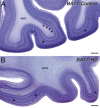
Neuronal loss in Brodmann's primary visual area 17 (BA17) in Huntington's disease (HD). A. Thick gallocyanin‐stained frontal tissue section through the caudal pole of the occipital lobe of a 50‐year‐old male control individual without neuropsychiatric diseases in his medical records showing Brodmann's primary visual area 17 (BA17) and its borders with the adjacent Brodmann's parastriate area 18 (broken lines). Note the prominent pale, cell‐poor and myelin‐rich Gennari stripe in the inner granular layer IVb of BA17 (asterisks) (4, 13, 70, 80). Arrowheads point to the basophilic multiform layer VI. B. BA17 of a 36‐year‐old female HD patient (HD case 8; Table 2). The striking pallor of and neuronal loss in (1) the outer pyramidal layer III and (2) inner granular layer IVc, as well as (3) the reduction of the multiform layer VI are detectable even at this low magnification. Broken lines mark the sharp boundaries with Brodmann's parastriate area 18 and asterisks Gennari stripe in the inner granular layer IVb of BA17. Bars in A and B indicate 1200 μm. (A, B: Nissl staining with gallocyanin).
Figure 2
Figure 2
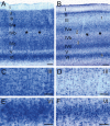
Predominant neuronal loss in layers III, IVa, IVc and VI in Brodmann's primary visual area 17 (BA17) in Huntington's disease (HD). A. Magnified section of a thick gallocyanin‐stained frontal tissue section through Brodmann's primary visual area 17 (BA17) of a 65‐year‐old female control individual. The “calcarine” cortex type of BA17 shows a strong reduction of the outer and inner pyramidal layers III and V, well‐developed granular layers II and IV with a very high neuronal density, a strongly basophilic multiform layer VI and a subdivision of the inner granular layer into layers IVa–c. Note the Gennari stripe in the inner granular layer IVb (asterisks) 4, 13, 70, 80. B. BA17 of a 36‐year‐old female HD patient (HD case 8; Table 2): (1) Severe neuronal loss and pallor of the outer pyramidal layer III of BA17. (2) Considerably reduced nerve cell density and pallor of the inner granular layer IVa of BA17. (3) Reduced nerve cell density and pallor of the inner granular layer IVc of BA17. (4) Narrowed multiform layer VI of BA17 owing to nerve cell loss. C. Picture detail of the outer pyramidal layer III of BA17 of the 65‐year‐old female control individual. D. Picture detail of the outer pyramidal layer III of BA17 of the 36‐year‐old female HD patient (HD case 8; Table 2) showing pallor and reduced neuronal density of this BA17 layer. E. Picture detail of the multiform layer VI of BA17 of the 65‐year‐old female control individual. F. Picture detail of the multiform layer VI of BA17 of the 36‐year‐old female HD patient (HD case 8; Table 2): narrowing of and neuronal loss in this BA17 layer. Asterisks indicate Gennari stripe. Bars in A and B indicate 150 μm, while bars in C–F indicate 75 μm (A–F: Nissl staining with gallocyanin).
Figure 3
Figure 3
The estimated absolute nerve cell number in Brodmann's primary visual area 17 (BA17) in Huntington's disease (HD). Estimated absolute nerve cell number in BA17 of age‐ and gender‐matched control individuals (filled circles) and Huntington's disease (HD) patients (filled squares). Horizontal lines represent arithmetic means. Estimation of the absolute neuronal numbers is based on the application of Cavalieri's principle for volume and the optical disector for nerve density estimations 7, 8, 18, 20, 21, 22, 48, 49, 61, 76, 77, 78 and revealed a reduction by 32% in the HD patients (71 044 037 ± 12 740 515 nerve cells) as compared with the control individuals (104 075 067 ± 9 424 491 nerve cells) (*** Mann–Whitney U‐test; P < 0.001) (Tables 1 and 2). In all seven HD patients studied BA17 nerve cell loss was concentrated in the outer pyramidal cell layer III, the inner granulary layers IVa and IVc, as well as in the multiform layer VI (Figures 1, 2).

References

    1. Akil M, Lewis DA (1993) The dopaminergic innervation of monkey entorhinal cortex. Cereb Cortex 3:533–550. - PubMed
    1. Alheid GF, Heimer L, Switzer RC (1990) Basal ganglia. In: The Human Nervous System. Paxinos G (ed.), pp. 483–582. Academic Press: San Diego.
    1. Braak H, Braak E (1992) Allocortical involvement in Huntington's disease. Neuropathol Appl Neurobiol 18:539–547. - PubMed
    1. Brodmann K (1909) Vergleichende Lokalisationslehre der Großhirnrinde. Johann Ambrosius Barth: Leipzig.
    1. Brouwers P, Cox C, Martin A, Chase T, Fedio P (1984) Differential perceptual‐spatial impairment in Huntington's and Alzheimer's dementias. Arch Neurol 41:1073–1076. - PubMed

Publication types