. 2014 Dec 13:14:944.
doi: 10.1186/1471-2407-14-944.

Identification of synthetic lethality of PRKDC in MYC-dependent human cancers by pooled shRNA screening

Affiliations

Identification of synthetic lethality of PRKDC in MYC-dependent human cancers by pooled shRNA screening

Zongxiang Zhou et al. BMC Cancer. .

Abstract

Background: MYC family members are among the most frequently deregulated oncogenes in human cancers, yet direct therapeutic targeting of MYC in cancer has been challenging thus far. Synthetic lethality provides an opportunity for therapeutic intervention of MYC-driven cancers.

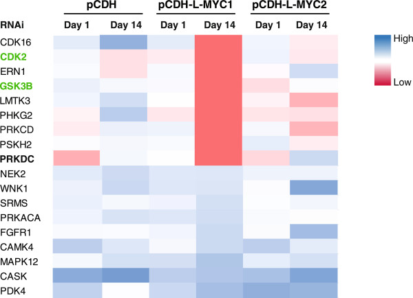
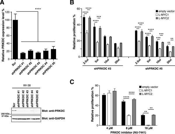
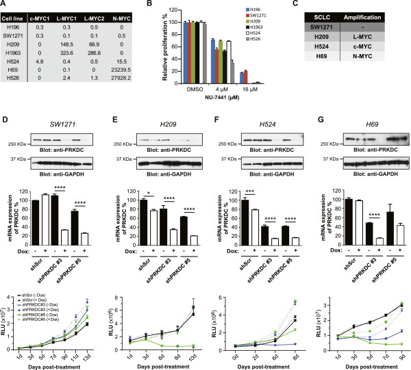
Methods: A pooled kinase shRNA library screen was performed and next-generation deep sequencing efforts identified that PRKDC was synthetically lethal in cells overexpressing MYC. Genes and proteins of interest were knocked down or inhibited using RNAi technology and small molecule inhibitors, respectively. Quantitative RT-PCR using TaqMan probes examined mRNA expression levels and cell viability was assessed using CellTiter-Glo (Promega). Western blotting was performed to monitor different protein levels in the presence or absence of RNAi or compound treatment. Statistical significance of differences among data sets were determined using unpaired t test (Mann-Whitney test) or ANOVA.

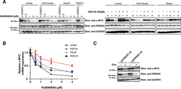
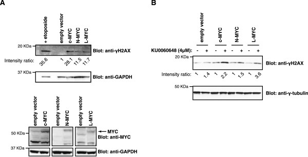
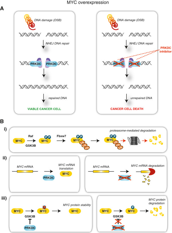
Results: Inhibition of PRKDC using RNAi (RNA interference) or small molecular inhibitors preferentially killed MYC-overexpressing human lung fibroblasts. Moreover, inducible PRKDC knockdown decreased cell viability selectively in high MYC-expressing human small cell lung cancer cell lines. At the molecular level, we found that inhibition of PRKDC downregulated MYC mRNA and protein expression in multiple cancer cell lines. In addition, we confirmed that overexpression of MYC family proteins induced DNA double-strand breaks; our results also revealed that PRKDC inhibition in these cells led to an increase in DNA damage levels.

Conclusions: Our data suggest that the synthetic lethality between PRKDC and MYC may in part be due to PRKDC dependent modulation of MYC expression, as well as MYC-induced DNA damage where PRKDC plays a key role in DNA damage repair.


Figures

Figure 1
Figure 1
Large-scale RNAi screen identifies PRKDC as a MYC synthetic lethal gene. Heat map of relative counts of 18 shRNAs for WI-38 cell lines stably-expressing empty vector (pCDH), L-MYC1 or L-MYC2, after infection with a pooled kinase shRNA library. Blue = high expression, red = low expression; intensity of color represents relative counts per million total reads. Highlighted in green (CDK2 and GSK3B) are previously published synthetic lethal partners of MYC.
Figure 2
Figure 2
PRKDC gene suppression in MYC-overexpressing human lung fibroblast cells decreases cell viability. A) Gene and protein knockdown efficiency with independent shRNA clones against PRKDC quantified by RT-PCR and immunoblotting, respectively, in WI-38 cells. Protein was analyzed via immunoblotting for PRKDC (anti-PRKDC) and GAPDH (anti-GAPDH). B) Stable WI-38 cell lines were exposed to increasing amounts of lentivirus expressing PRKDC shRNAs and subjected to a cell viability assay after 6 days. C) Stable WI-38 cell lines were treated with varying concentrations of a PRKDC inhibitor, NU-7441, for 3 days and subjected to a cell viability assay. Data are shown as mean ± SD. Statistical analysis using one-way ANOVA; ****P ≤0.0001; ***P ≤ 0.001; **P ≤ 0.01.
Figure 3
Figure 3
Inducible PRKDC knockdown decreases human SCLC cell proliferation and is dependent on high MYC expression levels. A) MYC family gene expression levels in non-isogenic SCLC cell lines (as compared to MYC family gene expression levels in 293 T cells). B) SCLC cell lines with different levels of MYC gene expression were treated with varying concentrations of a PRKDC inhibitor, NU-7441, for 3 days and subject to a cell viability assay. C) MYC gene amplification status in different SCLC cell lines. D-G) SCLC cell lines were subjected to inducible PRKDC downregulation with three independent shRNA clones and knockdown was confirmed via immunoblotting and RT-PCR after doxycycline exposure. Protein was analyzed via immunoblotting for PRKDC (anti-PRKDC) and GAPDH (anti-GAPDH). The SCLC cell lines were exposed to doxycycline for 6–13 days and then subjected to a cell viability assay. Data are shown as mean ± SD. Statistical analysis using one-way ANOVA; ****P ≤0.0001; ***P ≤ 0.001; **P ≤ 0.01; *P ≤ 0.05.
Figure 4
Figure 4
MYC mRNA and protein levels are negatively affected by PRKDC gene suppression in cancer cell lines. A) Different lymphoma cell lines were treated with increasing concentrations of the PRKDC inhibitor, KU0060648, and a proteasome inhibitor, MG132. After a 4 h drug exposure time, c-MYC protein levels were analyzed via immunoblotting with an anti-MYC antibody. PRKDC and GADPH protein levels were also monitored with anti-PRKDC and anti-GADPH antibodies, respectively. B) Cells from A were analyzed by RT-PCR for relative c-MYC mRNA expression levels. C) The H82 cell line expressing inducible PRKDC shRNA was exposed to doxycycline for 3 days. c-MYC, PRKDC and GADPH protein levels were analyzed by immunoblotting with anti-MYC antibody, anti-PRKDC and anti-GADPH antibodies, respectively. Gene knockdown efficiency with independent shRNA clones against PRKDC was quantified by RT-PCR as was relative c-MYC mRNA expression levels in these cell lines.
Figure 5
Figure 5
MYC overexpression induces double-strand breaks in DNA. A) A SV40-transformed human lung cell line, WI-38 VA13, stably-expressing c-MYC, L-MYC, or N-MYC, were lyzed and immunoblots were analyzed for phosphorylated histone H2AX (anti-γH2AX) and GADPH (anti-GADPH). As a positive control for DNA damage detection, parental cells were treated with etoposide and analyzed via immunoblotting. Cell lysates were also analyzed for overexpression of MYC variants. B) The same cells lines as in A were treated with the PRKDC inhibitor, KU0060648, for 8 h, lyzed and immunoblotted for phosphorylated histone H2AX (anti-γH2AX) and γ-tubulin (anti-γ-tubulin). Signal intensities of immunoblots were quantified using ImageJ.
Figure 6
Figure 6
Proposed model for synthetic lethality between MYC and PRKDC. A) In MYC-driven cancer cells, the overexpression of MYC leads to DNA damage and creates a dependence on DNA repair machinery for cancer cell survival. Double strand breaks (DSBs) induce non-homologous end joining (NHEJ) DNA repair mechanisms to correct for DNA insult. PRKDC is a major player during NHEJ repair, and along with other key components, will restore the impaired DNA allowing for cancer cell viability. In these same cells, exposure to anti-PRKDC drug treatments would ultimately lead to PRKDC inhibition, compromised NHEJ repair and cell death. B) PRKDC has also been implicated in MYC gene regulation. i) In a normal setting, MYC protein is phosphorylated through RAF- and AKT-mediated signaling cascades, resulting in its FBW7-mediated ubiquitination, and subsequent proteasomal degradation. ii) In cancer cells, at the gene level, the inhibition of PRKDC protein decreases MYC expression, potentially through a direct effect or epigenetic mechanism(s). iii) Additionally in cancer cells, PRKDC can phosphorylate AKT, which results in the inhibition of GSK3β and subsequent MYC degradation. Therefore, interference with PRKDC function(s) decreases the stability of MYC protein. This form of genotypic cytotoxicity represents synthetic lethality that selectively targets cancer cells while leaving normal cells unscathed, and offers a potential for wider therapeutic windows for cancer therapies.

Similar articles

Cited by

References

    1. Roberts PJ, Der CJ. Targeting the Raf-MEK-ERK mitogen-activated protein kinase cascade for the treatment of cancer. Oncogene. 2007;26(22):3291–3310. doi: 10.1038/sj.onc.1210422. - DOI - PubMed
    1. Karnoub AE, Weinberg RA. Ras oncogenes: split personalities. Nat Rev Mol Cell Biol. 2008;9(7):517–531. doi: 10.1038/nrm2438. - DOI - PMC - PubMed
    1. Beroukhim R, Mermel CH, Porter D, Wei G, Raychaudhuri S, Donovan J, Barretina J, Boehm JS, Dobson J, Urashima M, Mc Henry K, Pinchback R, Ligon A, Cho Y, Haery L, Greulich H, Reich M, Winckler W, Lawrence M, Weir B, Tanaka K, Chiang D, Bass A, Loo A, Hoffman C, Prensner J, Liefeld T, Gao Q, Yecies D, Signoretti S. The landscape of somatic copy-number alteration across human cancers. Nature. 2010;463(7283):899–905. doi: 10.1038/nature08822. - DOI - PMC - PubMed
    1. Dang CV. MYC on the path to cancer. Cell. 2012;149(1):22–35. doi: 10.1016/j.cell.2012.03.003. - DOI - PMC - PubMed
    1. Larsson LG, Henriksson MA. The Yin and Yang functions of the Myc oncoprotein in cancer development and as targets for therapy. Exp Cell Res. 2010;316(8):1429–1437. doi: 10.1016/j.yexcr.2010.03.025. - DOI - PubMed
Pre-publication history
    1. The pre-publication history for this paper can be accessed here:http://www.biomedcentral.com/1471-2407/14/944/prepub

Publication types

MeSH terms

LinkOut - more resources