

Pseudomonas aeruginosa keratitis misdiagnosed as fungal keratitis by in vivo confocal microscopy: a case report
- PMID: 25495791
- PMCID: PMC4417546
- DOI: 10.1186/1756-0500-7-907
Pseudomonas aeruginosa keratitis misdiagnosed as fungal keratitis by in vivo confocal microscopy: a case report
Abstract
Background: To report a case of non-typical Pseudomonas aeruginosa keratitis that was misdiagnosed as fungal keratitis by in vivo confocal microscopy.
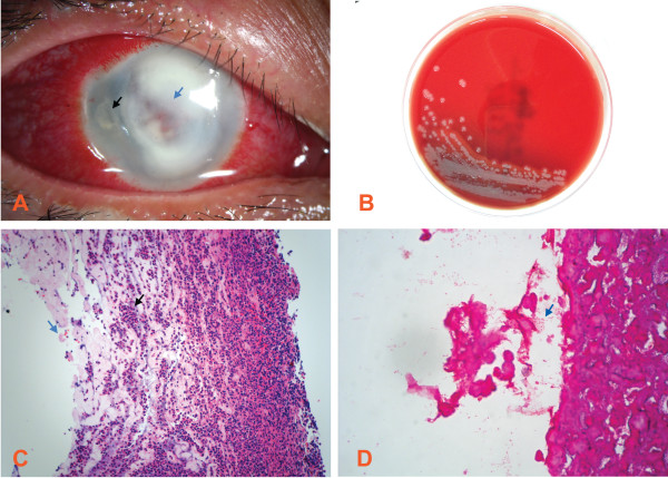
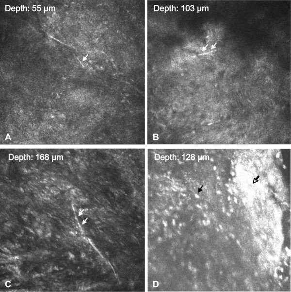
Case presentation: A 37-year-old Chinese woman presented with a 2-week history of increasing pain and redness of the right eye. She was started on hourly topical fortified tobramycin and levofloxacin by the referring doctor without improvement. She denied any improvement of her symptoms and signs. On examination, she had a large central corneal ulcer extending to the peripheral cornea. Further symptoms included a satellite lesion, intense conjunctival injection and marked corneal oedema. The corneal scrape was not performed initially because of the deep infiltrate in the stroma. The patient was examined by in vivo confocal microscopy. Confocal microscopy images showed hyper-reflective, thin, and branching interlocking linear structures in the stroma that were 5-8 μm in width and 200-400 μm in length. The morphology was consistent with that of fungus. However, the histopathological examination, Gram stain, and culture of the cornea only confirmed the presence of a Pseudomonas species within the deep strom. No fungal element was found. The pathogen was sensitive to ciprofloxacin, gentamicin, levofloxacin, tobramycin and amikacin.
Conclusion: This case reports the potential for a false positive finding of fungus in Pseudomonas aeruginosa keratitis and emphasizes the importance of bacterial culture and antibiotic susceptibility testing in the management of microbial keratitis.
Figures
References
-
- de Rojas Silva MV, Abraldes MJ, Díez-Feijóo E, Yáñez PM, Javaloy J, Sánchez-Salorio M. Confocal microscopy and histopathological examination of diffuse lamellar keratitis in an experimental animal model. J Refract Surg. 2007;23(3):299–304. - PubMed
Publication types
MeSH terms
LinkOut - more resources
Full Text Sources
Other Literature Sources
