

Thioredoxin reductase activity may be more important than GSH level in protecting human lens epithelial cells against UVA light
- PMID: 25495870
- PMCID: PMC4355078
- DOI: 10.1111/php.12404
Thioredoxin reductase activity may be more important than GSH level in protecting human lens epithelial cells against UVA light
Abstract
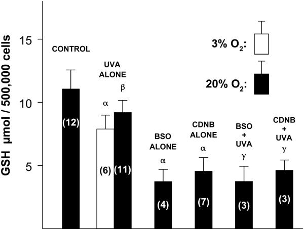
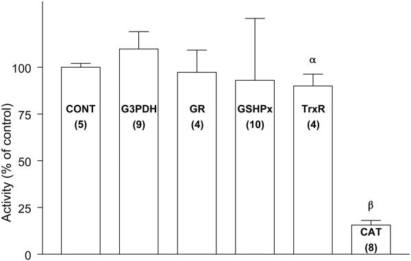
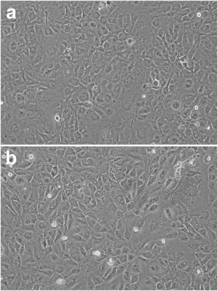
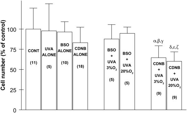
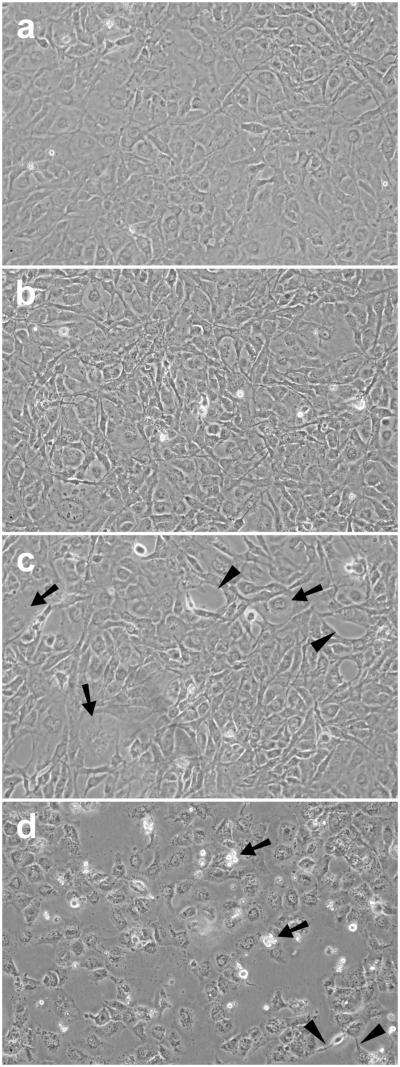
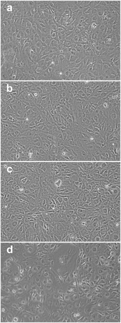
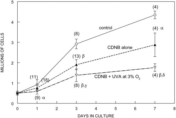
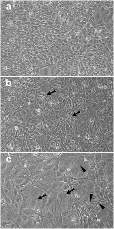
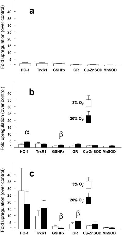
This study compares the abilities of the glutathione (GSH) and thioredoxin (Trx) antioxidant systems in defending cultured human lens epithelial cells (LECs) against UVA light. Levels of GSH were depleted with either L-buthionine-(S,R)-sulfoximine (BSO) or 1-chloro-2,4-dinitrobenzene (CDNB). CDNB treatment also inhibited the activity of thioredoxin reductase (TrxR). Two levels of O2 , 3% and 20%, were employed during a 1 h exposure of the cells to 25 J cm(-2) of UVA radiation (338-400 nm wavelength, peak at 365 nm). Inhibition of TrxR activity by CDNB, combined with exposure to UVA light, produced a substantial loss of LECs and cell damage, with the effects being considerably more severe at 20% O2 compared to 3%. In contrast, depletion of GSH by BSO, combined with exposure to UVA light, produced only a slight cell loss, with no apparent morphological effects. Catalase was highly sensitive to UVA-induced inactivation, but was not essential for protection. Although UVA light presented a challenge for the lens epithelium, it was well tolerated under normal conditions. The results demonstrate an important role for TrxR activity in defending the lens epithelium against UVA light, possibly related to the ability of the Trx system to assist DNA synthesis following UVA-induced cell damage.
© 2014 The American Society of Photobiology.
Figures
Similar articles
-
Protection from oxidative insult in glutathione depleted lens epithelial cells.Exp Eye Res. 1999 Jan;68(1):117-27. doi: 10.1006/exer.1998.0606. Exp Eye Res. 1999. PMID: 9986749
-
Regulation of thioltransferase expression in human lens epithelial cells.Invest Ophthalmol Vis Sci. 2001 Apr;42(5):1002-8. Invest Ophthalmol Vis Sci. 2001. PMID: 11274078
-
Depletion of glutathione by L-buthionine sulfoximine does not promote inactivation of myo-inositol transport in cultured bovine lens epithelial cells.Curr Eye Res. 1991 Apr;10(4):321-30. doi: 10.3109/02713689108996338. Curr Eye Res. 1991. PMID: 1676962
-
The thioredoxin antioxidant system.Free Radic Biol Med. 2014 Jan;66:75-87. doi: 10.1016/j.freeradbiomed.2013.07.036. Epub 2013 Jul 27. Free Radic Biol Med. 2014. PMID: 23899494 Review.
-
Effects of intermittent UVA exposure on cultured lens epithelial cells.Curr Eye Res. 2000 Feb;20(2):95-100. Curr Eye Res. 2000. PMID: 10617909
Cited by
-
PARP-1/PAR Activity in Cultured Human Lens Epithelial Cells Exposed to Two Levels of UVB Light.Photochem Photobiol. 2018 Jan;94(1):126-138. doi: 10.1111/php.12814. Epub 2017 Sep 15. Photochem Photobiol. 2018. PMID: 28756616 Free PMC article.
-
UV light and the ocular lens: a review of exposure models and resulting biomolecular changes.Front Ophthalmol (Lausanne). 2024 Sep 5;4:1414483. doi: 10.3389/fopht.2024.1414483. eCollection 2024. Front Ophthalmol (Lausanne). 2024. PMID: 39301012 Free PMC article. Review.
-
Reactive oxygen species prevent lysosome coalescence during PIKfyve inhibition.PLoS One. 2021 Nov 23;16(11):e0259313. doi: 10.1371/journal.pone.0259313. eCollection 2021. PLoS One. 2021. PMID: 34813622 Free PMC article.
-
Understanding cataract development in axial myopia: The contribution of oxidative stress and related pathways.Redox Biol. 2025 Mar;80:103495. doi: 10.1016/j.redox.2025.103495. Epub 2025 Jan 10. Redox Biol. 2025. PMID: 39813957 Free PMC article. Review.
-
The role of sodium hydrosulfide in attenuating the aging process via PI3K/AKT and CaMKKβ/AMPK pathways.Redox Biol. 2017 Aug;12:987-1003. doi: 10.1016/j.redox.2017.04.031. Epub 2017 Apr 25. Redox Biol. 2017. PMID: 28499253 Free PMC article.
References
-
- Zigman S. Environmental near-UV radiation and cataracts. Optometry and vision science : official publication of the American Academy of Optometry. 1995;72:899–901. - PubMed
-
- Dillon J, Zheng L, Merriam JC, Gaillard ER. The optical properties of the anterior segment of the eye: implications for cortical cataract. Experimental eye research. 1999;68:785–95. - PubMed
-
- Tyrrell R. UVA (320-380nm) radiation as an oxidative stress. In: Sies H, editor. Oxidative stress: oxidants and antioxidants. Academic Press; San Diego, CA: 1991. pp. 57–83.
-
- McMillan TJ, Leatherman E, Ridley A, Shorrocks J, Tobi SE, Whiteside JR. Cellular effects of long wavelength UV light (UVA) in mammalian cells. The Journal of pharmacy and pharmacology. 2008;60:969–76. - PubMed
-
- Giblin FJ, Reddy VN. Pyridine nucleotides in ocular tissues as determined by the cycling assay. Experimental eye research. 1980;31:601–9. - PubMed
Publication types
MeSH terms
Substances
Grants and funding
LinkOut - more resources
Full Text Sources
Other Literature Sources
Miscellaneous
