

Expression of bioactive lysophospholipids and processing enzymes in the vitreous from patients with proliferative diabetic retinopathy
- PMID: 25496321
- PMCID: PMC4293108
- DOI: 10.1186/1476-511X-13-187
Expression of bioactive lysophospholipids and processing enzymes in the vitreous from patients with proliferative diabetic retinopathy
Abstract
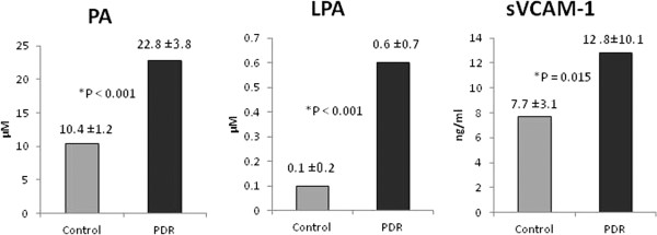
Background: The bioactive lysophospholipids phosphatidic acid (PA), lysophosphatidic acid (LPA) and sphingosine-1-phosphate (S1P) have been implicated in mediating cell migration, proliferation and apoptosis, inflammation, angiogenesis and fibrosis. This study was conducted to measure the levels of PA, LPA, LPA-producing enzymes phospholipase A1/A2 (PLA1A/PLA2, respectively) and acylgylycerol kinase (AGK), the S1P receptor S1PR1, the S1P catabolising enzyme S1P lyase (SPL) and 5-lipoxygenase in the vitreous fluid from patients with proliferative diabetic retinopathy (PDR). In addition, we investigated the correlations between the levels of PA and LPA and the levels of the inflammatory and endothelial dysfunction biomarker soluble vascular cell adhesion molecule-1 (sVCAM-1).
Methods: Vitreous samples from 34 PDR and 29 nondiabetic patients were studied by biochemical and enzyme-linked immunosorbent assays and Western blot analysis.
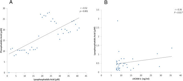
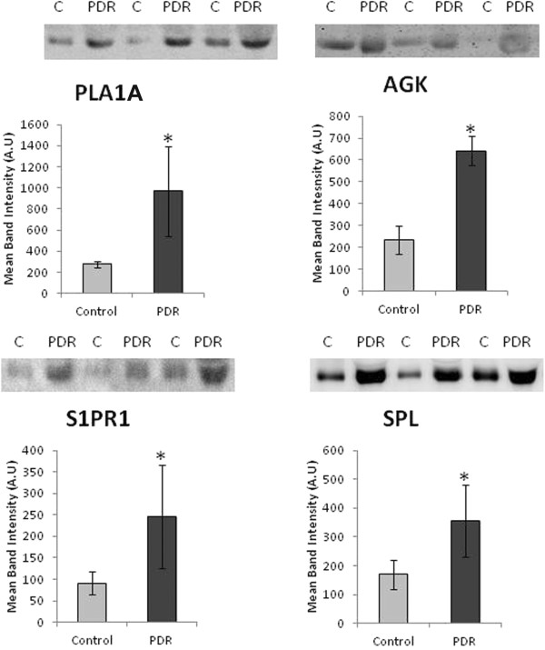
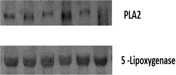
Results: PA, LPA and sVCAM-1 levels in vitreous samples from PDR patients were significantly higher than those in nondiabetic patients. Significant correlations were observed between levels of LPA and levels of PA and sVCAM-1. Western blot analysis revealed a significant increase in the expression of PLA1A, AGK, S1PR1 and SPL in vitreous samples from PDR patients compared to nondiabetic controls, whereas PLA2 and 5-lipoxygenase were not detected.
Conclusions: Our findings suggest that the enzymatic activities of PLA1A and AGK might be responsible for increased synthesis of LPA in PDR and that PLA1A, but not PLA2 is responsible for deacylation of PA to generate LPA. S1PR1 and SPL might regulate inflammatory, angiogenic and fibrogenic responses in PDR.
Figures
References
-
- Abu El-Asrar AM, Struyf S, Kangave D, Geboes K, Van Damme J. Chemokines in proliferative diabetic retinopathy and proliferative vitreoretinopathy. Eur Cytokine Netw. 2006;17:155–165. - PubMed
-
- Abu El-Asrar AM, Mohammad G, Nawaz MI, Siddiquei MM, Van den Eynde K, Mousa A, De Hertogh G, Opdenakker G. Relationship between vitreous levels of matrix metalloproteinases and vascular endothelial growth factor in proliferative diabetic retinopathy. PLoS One. 2013;8:e85857. doi: 10.1371/journal.pone.0085857. - DOI - PMC - PubMed
Publication types
MeSH terms
Substances
LinkOut - more resources
Full Text Sources
Other Literature Sources
Medical
Miscellaneous
