

Color-coded digital subtraction angiography in the management of a rare case of middle cerebral artery pure arterial malformation. A technical and case report
- PMID: 25496681
- PMCID: PMC4295243
- DOI: 10.15274/INR-2014-10086
Color-coded digital subtraction angiography in the management of a rare case of middle cerebral artery pure arterial malformation. A technical and case report
Abstract
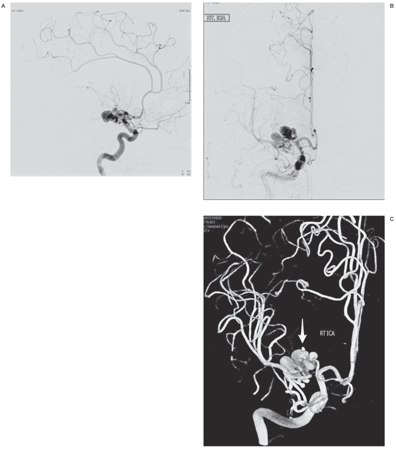
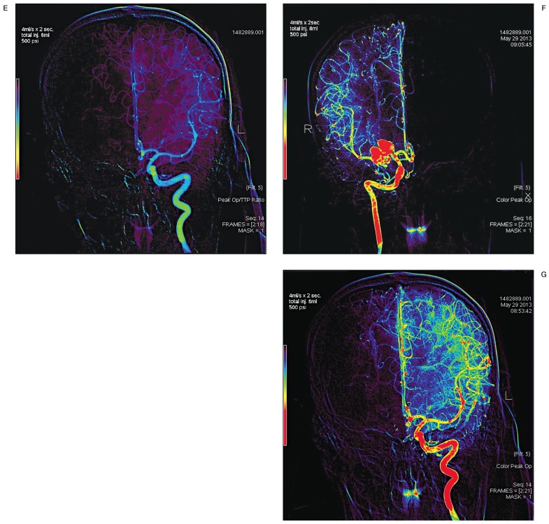
The advent of flow dynamics and the recent availability of perfusion analysis software have provided new diagnostic tools and management possibilities for cerebrovascular patients. To this end, we provide an example of the use of color-coded angiography and its application in a rare case of a patient with a pure middle cerebral artery (MCA) malformation. A 42-year-old male chronic smoker was evaluated in the emergency room due to sudden onset of severe headache, nausea, vomiting and left-sided weakness. Head computed tomography revealed a right basal ganglia hemorrhage. Cerebral digital subtraction angiography (DSA) showed a right middle cerebral artery malformation consisting of convoluted and ectatic collateral vessels supplying the distal middle cerebral artery territory-M1 proximally occluded. An associated medial lenticulostriate artery aneurysm was found. Brain single-photon emission computed tomography with and without acetazolamide failed to show problems in vascular reserve that would indicate the need for flow augmentation. Twelve months after discharge, the patient recovered from the left-sided weakness and did not present any similar events. A follow-up DSA and perfusion study using color-coded perfusion analysis showed perforator aneurysm resolution and adequate, albeit delayed perfusion in the involved vascular territory. We propose a combined congenital and acquired mechanism involving M1 occlusion with secondary dysplastic changes in collateral supply to the distal MCA territory. Angiographic and cerebral perfusion work-up was used to exclude the need for flow augmentation. Nevertheless, the natural course of this lesion remains unclear and long-term follow-up is warranted.
Keywords: arterial malformation; color-coded angiography; intracerebral hemorrhage.
Figures


Similar articles
-
Revascularization for Aplastic or Twiglike Middle Cerebral Artery: A Case Report.J Stroke Cerebrovasc Dis. 2018 May;27(5):e78-e79. doi: 10.1016/j.jstrokecerebrovasdis.2017.12.004. Epub 2018 Jan 5. J Stroke Cerebrovasc Dis. 2018. PMID: 29310957
-
[A case of spontaneous middle cerebral artery occlusion associated with a cerebral aneurysm angiographically disappearing after STA-MCA anastomosis].No Shinkei Geka. 1997 Aug;25(8):727-32. No Shinkei Geka. 1997. PMID: 9266566 Japanese.
-
Nonaneurysmal Subarachnoid Hemorrhage Due to Unfused or Twiglike Middle Cerebral Artery Rupture: Two Case Reports.J Stroke Cerebrovasc Dis. 2016 Jun;25(6):e77-8. doi: 10.1016/j.jstrokecerebrovasdis.2016.03.029. Epub 2016 Apr 8. J Stroke Cerebrovasc Dis. 2016. PMID: 27068778
-
Rete middle cerebral artery anomalies: a unifying name, case series, and literature review.J Neurosurg. 2018 Aug 3;131(2):453-461. doi: 10.3171/2018.2.JNS1832. Print 2019 Aug 1. J Neurosurg. 2018. PMID: 30074465 Review.
-
Real-Time Distal, Multifocal, Repeated Lenticulostriate Bleeding Points during Thrombectomy in a Patient with Acute Variable M1 Occlusion: A Case Report and a Literature Review.J Stroke Cerebrovasc Dis. 2017 Oct;26(10):2082-2086. doi: 10.1016/j.jstrokecerebrovasdis.2017.04.026. Epub 2017 Jun 1. J Stroke Cerebrovasc Dis. 2017. PMID: 28579509 Review.
Cited by
-
Pure arterial malformation with associated aneurysmal subarachnoid hemorrhage: Two case reports and literature review.Zhong Nan Da Xue Xue Bao Yi Xue Ban. 2021 Feb 28;46(2):200-206. doi: 10.11817/j.issn.1672-7347.2021.190673. Zhong Nan Da Xue Xue Bao Yi Xue Ban. 2021. PMID: 33678659 Free PMC article. Review. Chinese, English.
-
Reliability and Accuracy of Peri-Interventional Stenosis Grading in Peripheral Artery Disease Using Color-Coded Quantitative Fluoroscopy: A Phantom Study Comparing a Clinical and Scientific Postprocessing Software.Biomed Res Int. 2018 Jul 24;2018:6180138. doi: 10.1155/2018/6180138. eCollection 2018. Biomed Res Int. 2018. PMID: 30140698 Free PMC article.
-
Diagnosis and treatment of pure arterial malformation: Three case reports and literature review.Medicine (Baltimore). 2020 May 22;99(21):e20229. doi: 10.1097/MD.0000000000020229. Medicine (Baltimore). 2020. PMID: 32481296 Free PMC article. Review.
-
Microneurosurgical treatment of a small perimesencephalic pure pial arterial malformation: an under-recognized etiology of angiographically occult subarachnoid hemorrhage. Illustrative case.J Neurosurg Case Lessons. 2023 Jul 31;6(5):CASE23246. doi: 10.3171/CASE23246. Print 2023 Jul 31. J Neurosurg Case Lessons. 2023. PMID: 37548523 Free PMC article.
-
Endovascular treatment of a ruptured pure arterial malformation and associated dysplastic middle cerebral artery dissecting aneurysm: illustrative case.J Neurosurg Case Lessons. 2023 May 22;5(21):CASE23150. doi: 10.3171/CASE23150. Print 2023 May 22. J Neurosurg Case Lessons. 2023. PMID: 37218731 Free PMC article.
References
-
- Borghammer P. Perfusion and metabolism imaging studies in Parkinson's disease. Dan Med J. 2012;59:B4466. - PubMed
-
- Hochberg AR, Young GS. Cerebral perfusion imaging. Semin Neurol. 2012;32:454–465. doi: 10.1055/s-0032-1331815. - DOI - PubMed
-
- Kim DJ, Krings T. Whole-brain perfusion CT patterns of brain arteriovenous malformations: a pilot study in 18 patients. Am J Neuroradiol. 2011;32:2061–2066. doi: 10.3174/ajnr.A2659. - DOI - PMC - PubMed
-
- Peet AC, Arvanitis TN, Leach MO, et al. Functional imaging in adult and paediatric brain tumours. Nat Rev Clin Oncol. 2012;9:700–711. doi: 10.1038/nrclinonc.2012.187. - DOI - PubMed
-
- Golitz P, Struffert T, Lucking H, et al. Parametric color coding of digital subtraction angiography in the evaluation of carotid cavernous fistulas. Clin Neuroradiol. 2013;23:113–120. doi: 10.1007/s00062-012-0184-8. - DOI - PubMed
Publication types
MeSH terms
Substances
LinkOut - more resources
Full Text Sources
Other Literature Sources
