

Case Reports
doi: 10.15274/INR-2014-10071.
Epub 2014 Dec 5.
Endovascular treatment of a ruptured flow aneurysm of the heubner artery as part of a moyamoya collateral network in a young patient with an occluded middle cerebral artery
Affiliations
- PMID: 25496692
- PMCID: PMC4295254
- DOI: 10.15274/INR-2014-10071
Item in Clipboard
Case Reports
Endovascular treatment of a ruptured flow aneurysm of the heubner artery as part of a moyamoya collateral network in a young patient with an occluded middle cerebral artery
Interv Neuroradiol.
2014 Dec.
Abstract
A young woman with an occluded middle cerebral artery presented with a ruptured flow aneurysm distal on a Heubner artery as part of a moyamoya collateral network. Leptomeningeal collateral supply was tested by occlusion of the A1 origin of the Heubner artery. This test occlusion demonstrated ample collateral leptomeningeal supply over the hemispheres to the M2. Subsequently, the Heubner artery harbouring the aneurysm could be safely proximally occluded with coils.
Keywords: Heubner artery; leptomeningeal collateral circulation; moyamoya; test occlusion.
Figures
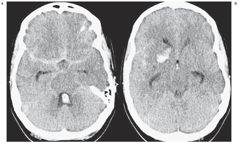
CT scan with subarachnoid, intraventricular and intraparenchymal blood.
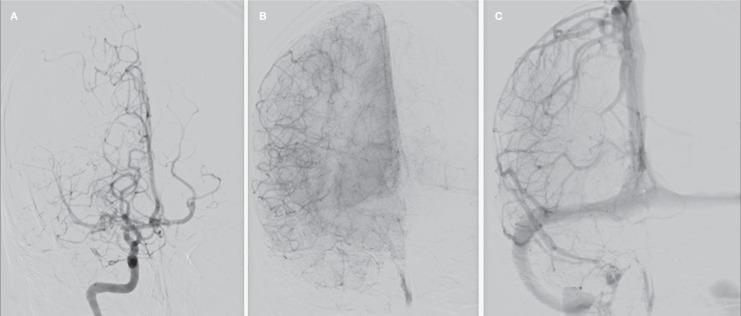
Frontal right internal carotid angiogram in early (A), mid (B) and late (C) phase showing M1 occlusion and perfusion of right hemisphere by the basal collateral moyamoya network and leptomeningeal collateral vessels.
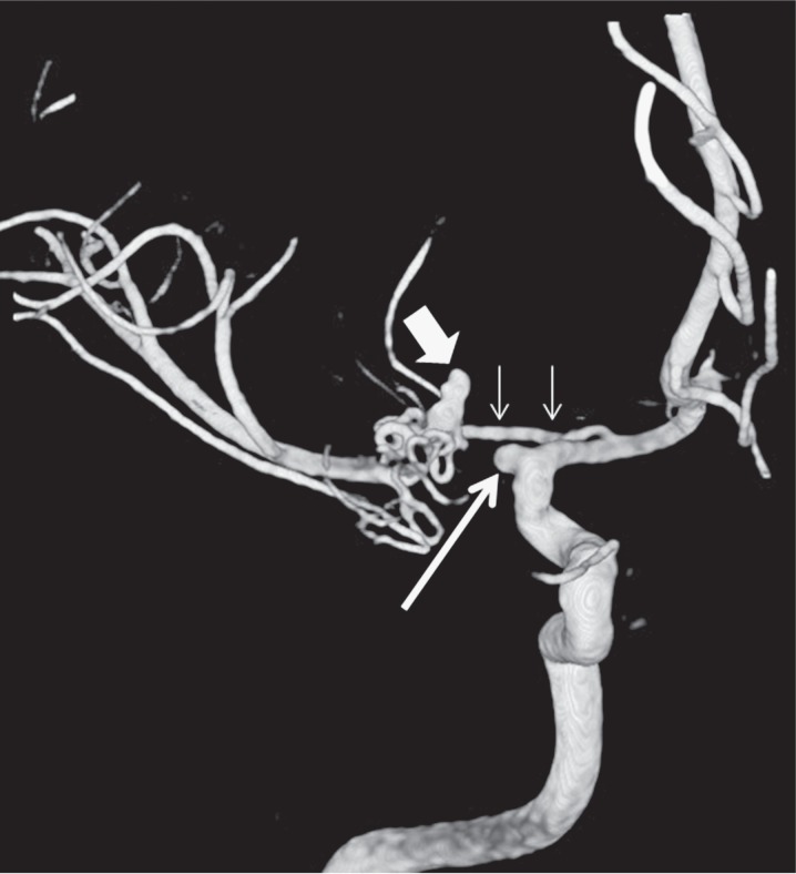
3D angiogram of the right internal carotid artery shows the occluded middle cerebral artery at its origin (long arrow), hypertrophied Heubner artery as part of a collateral network (small arrows) and ruptured aneurysm on the distal Heubner artery (broad arrow).
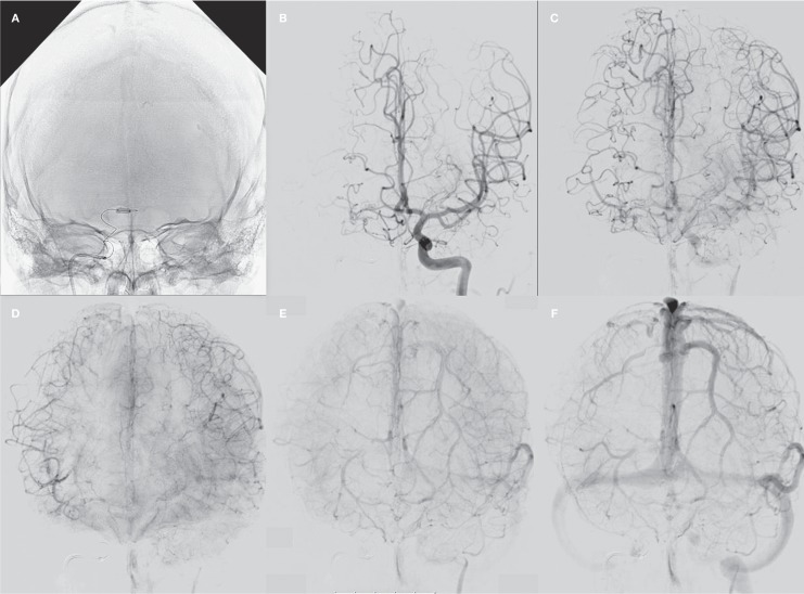
Test occlusion of the right A1. A) Balloon inflated in the right A1 occluding the origin of the Heubner artery. B-F) Arterial, parenchymal and venous phases of left internal carotid angiogram during test occlusion: abundant leptomeningeal collateral supply over the hemispheres with synchronous venous phase filling.
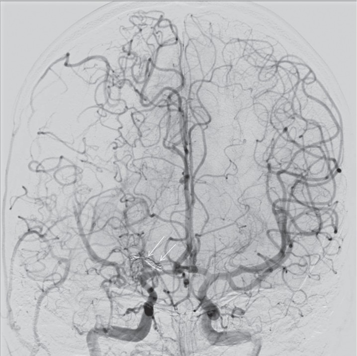
Bilateral internal carotid angiogram after proximal coil occlusion of the Heubner artery (arrows).
Similar articles
-
Mechanism of moyamoya vessels secondary to intracranial atherosclerotic disease: angiographic findings in patients with middle cerebral artery occlusion.J Stroke Cerebrovasc Dis. 2012 Jul;21(5):373-8. doi: 10.1016/j.jstrokecerebrovasdis.2010.10.001. Epub 2010 Nov 26. J Stroke Cerebrovasc Dis. 2012. PMID: 21111634
-
Microsurgical Repair of Ruptured Aneurysms Associated with Moyamoya-Pattern Collateral Vessels of the Middle Cerebral Artery: A Report of Two Cases.World Neurosurg. 2017 Sep;105:1042.e5-1042.e10. doi: 10.1016/j.wneu.2017.06.166. Epub 2017 Jul 8. World Neurosurg. 2017. PMID: 28698088
-
A ruptured aneurysm arising at the leptomeningeal collateral circulation from the extracranial vertebral artery to the posterior inferior cerebellar artery associated with bilateral vertebral artery occlusion.J Stroke Cerebrovasc Dis. 2014 Feb;23(2):e135-9. doi: 10.1016/j.jstrokecerebrovasdis.2013.08.028. Epub 2013 Dec 8. J Stroke Cerebrovasc Dis. 2014. PMID: 24321776
-
Prevention of the Rerupture of Collateral Artery Aneurysms on the Ventricular Wall by Early Surgical Revascularization in Moyamoya Disease: Report of Two Cases and Review of the Literature.World Neurosurg. 2018 Jan;109:393-397. doi: 10.1016/j.wneu.2017.10.059. Epub 2017 Oct 20. World Neurosurg. 2018. PMID: 29061453 Review.
-
[Endovascular treatment of a ruptured aneurysm associated with unilateral moyamoya disease].No Shinkei Geka. 2004 Feb;32(2):167-71. No Shinkei Geka. 2004. PMID: 15031978 Review. Japanese.
Cited by
-
Endovascular glue embolization of dissecting aneurysm of type-3 accessory middle cerebral artery: A contralateral approach.Interv Neuroradiol. 2015 Dec;21(6):664-8. doi: 10.1177/1591019915609780. Epub 2015 Oct 27. Interv Neuroradiol. 2015. PMID: 26508091 Free PMC article.
-
Spontaneous Obliteration of a Dissecting Aneurysm of Recurrent Artery of Heubner Monitored by Serial Magnetic Resonance Vessel Wall Imaging.Asian J Neurosurg. 2022 Aug 25;17(2):331-336. doi: 10.1055/s-0042-1750712. eCollection 2022 Jun. Asian J Neurosurg. 2022. PMID: 36120605 Free PMC article.
-
Spontaneous thrombosis of an unruptured recurrent artery of Heubner aneurysm within a few months: illustrative case.J Neurosurg Case Lessons. 2025 Jul 28;10(4):CASE25295. doi: 10.3171/CASE25295. Print 2025 Jul 28. J Neurosurg Case Lessons. 2025. PMID: 40720898 Free PMC article.
-
Western Moyamoya Phenotype: A Scoping Review.Cureus. 2021 Nov 22;13(11):e19812. doi: 10.7759/cureus.19812. eCollection 2021 Nov. Cureus. 2021. PMID: 34956795 Free PMC article.
References
-
- Fukawa O, Aihara H, Ishii K, et al. Middle cerebral artery occlusion with moyamoya phenomenon. First report: clinical course and angiographic findings.; Proceedings of the 10th Conference of Surgery for Cerebral Stroke; Tokyo. 1981. pp. 36–41.
-
- Yasargil MG, Smith RD. Association of middle cerebral artery anomalies with saccular aneurysms and Moyamoya disease. Surg Neurol. 1976;6(1):39–43. - PubMed
-
- Seki Y, Fujita M, Mizutani N, et al. Spontaneous middle cerebral artery occlusion leading to moyamoya phenomenon and aneurysm formation on collateral arteries. Surg Neurol. 2001;55:58–62. Discussion 62. - PubMed
-
- Yeon JY, Shin HJ, Seol HJ, et al. Unilateral intracranial arteriopathy in pediatric stroke: Course, outcome, and prediction of reversible arteriopathy. Stroke. 2014;5(4):1173–6. doi: 10.1161/STROKEAHA.113.004125. - DOI - PubMed
-
- Yamashita M, Oka K, Tanaka K. Histopathology of the brain vascular network in moyamoya disease. Stroke. 1983;14(1):50–58. doi: 10.1161/01.STR.14.1.50. - DOI - PubMed
Publication types
MeSH terms
LinkOut - more resources
Full Text Sources
Other Literature Sources
