Review
. 2015 Jan;14(1):81-91.
doi: 10.1016/S1474-4422(14)70193-0.

Functional MRI of migraine

Affiliations
Review

Functional MRI of migraine

Todd J Schwedt et al. Lancet Neurol. 2015 Jan.

Abstract

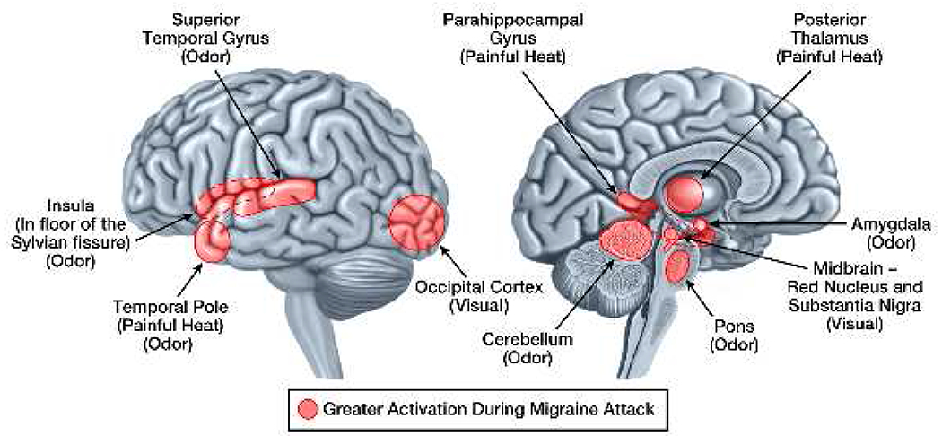
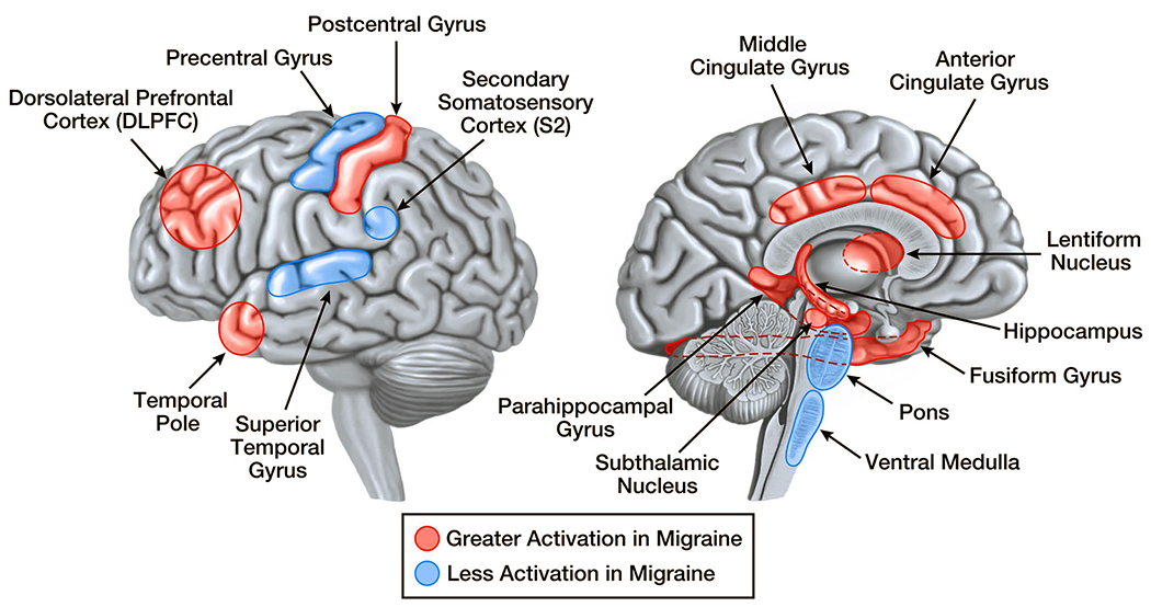
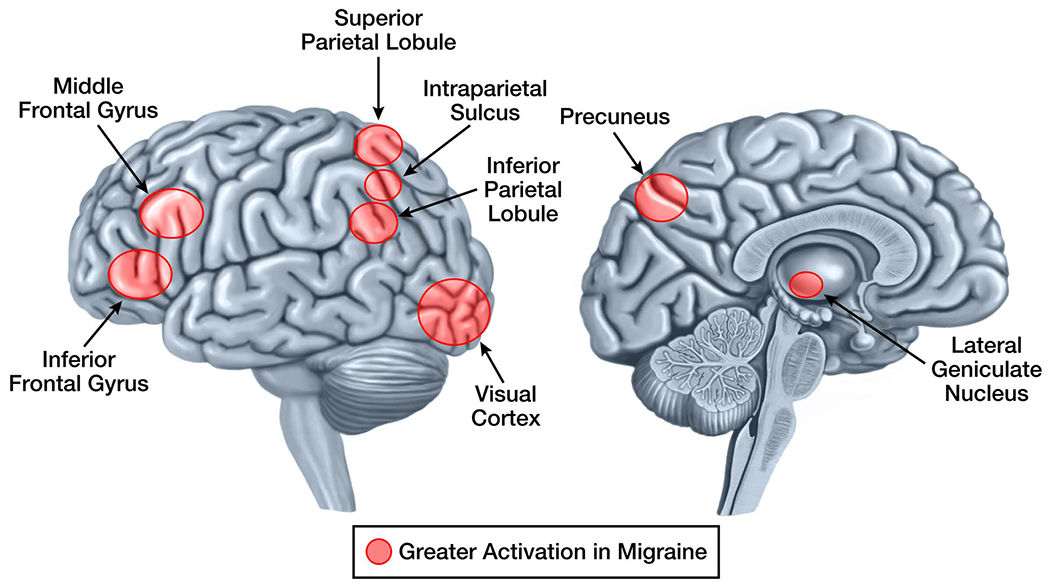
Migraine is a disabling neurological condition manifesting with attacks of headache, hypersensitivities to visual, auditory, olfactory and somatosensory stimuli, nausea, and vomiting. Exposure to sensory stimuli, such as odours, visual stimuli, and sounds, commonly triggers migraine attacks, and hypersensitivities to sensory stimuli are prominent during migraine attacks, but can persist with less magnitude between attacks. Functional MRI (fMRI) has been used to investigate the mechanisms that lead to migraine sensory hypersensitivities by measuring brain responses to visual, olfactory, and painful cutaneous stimulation, and functional connectivity analyses have investigated the functional organisation of specific brain regions and networks responsible for sensory processing. These studies have consistently shown atypical brain responses to sensory stimuli, absence of the normal habituating response between attacks, and atypical functional connectivity of sensory processing regions. Identification of the mechanisms that lead to migraine sensory hypersensitivities and that trigger migraine attacks in response to sensory stimuli might help to better understand neural dysfunction in migraine and provide new targets for migraine prevention, and could provide fMRI biomarkers that indicate early responses to preventive therapy.


Conflict of interest statement

Conflicts of Interest: All authors state no potential conflicts of interest regarding the contents of this manuscript.

Figures

Figure 1:
Figure 1:. Pain-Induced Brain Activations that Differ in Migraineurs vs. Controls.
Brain regions that have pain-induced activations in interictal migraineurs that differ from activations in healthy controls are demonstrated on the lateral and medial brain surfaces. Areas shaded red are those that have greater activation in migraine compared to controls. Areas shaded blueare those that have less activation in migraine compared to controls. Although shaded areas are within regions of the brain that show differential activation between migraineurs and controls, their placement does not represent the exact location, size, or extent of differential activation.
Figure 2:
Figure 2:. Stimulus-Induced Brain Activations that Differ in Ictal vs. Interictal Migraineurs.
Brain regions that have pain, visual, and odor-induced activations that differ in migraineurs during a migraine vs. between migraines are demonstrated on the lateral and medial brain surfaces. Areas shaded red are those that have greater activation during a migraine attack compared to between migraine attacks. There are no areas that have less activation during a migraine compared to between migraines.Although shaded areas are within regions of the brain that show differential activation between migraineurs during migraine attacks vs. migraineurs between migraine attacks, their placement does not represent the exact location, size, or extent of differential activation.
Figure 3:
Figure 3:. Visual Stimuli-Induced Brain Activations that Differ in Migraineurs vs. Controls.
Brain regions that have visual stimuli-induced activations in interictal migraineurs that differ from activations in healthy controls are demonstrated on the lateral and medial brain surfaces. Areas shaded red are those that have greater activation in migraine compared to controls. There are no areas that have less activation in migraine compared to controls. Although shaded areas are within regions of the brain that show differential activation between migraineurs and controls, their placement does not represent the exact location, size, or extent of differential activation.

References

    1. Lipton RB, Bigal ME, Diamond M, Freitag F, Reed ML, Stewart WF. Migraine prevalence, disease burden, and the need for preventive therapy. Neurology. 2007; 68(5): 343–9. - PubMed
    1. The International Classification of Headache Disorders, 3rd edition (beta version). Cephalalgia. 2013; 33(9): 629–808. - PubMed
    1. Schwedt TJ. Multisensory integration in migraine. Curr Opin Neurol. 2013; 26(3): 248–53. - PMC - PubMed
    1. Launer LJ, Terwindt GM, Ferrari MD. The prevalence and characteristics of migraine in a population-based cohort: the GEM study. Neurology. 1999; 53(3): 537–42. - PubMed
    1. Russell MB, Olesen J. A nosographic analysis of the migraine aura in a general population. Brain. 1996; 119 (Pt 2): 355–61. - PubMed

Publication types

MeSH terms