. 2014 Dec 24;9(6):2219-32.
doi: 10.1016/j.celrep.2014.11.033. Epub 2014 Dec 11.

Rfx6 maintains the functional identity of adult pancreatic β cells

Affiliations

Rfx6 maintains the functional identity of adult pancreatic β cells

Julie Piccand et al. Cell Rep. .

Abstract

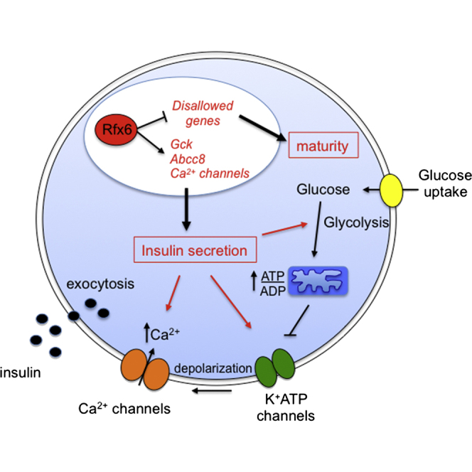
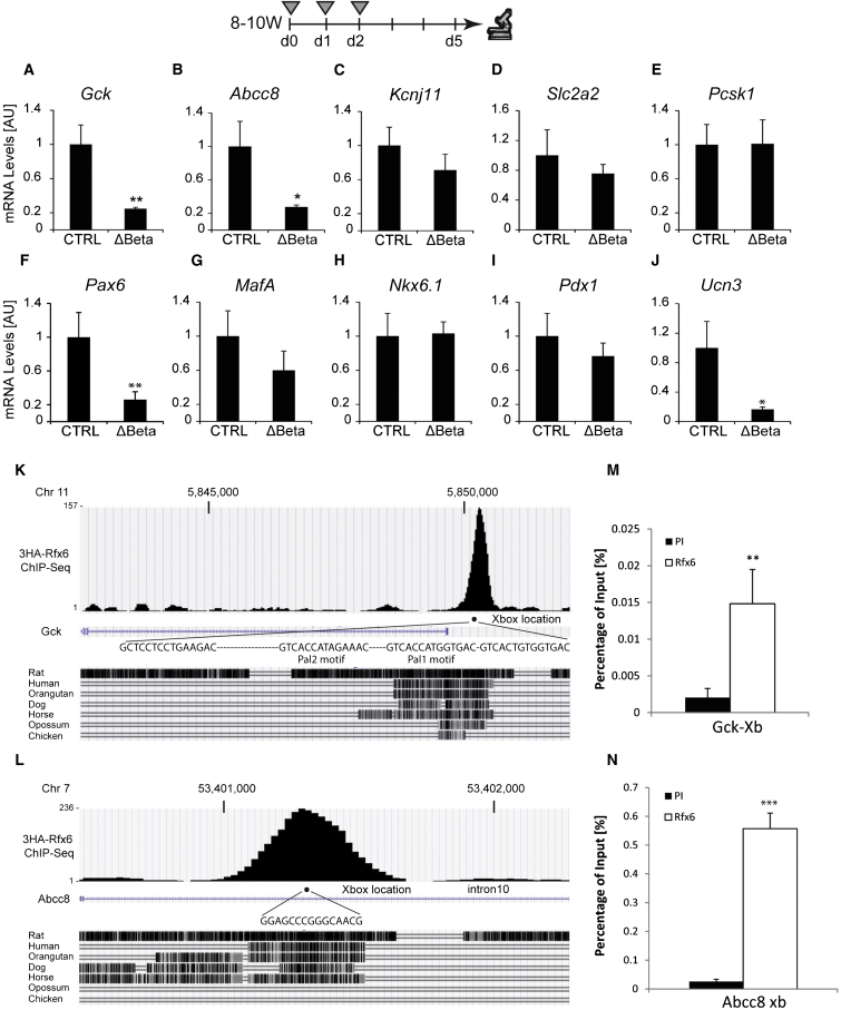
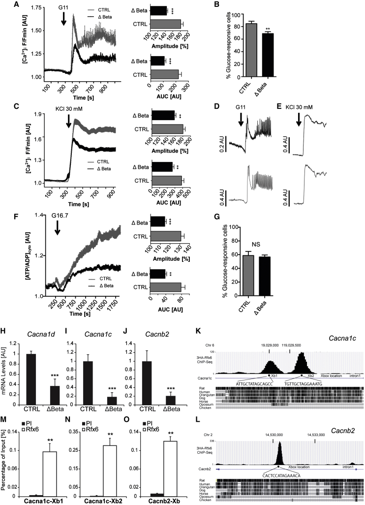
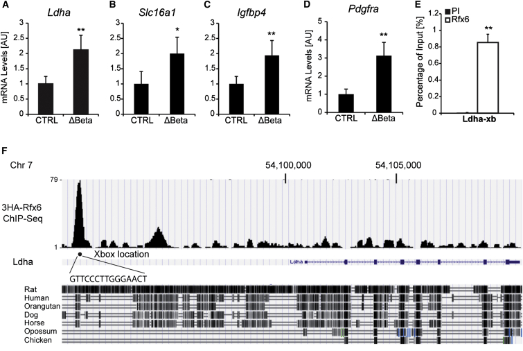
Increasing evidence suggests that loss of β cell characteristics may cause insulin secretory deficiency in diabetes, but the underlying mechanisms remain unclear. Here, we show that Rfx6, whose mutation leads to neonatal diabetes in humans, is essential to maintain key features of functionally mature β cells in mice. Rfx6 loss in adult β cells leads to glucose intolerance, impaired β cell glucose sensing, and defective insulin secretion. This is associated with reduced expression of core components of the insulin secretion pathway, including glucokinase, the Abcc8/SUR1 subunit of KATP channels and voltage-gated Ca(2+) channels, which are direct targets of Rfx6. Moreover, Rfx6 contributes to the silencing of the vast majority of "disallowed" genes, a group usually specifically repressed in adult β cells, and thus to the maintenance of β cell maturity. These findings raise the possibility that changes in Rfx6 expression or activity may contribute to β cell failure in humans.


Figures

None
Graphical abstract
Figure 1
Figure 1
Deletion of Rfx6 Downstream of Ngn3 Results in the Loss of Insulin-, Glucagon-, Somatostatin-, and Ghrelin-Producing Islet Cells in Newborn Mice (A–F) Immunofluorescence experiments on pancreata from controls and Rfx6ΔEndo pups at postnatal day 0 (P0). Staining for insulin and glucagon (green) and Rfx6 (red) showing efficient deletion of Rfx6 and strong reduction of insulin- and glucagon-expressing cells in Rfx6ΔEndo mutants (A and B). (B) and (F) show very rare sections where hormone-positive cells were found. Staining for PP (green) and Rfx6 (red) revealed that PP is not dependent on Rfx6 (C and D). Staining for hormones (insulin, glucagon, PP, somatostatin) in green and the panendocrine marker chromogranin A in red show that chromogranin A-positive endocrine cells, which do not express any of the islet hormones, are found in the pancreas of Rfx6ΔEndo pups (E and F). (G–W) qRT-PCR experiments for Rfx6, hormones, and transcription factors controlling islet cell development in Rfx6ΔEndo pups and wild-type controls at P0. Scale bars, 50 μM. Data are presented as mean ± SD on n = 4 samples; ∗∗∗p < 0.001, ∗∗p < 0.01, p < 0.05.
Figure 2
Figure 2
Insulin Is Produced in Adult β Cells Lacking Rfx6 (A–F) Immunofluorescence staining on controls and Rfx6ΔBeta (Rfx6fl/fl; Ins1-CreERT2) adult mice, 3 weeks after the first day of tamoxifen injections (8- to 10-week-old mice were injected once a day during 3 consecutive days). Staining for insulin (green, A and B) and Rfx6 (red, A and B) reveal insulin expression despite efficient deletion of Rfx6 in β cells of Rfx6ΔBeta mice (white arrows in B). Staining for c-peptide1 (red, C and D) and c-peptide2 (red, E and F) supports efficient insulin synthesis. (G–L) qRT-PCR experiments on islets purified from controls and Rfx6ΔBeta adult (8- to 10-week-old) mice 5 days after tamoxifen injections revealing rapid and specific deletion of Rfx6 (G) in β cells and decreased Ins1 transcription (H), while the expression of Gcg, Ppy Sst and ChgA is unaltered. Grey triangles indicate the days of tamoxifen injections. Yellow and red arrows point to examples of β cells expressing Rfx6 in controls and insulin-negative/Rfx6-positive cells in Rfx6ΔBeta mice, respectively. Scale bars, 50 μM. Data are presented as mean ± SD on n = 4 samples; ∗∗∗p < 0.001, ∗∗p < 0.01, p < 0.05.
Figure 3
Figure 3
The Deletion of Rfx6 in β Cells Causes Glucose Intolerance due to Defective Insulin Secretion (A–C) Exploration of glucose metabolism in adult (12- to 14-week-old) controls (n = 8) and Rfx6ΔBeta (n = 7) males under normal diet 4 weeks after tamoxifen injections. Blood glucose levels are measured in overnight (16 hr)-fasted and ad libitum-fed animals 1 month after tamoxifen injections (A). Intraperitoneal glucose tolerance test (IPGTT) after 16 hr fasting in male mice 1 month after tamoxifen injections (B). Oral glucose tolerance test (OGTT) after 16 hr fasting in male mice 1 month after tamoxifen injections (C). (D) Histogram representing the plasma insulin levels of control and Rfx6ΔBeta mice (9–11 weeks old) during an in vivo glucose-stimulated insulin secretion test (n = 6) performed 5 days after tamoxifen injections. (E) Histogram representing the insulin released during an ex vivo glucose- and KCl-stimulated insulin secretion tests on islets purified from controls and Rfx6ΔBeta (n = 4) mice (9–11 weeks old), 5 days after tamoxifen injections. Data are presented as mean ± SD; p < 0.05, ∗∗p < 0.01, ∗∗∗p < 0.001.
Figure 4
Figure 4
Rfx6 Regulates a Subset of Genes Controlling β Cell Function and Directly Targets Gck and Abcc8 (A–J) qRT-PCR experiments for Gck, Kcnj11, Abcc8, Slc2a2, Pcsk1, Pax6, MafA, Nkx6.1, Pdx1, and Ucn3 on islets purified from controls and Rfx6ΔBeta adult mice (9–11 weeks old), 5 days after tamoxifen injections. Grey triangles indicate the days of tamoxifen injections. (K and L) ChIP-seq (anti-HA) data showing Rfx6 binding peaks in Abcc8 and Gck genes in 3HA-Rfx6 transfected Min6B1 cells. (M and N) Quantitative ChIP (anti-Rfx6 antibody) in Min6B1 cells illustrating the binding of Rfx6 to X-boxes indicated in (K) and (L). PI and Rfx6 stand for preimmune and anti-Rfx6 serum. Data are presented as mean ± SD on n = 4–5 samples; ∗∗p < 0.01, p < 0.05.
Figure 5
Figure 5
Impaired ATP/ADP Ratio and Calcium Trafficking in Glucose Stimulated β Cells in Rfx6ΔBeta Islets (A) Mean (±SEM) Ca2+ traces following elevation of glucose from 3 mM to 11 mM (n = 16 islets from five mutants and six controls). Insets are the amplitude and area under the curves. (B) Proportion of fluo2-loaded cells that respond to 11 mM glucose. (C) Mean (±SEM) Ca2+ traces following application of the depolarizing stimulus 30 mM KCl (n = 18 islets from same animals as A). (D) Representative Ca2+ responses to 11mM glucose in a single islet (average of about 50 responsive cells per islet). (E) Representative Ca2+ responses to 30 mM KCl in a single islet. (F) Mean (±SEM) Perceval traces recording ATP dynamics following elevation of glucose from 3 mM to 16.7 mM (∼250 cells from n = 12 islets from four mutants and four controls). Insets are the amplitude and area under the curves of cytosolic ATP/ADP rises ([ATP/ADP]cyto). (G) Proportion of Perceval-expressing cells, shown to represent beta cells (Hodson et al., 2014a) that respond to 16.7 mM glucose. (H–J) qRT-PCR revealing the decrease in the transcription of VDCCs in Rfx6ΔBeta islets (n = 4). (K–O) ChIP-seq (K and L) and quantitative ChIP (M–O) showing the binding of Rfx6 on X-boxes of Cacna1c and Cacnb2 genes in Min6B1 cells (n = 3). PI and Rfx6 in (M) and (N) stand for preimmune and anti-Rfx6 serum. Data are presented as mean ± SD; ∗∗∗p < 0.001, ∗∗p < 0.01.
Figure 6
Figure 6
Rfx6 Targets and Represses Disallowed Genes in β Cells (A–D) qRT-PCR showing the downregulation of Ldha, Slc16a1, Igfbp4, and Pdgfra in islet cells from Rfx6ΔBeta mice (9–11 weeks old), 5 days after tamoxifen injections compared to controls (n = 4). (E and F) ChIP-PCR and ChIP-seq revealing the binding of Rfx6 on one X-box in Ldha gene in Min6B1 cells (n = 3). Data are presented as mean ± SD; ∗∗p < 0.01, p < 0.05.

References

    1. Aftab S., Semenec L., Chu J.S., Chen N. Identification and characterization of novel human tissue-specific RFX transcription factors. BMC Evol. Biol. 2008;8:226. - PMC - PubMed
    1. Ainscow E.K., Zhao C., Rutter G.A. Acute overexpression of lactate dehydrogenase-A perturbs beta-cell mitochondrial metabolism and insulin secretion. Diabetes. 2000;49:1149–1155. - PubMed
    1. Ait-Lounis A., Bonal C., Seguín-Estévez Q., Schmid C.D., Bucher P., Herrera P.L., Durand B., Meda P., Reith W. The transcription factor Rfx3 regulates beta-cell differentiation, function, and glucokinase expression. Diabetes. 2010;59:1674–1685. - PMC - PubMed
    1. Artuso R., Provenzano A., Mazzinghi B., Giunti L., Palazzo V., Andreucci E., Blasetti A., Chiuri R.M., Gianiorio F.E., Mandich P. Therapeutic implications of novel mutations of the RFX6 gene associated with early-onset diabetes. Pharmacogenomics J. 2014 Published online July 22, 2014. - PubMed
    1. Blum B., Hrvatin S.S., Schuetz C., Bonal C., Rezania A., Melton D.A. Functional beta-cell maturation is marked by an increased glucose threshold and by expression of urocortin 3. Nat. Biotechnol. 2012;30:261–264. - PMC - PubMed

Publication types

MeSH terms

Associated data