. 2014 Dec 24;9(6):2071-83.
doi: 10.1016/j.celrep.2014.11.021. Epub 2014 Dec 11.

Hand2 is an essential regulator for two Notch-dependent functions within the embryonic endocardium

Affiliations

Hand2 is an essential regulator for two Notch-dependent functions within the embryonic endocardium

Nathan J VanDusen et al. Cell Rep. .

Abstract

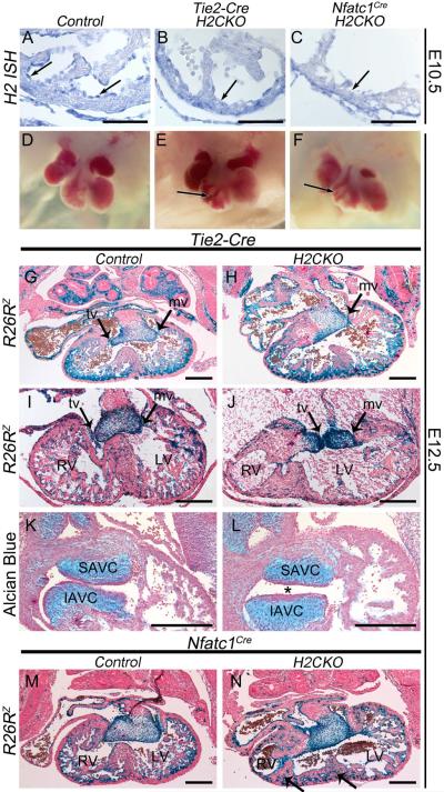
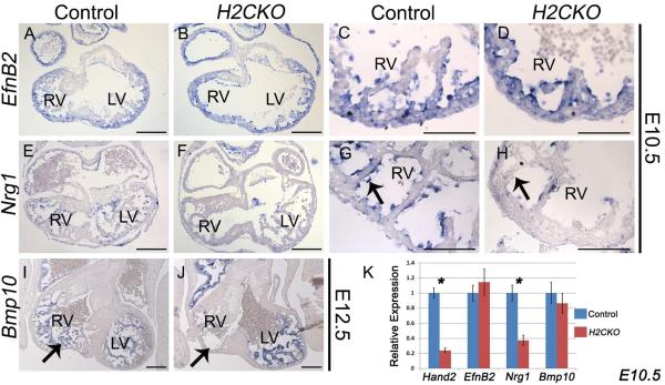
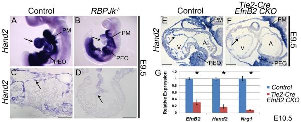
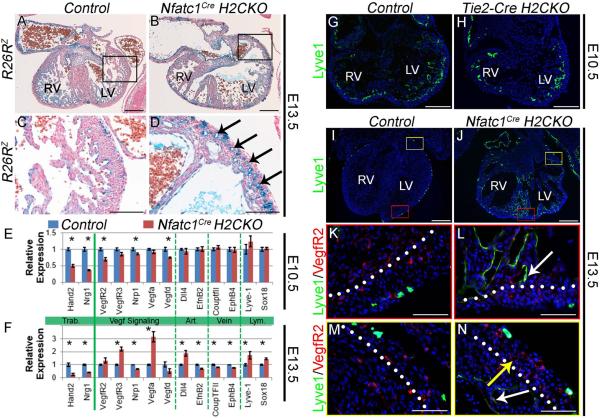
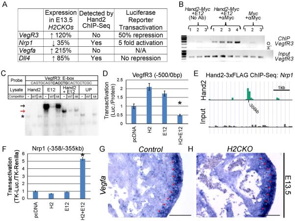
The basic-helix-loop-helix (bHLH) transcription factor Hand2 plays critical roles during cardiac morphogenesis via expression and function within myocardial, neural crest, and epicardial cell populations. Here, we show that Hand2 plays two essential Notch-dependent roles within the endocardium. Endocardial ablation of Hand2 results in failure to develop a patent tricuspid valve, intraventricular septum defects, and hypotrabeculated ventricles, which collectively resemble the human congenital defect tricuspid atresia. We show endocardial Hand2 to be an integral downstream component of a Notch endocardium-to-myocardium signaling pathway and a direct transcriptional regulator of Neuregulin1. Additionally, Hand2 participates in endocardium-to-endocardium-based cell signaling, with Hand2 mutant hearts displaying an increased density of coronary lumens. Molecular analyses further reveal dysregulation of several crucial components of Vegf signaling, including VegfA, VegfR2, Nrp1, and VegfR3. Thus, Hand2 functions as a crucial downstream transcriptional effector of endocardial Notch signaling during both cardiogenesis and coronary vasculogenesis.


Figures

Figure 1
Figure 1. Endocardial deletion of Hand2 results in a VSD, hypotrabeculation, hypoplastic RV, IVS defects and TA
Hand2 ISH of RV section from E10.5 control, Tie2-Cre H2CKO, and Nfatc1CreH2CKO, respectively (A–C). Wholemount view of E12.5 control heart, Tie2-Cre H2CKO, and Nfatc1Cre H2CKO, respectively (D–F). R26RlacZ stained sections from E12.5 Tie2-Cre(+) control embryo (G), Tie2-Cre H2CKO with TA (H). R26RlacZ stained E12.5 Tie2-Cre(+) control embryo (I), and Tie2-Cre H2CKO with DILV (J). R26RlacZ stained sections from E12.5 Nfatc1Cre control embryo (M), and Nfatc1Cre H2CKO (N; arrows denote multiple IVSs). Alcian blue staining of E12.5 Tie2-Cre(+) control AV cushions (K), and Tie2-Cre H2CKO (L). Asterisk denotes abnormalities in AV cushion shape and position. SAVC, superior atrioventricular cushion; IAVC, inferior atrioventricular cushion; tv, tricuspid valve; mv, mitral valve. Scale bars in A – C represent 100μm; scale bars in G – N represent 250μm.
Figure 2
Figure 2. H2CKOs exhibit down-regulation of Nrg1, and loss of Bmp10 expressing trabecular myocardium
Expression of the Notch1 target EfnB2 is comparable between H2CKOs at E10.5 with controls (A–D). Nrg1 expression at E10.5 is markedly decreased in H2CKOs when compared to controls (E–H arrows in G and H denote endocardium). H2CKOs have less Bmp10 expressing trabecular myocardium in the RV at E12.5 (I, J arrows denote trabecular tissue). qPCR on isolated ventricles confirms the significant reduction (* p≤0.05) of Nrg1 in E10.5 H2CKOs. Scale bars in A, B, E, F, I, and J represent 200μm; scale bars in C, D, G, and H represent 100μm.
Figure 3
Figure 3. Hand2 functions downstream of Notch1 and the direct Notch1 target EphrinB2 during trabeculation
Wholemount Hand2 ISH in wild type (A) and RBPJk knockout embryos (B). Arrows indicate endocardial Hand2 expression in control (A) and loss of expression in RBPJk knockouts (B). Hand2 ISH (C) shows robust endocardial Hand2 expression that is downregulated within RBPJk−/− endocardium (D). Hand2 ISH (E) revealed a similar downregulation of Hand2 within the endocardium of Tie2-Cre EfnB2CKO hearts (F Arrows denote endocardium). PM, pharyngeal mesenchyme; PEO, proepicardial organ. qPCR on RNA isolated from ventricles confirms significant (* p≤0.05) downregulation of Hand2 and Nrg1 in Tie2-Cre EfnB2CKO hearts (G). Scale bars in C and D represent 50μm; scale bars in E and F represent 100μm.
Figure 4
Figure 4. Hand2 directly regulates Nrg1
Sequence alignments reveal three Hand2 consensus sites that are highly conserved among mammals (A). ChIP of the NRG1 promoter (B). In-vitro transcribed and translated Hand2 and E12 used in EMSAs (C). EMSAs for D-box 1 (D) and E-box 3 (E) show robust and specific binding of Hand2-E12 heterodimers. No binding is observed for site 2. Luciferase reporter assays with a deletion series of the Nrg1 promoter. Red numbers denote positions of the Hand2 consensus sites; green arrows denote positions of the transcription start sites; black arrows, location of ChIP primers (F). Transactivation of 500bp Nrg1 promoter reporter (G; *p≤0.05).
Figure 5
Figure 5. Conditional CAG-CAT-Hand2 transgene expression in Nfatc1Cre EfnB2 CKOs partially improves trabeculation and Nrg1 expression
Wholemount images of E10.5 control, CC-H2(+) control, EfnB2 CKO, and CC-H2(+); EfnB2 CKO embryos (A–D). Hand2 section ISH (E–H). Arrow in G indicates loss of Hand2; arrow in H indicates restoration of endocardial Hand2 via CC-H2. Nrg1 section ISH (I–L). Arrow in K indicates loss of Nrg1 in EfnB2 CKOs; arrow in L indicates restored Nrg1 expression in EfnB2 CKOs that are also CC-H2(+).Bmp10 section ISH (M–P). Arrow in O indicates Bmp10 expressing myocardium and lack of trabeculation; arrow in P indicates restoration of Bmp10 expressing trabeculations. qPCR analysis of isolated E10.5 ventricles (Q). Asterisks denote significant difference (p≤0.05). CC-H2, CAG-CAT-Hand2. Scale bars in E – P represent 100μm.
Figure 6
Figure 6. Hand2 controls coronary development and endocardial maturation via regulation of Vegf signaling
R26RlacZ stained E13.5 hearts (A, B). LV outer curvature (C, D). Nfatc1Cre H2CKOs display hyper-vascularization (arrows in D). qPCR analysis of E10.5 and E13.5 gene expression (n = 4) in Nfatc1Cre H2CKOs (E, F; Asterisks denote significant difference (p≤0.05). Lyve-1 immunostaining in E10.5 Control and Tie2-CreH2CKO hearts (G, H). Lyve-1 immunostaining in E13.5 Control and Nfatc1Cre H2CKO hearts (I–N). In E13.5 control hearts Lyve-1 expression is restricted to endothelial lymphatic precursors (K), while H2CKOs continue to express Lvye-1 within ventricular endocardium (L white arrow). Persistent Lyve-1 expression marks ventricular endocardium (N white arrow) but not coronary endothelium (N yellow arrow). Dotted lines denote the border between compact and trabecular myocardium. Scale bars in A and B represent 250μm, 100μm for C and D, 200μm for G and H, 250μm for I and J, and 50μm for K–N.
Figure 7
Figure 7. Hand2 regulates Vegf signaling
Differentially expressed genes that were analyzed for direct regulation by Hand2 (A). ChIP of a region within the 500bp VegfR3 promoter (B). EMSA demonstrates Hand2-E12 heterodimers specifically bind an oligo corresponding to an E-box within the ChIP amplicon of the VegfR3 promoter (C). Black arrow indicates E12 homodimer binding, red arrow indicates Hand2-E12 heterodimer binding, and asterisk indicates nonspecific binding. Hand2-E12 heterodimers repress significantly (p≤0.05) a luciferase reporter containing the 500bp VegfR3 promoter (D). Hand2-3xFLAG ChIP-seq demonstrates a prominent region of enrichment approximately 356kb upstream of the Nrp1 coding region (E). A luciferase reporter containing this potential enhancer region is significantly (p≤0.05) trans-activated 5-fold by H2-E12 heterodimers (F). ISH demonstrates strong myocardial expression of VegfA within the compact zone of control E13.5 hearts (G). Red dots denote the border between compact and trabecular myocardium. In H2CKOs, the distinction between VegfA expression levels in compact versus trabecular myocardium is more poorly defined (H). Scale bars in G and H represent 100μm.

References

    1. Anderson R, Becker A, Macartney F, Shinebourne E, Wilkinson J, Tynan M. Is “tricuspid atresia” a univentricular heart? Pediatric Cardiology. 1979;1:51–56.
    1. Barnes RM, Firulli BA, VanDusen NJ, Morikawa Y, Conway SJ, Cserjesi P, Vincentz JW, Firulli AB. Hand2 Loss-of-Function in Hand1-Expressing Cells Reveals Distinct Roles in Epicardial and Coronary Vessel Development. Circulation Research. 2011;108:940–949. - PMC - PubMed
    1. Benedito R, Rocha SF, Woeste M, Zamykal M, Radtke F, Casanovas O, Duarte A, Pytowski B, Adams RH. Notch-dependent VEGFR3 upregulation allows angiogenesis without VEGF-VEGFR2 signalling. Nature. 2012;484:110–114. - PubMed
    1. Chen H, Shi S, Acosta L, Li W, Lu J, Bao S, Chen Z, Yang Z, Schneider MD, Chien KR, et al. BMP10 is essential for maintaining cardiac growth during murine cardiogenesis. Development. 2004;131:2219–2231. - PMC - PubMed
    1. Connerney J, Andreeva V, Leshem Y, Muentener C, Mercado MA, Spicer DB. Twist1 dimer selection regulates cranial suture patterning and fusion. Dev Dyn. 2006;235:1345–1357. - PubMed

Publication types

MeSH terms

Substances

LinkOut - more resources