

Temporal characteristics of melanopsin inputs to the human pupil light reflex
- PMID: 25497360
- PMCID: PMC4308541
- DOI: 10.1016/j.visres.2014.12.001
Temporal characteristics of melanopsin inputs to the human pupil light reflex
Abstract
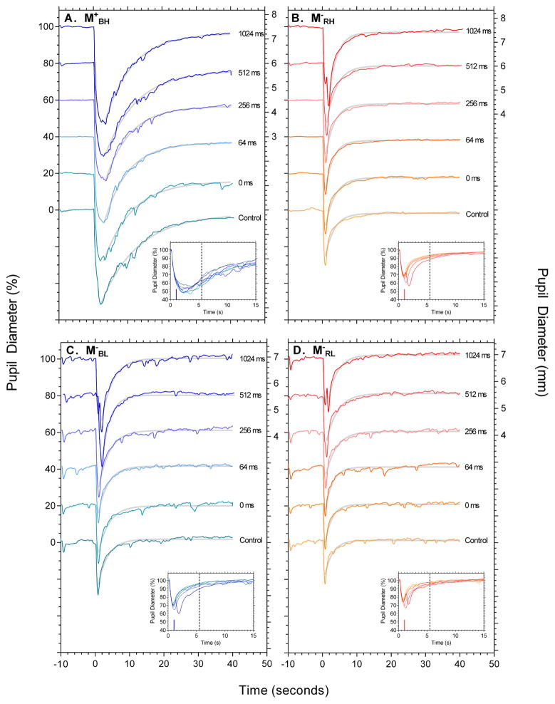
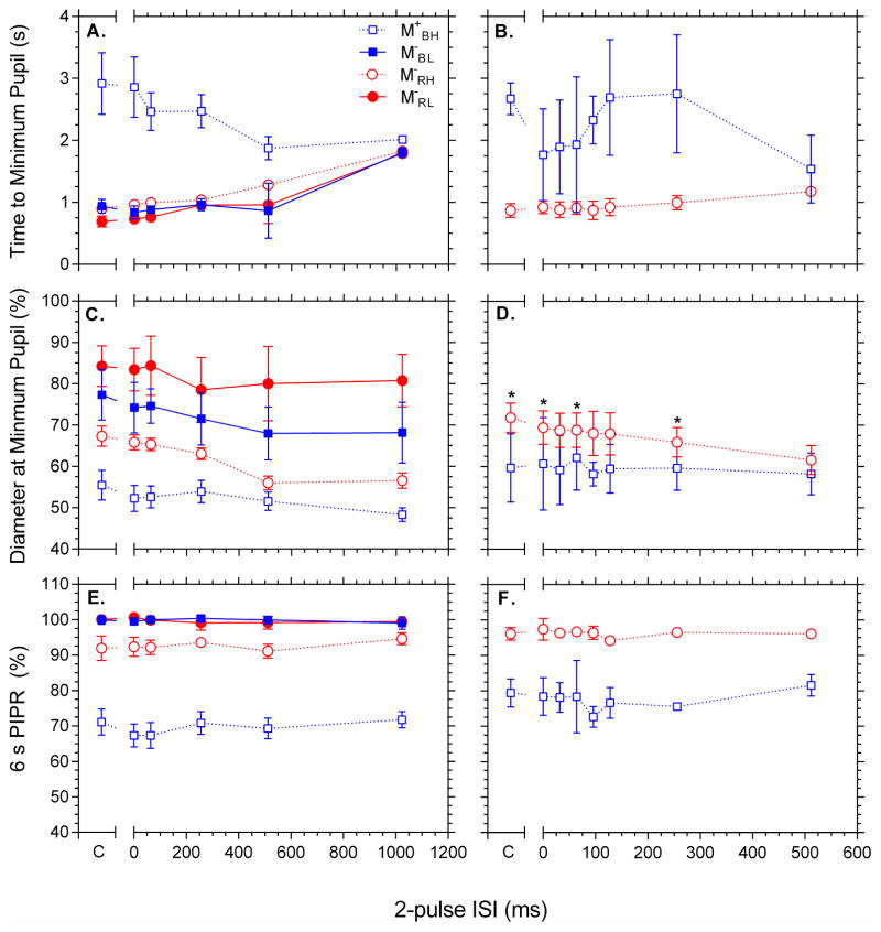
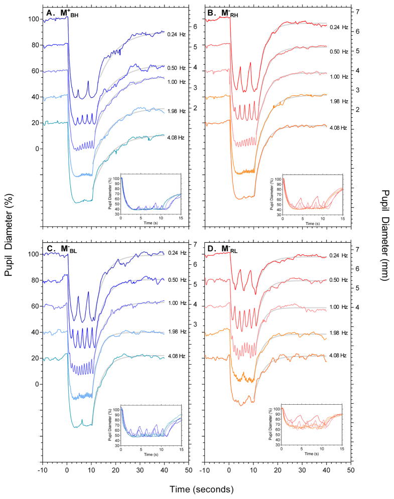
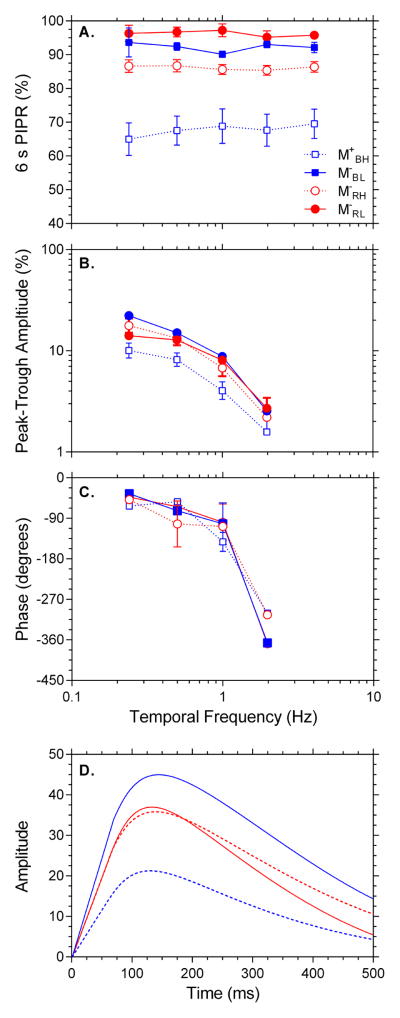
Rods, cones and melanopsin containing intrinsically photosensitive retinal ganglion cells (ipRGCs) operate in concert to regulate pupil diameter. The temporal properties of intrinsic ipRGC signalling are distinct to those of rods and cones, including longer latencies and sustained signalling after light offset. We examined whether the melanopsin mediated post-illumination pupil response (PIPR) and pupil constriction were dependent upon the inter-stimulus interval (ISI) between successive light pulses and the temporal frequency of sinusoidal light stimuli. Melanopsin excitation was altered by variation of stimulus wavelength (464 nm and 638 nm lights) and irradiance (11.4 and 15.2 log photons cm(-2) s(-1)). We found that 6s PIPR amplitude was independent of ISI and temporal frequency for all melanopsin excitation levels, indicating complete summation. In contrast to the PIPR, the maximum pupil constriction increased with increasing ISI with high and low melanopsin excitation, but time to minimum diameter was slower with high melanopsin excitation only. This melanopsin response to briefly presented pulses (16 and 100 ms) slows the temporal response of the maximum pupil constriction. We also demonstrate that high melanopsin excitation attenuates the phasic peak-trough pupil amplitude compared to conditions with low melanopsin excitation, indicating an interaction between inner and outer retinal inputs to the pupil light reflex. We infer that outer retina summation is important for rapidly controlling pupil diameter in response to short timescale fluctuations in illumination and may occur at two potential sites, one that is presynaptic to extrinsic photoreceptor input to ipRGCs, or another within the pupil control pathway if ipRGCs have differential temporal tuning to extrinsic and intrinsic signalling.
Keywords: Intrinsically photosensitive retinal ganglion cells (ipRGCs); Melanopsin; Post-illumination pupil response (PIPR); Pupil light reflex; Summation.
Copyright © 2014 Elsevier Ltd. All rights reserved.
Figures
Similar articles
-
Rhodopsin and Melanopsin Contributions to the Early Redilation Phase of the Post-Illumination Pupil Response (PIPR).PLoS One. 2016 Aug 22;11(8):e0161175. doi: 10.1371/journal.pone.0161175. eCollection 2016. PLoS One. 2016. PMID: 27548480 Free PMC article.
-
Melanopsin-mediated post-illumination pupil response in the peripheral retina.J Vis. 2016 Jun 1;16(8):5. doi: 10.1167/16.8.5. J Vis. 2016. PMID: 27271992
-
Melanopsin-Mediated Post-Illumination Pupil Response in Early Age-Related Macular Degeneration.Invest Ophthalmol Vis Sci. 2015 Oct;56(11):6906-13. doi: 10.1167/iovs.15-17357. Invest Ophthalmol Vis Sci. 2015. PMID: 26505464
-
Intrinsically photosensitive retinal ganglion cells.J Neuroophthalmol. 2007 Sep;27(3):195-204. doi: 10.1097/WNO.0b013e31814b1df9. J Neuroophthalmol. 2007. PMID: 17895821 Review.
-
[Pupil and melanopsin photoreception].Nippon Ganka Gakkai Zasshi. 2013 Mar;117(3):246-68; discussion 269. Nippon Ganka Gakkai Zasshi. 2013. PMID: 23631256 Review. Japanese.
Cited by
-
The effect of lens aging and cataract surgery on circadian rhythm.Int J Ophthalmol. 2016 Jul 18;9(7):1066-74. doi: 10.18240/ijo.2016.07.21. eCollection 2016. Int J Ophthalmol. 2016. PMID: 27500118 Free PMC article. Review.
-
Current Insights into Optimal Lighting for Promoting Sleep and Circadian Health: Brighter Days and the Importance of Sunlight in the Built Environment.Nat Sci Sleep. 2022 Jan 6;14:25-39. doi: 10.2147/NSS.S251712. eCollection 2022. Nat Sci Sleep. 2022. PMID: 35023979 Free PMC article. Review.
-
Diverse Cell Types, Circuits, and Mechanisms for Color Vision in the Vertebrate Retina.Physiol Rev. 2019 Jul 1;99(3):1527-1573. doi: 10.1152/physrev.00027.2018. Physiol Rev. 2019. PMID: 31140374 Free PMC article. Review.
-
Duration invariance and intensity dependence of the human circadian system phase shifting response to brief light flashes.Proc Biol Sci. 2022 Mar 9;289(1970):20211943. doi: 10.1098/rspb.2021.1943. Epub 2022 Mar 9. Proc Biol Sci. 2022. PMID: 35259981 Free PMC article.
-
A five-primary photostimulator suitable for studying intrinsically photosensitive retinal ganglion cell functions in humans.J Vis. 2015 Jan 26;15(1):15.1.27. doi: 10.1167/15.1.27. J Vis. 2015. PMID: 25624466 Free PMC article.
References
-
- Baker FH. Pupillary response to double-pulse stimulation; a study of nonlinearity in the human pupil system. Journal of the Optical Society of America. 1963;53(12):1430–1436. - PubMed
-
- Beer RD, MacLeod DIA, Miller TP. The extended maxwellian view (BIGMAX): A high-intensity, high-saturation color display for clinical diagnosis and vision research. Behavior Research Methods. 2005;37(3):513–521. - PubMed
-
- Berson DM, Dunn FA, Takao M. Phototransduction by retinal ganglion cells that set the circadian clock. Science. 2002;295:1070–1073. - PubMed
Publication types
MeSH terms
Substances
Grants and funding
LinkOut - more resources
Full Text Sources
Other Literature Sources
