Comparative Study
. 2014 Dec 14:11:67.
doi: 10.1186/s12989-014-0067-z.

Nanometer-long Ge-imogolite nanotubes cause sustained lung inflammation and fibrosis in rats

Affiliations
Comparative Study

Nanometer-long Ge-imogolite nanotubes cause sustained lung inflammation and fibrosis in rats

Sybille van den Brule et al. Part Fibre Toxicol. .

Abstract

Background: Ge-imogolites are short aluminogermanate tubular nanomaterials with attractive prospected industrial applications. In view of their nano-scale dimensions and high aspect ratio, they should be examined for their potential to cause respiratory toxicity. Here, we evaluated the respiratory biopersistence and lung toxicity of 2 samples of nanometer-long Ge-imogolites.

Methods: Rats were intra-tracheally instilled with single wall (SW, 70 nm length) or double wall (DW, 62 nm length) Ge-imogolites (0.02-2 mg/rat), as well as with crocidolite and the hard metal particles WC-Co, as positive controls. The biopersistence of Ge-imogolites and their localization in the lung were assessed by ICP-MS, X-ray fluorescence, absorption spectroscopy and computed micro-tomography. Acute inflammation and genotoxicity (micronuclei in isolated type II pneumocytes) was assessed 3 d post-exposure; chronic inflammation and fibrosis after 2 m.

Results: Cytotoxic and inflammatory responses were shown in bronchoalveolar lavage 3 d after instillation with Ge-imogolites. Sixty days after exposure, a persistent dose-dependent inflammation was still observed. Total lung collagen, reflected by hydroxyproline lung content, was increased after SW and DW Ge-imogolites. Histology revealed lung fibre reorganization and accumulation in granulomas with epithelioid cells and foamy macrophages and thickening of the alveolar walls. Overall, the inflammatory and fibrotic responses induced by SW and DW Ge-imogolites were more severe (on a mass dose basis) than those induced by crocidolite. A persistent fraction of Ge-imogolites (15% of initial dose) was mostly detected as intact structures in rat lungs 2 m after instillation and was localized in fibrotic alveolar areas. In vivo induction of micronuclei was significantly increased 3 d after SW and DW Ge-imogolite instillation at non-inflammatory doses, indicating the contribution of primary genotoxicity.

Conclusions: We showed that nm-long Ge-imogolites persist in the lung and promote genotoxicity, sustained inflammation and fibrosis, indicating that short high aspect ratio nanomaterials should not be considered as innocuous materials. Our data also suggest that Ge-imogolite structure and external surface determine their toxic activity.


Figures
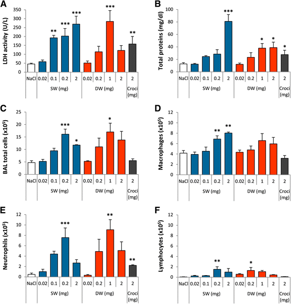
Figure 1
Figure 1
Ge-imogolites induce a strong inflammation in rat lungs 3 d after intra-tracheal instillation. Wistar rats were intra-tracheally instilled with NaCl (controls), 2 mg crocidolite, 0.02 to 2 mg SW and DW Ge-imogolites. Inflammation was investigated in the BAL after 3 d. (A) LDH activity and (B) total proteins measured in BALF. BAL number of (C) total cells, (D) macrophages, (E) neutrophils and (F) lymphocytes. *P < 0.05, **P < 0.01 and ***P < 0.001 relative to NaCl-treated rats (Dunnett multiple comparisons test between NaCl and Ge-imogolite-treated rats or t-test between NaCl and crocidolite-treated rats, n = 3–6, means ± SEM).
Figure 2
Figure 2
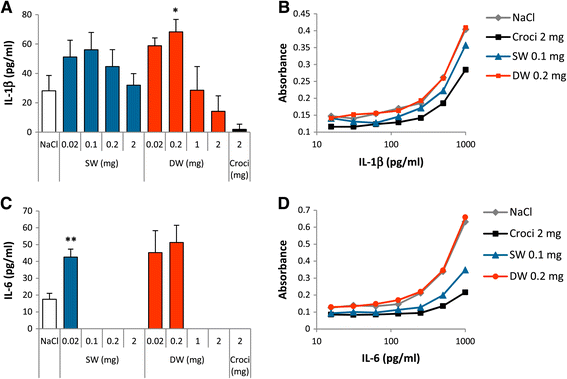
Ge-imogolites induce inflammatory cytokine secretion in rat alveoli 3 d after intra-tracheal instillation. Wistar rats were intra-tracheally instilled with NaCl (controls), 2 mg crocidolite, 0.02 to 2 mg SW and DW Ge-imogolites. Inflammatory cytokines were quantified by ELISA in the BALF after 3 d. BALF (A) IL-1β and (C) IL-6. (B) IL-1β and (D) IL-6 curves obtained from cytokine standards diluted with BALF from NaCl, 2 mg crocidolite, 0.1 mg SW and 0.2 mg DW Ge-imogolite-exposed rats. *P < 0.05 and **P < 0.01 relative to control rats (Dunnett multiple comparisons test between NaCl and Ge-imogolite-treated rats or t-test between NaCl and crocidolite-treated rats, n = 4, means ± SEM).
Figure 3
Figure 3
Ge-imogolites induce a persistent inflammatory and fibrotic lung response. Wistar rats were intra-tracheally instilled with NaCl (controls), 2 mg crocidolite, 0.2 and 1 mg SW and DW Ge-imogolites. Inflammatory and fibrotic parameters were measured after 60 d. (A) Total proteins in BALF. BAL number of (B) total cells, (C) macrophages, (D) neutrophils and (E) lymphocytes. (F) OH-proline lung content, measured in lung homogenates. *P < 0.05, **P < 0.01 and ***P < 0.001 relative to control rats (Dunnett multiple comparisons test between NaCl and Ge-imogolite-treated rats or t-test between NaCl and crocidolite-treated rats, n = 4, means ± SEM).
Figure 4
Figure 4
Ge-imogolites induce a fibrotic lung response. Wistar rats were intra-tracheally instilled with NaCl (controls), 2 mg crocidolite, 1 mg SW and DW Ge-imogolites. Lung histology was examined 60 d after exposure. (A-F) NaCl, (G-L) SW Ge-imogolites, (M-R) DW Ge-imogolites and (S-X) crocidolite. Sections stained with H&E (A-B,G-H,M-N,S-T), Trichrome blue (C,I,O,U), Sirius Red (D,J,P,V), reticulin (E,K,Q,W), PAS (F,L,R,X). Black and blue arrows indicate asbestos fibres and glycoprotein-rich areas, respectively.
Figure 5
Figure 5
Ge-imogolites with intact local atomic structure are localized in fibrotic lung zones. Wistar rats were intra-tracheally instilled with 1 mg SW and DW Ge-imogolites (Ge-imo) or only with their vehicle (NaCl) for control lung. All analyses were performed on paraffin-embedded lung cross sections obtained from animals sacrificed after 60 d. (A) micro-XRF sum spectra of lungs from rats instilled with NaCl, DW and SW Ge-imogolites. Sum spectra are extracted from hyperspectral mapping of lung regions (shown in D-E, H-I, L-M). Spectra are intentionally shifted along y-axis expressed in arbitrary unit. (B) XAS at Ge K-edge: radial distribution functions (RDF) of lungs from rats instilled with SW and DW Ge-imogolites compared to RDF of a DW Ge-imogolite reference sample. (C) Theoretical structure of Ge-imogolite showing the coordination environment around Ge: the first atomic shell is attributed to 4 oxygen atoms surrounding the Ge atom at 1,75 Å and the second coordination sphere of Ge corresponds to Ge-Al atomic pairs (theoretical number of 6 Al neighbors around 3,27 Å). Micro-XRF maps (pixel size of 104 μm) showing the distribution of S (D, H, L) and Ge (E, I, M) in NaCl (D-E), SW (H-I) and DW (L-M) treated lungs. Pulmonary zones are S positive. Scans of lung upper layer sections stained with H&E from NaCl (F), SW (J) and DW (N) Ge-imogolite samples. Squares delimitate Ge-rich areas selected for micro-CT. 2D virtual slices extracted from reconstructed micro-CT volume of NaCl (G), SW (K) and DW (O) Ge-imogolite treated samples. White arrows indicate brilliant areas with high X-ray attenuation (attributed to the presence of Ge-imogolites).
Figure 6
Figure 6
Ge-imogolites induce genotoxicity in rat lungs. Wistar rats were intra-tracheally instilled with NaCl (controls), 5 mg WC-Co, 0.02 and 1 mg SW and DW Ge-imogolites. Micronuclei were assessed in isolated AEC-II 3 d after exposure. (A) BALF LDH activity, (B) % of necrotic and apoptotic cells and (C) ‰ micronucleated cells. **P < 0.01 and ***P < 0.001 relative to control rats (Dunnett multiple comparisons test between NaCl and Ge-imogolite-treated rats or t-test between NaCl and WC-Co-treated rats, n = 3, means ± SEM).

References

    1. Poland CA, Duffin R, Kinloch I, Maynard A, Wallace WA, Seaton A, Stone V, Brown S, Macnee W, Donaldson K. Carbon nanotubes introduced into the abdominal cavity of mice show asbestos-like pathogenicity in a pilot study. Nat Nanotechnol. 2008;3:423–428. doi: 10.1038/nnano.2008.111. - DOI - PubMed
    1. Schinwald A, Murphy FA, Prina-Mello A, Poland CA, Byrne F, Movia D, Glass JR, Dickerson JC, Schultz DA, Jeffree CE, Macnee W, Donaldson K. The threshold length for fiber-induced acute pleural inflammation: shedding light on the early events in asbestos-induced mesothelioma. Toxicol Sci. 2012;128:461–470. doi: 10.1093/toxsci/kfs171. - DOI - PubMed
    1. Schinwald A, Chernova T, Donaldson K. Use of silver nanowires to determine thresholds for fibre length-dependent pulmonary inflammation and inhibition of macrophage migration in vitro. Part Fibre Toxicol. 2012;9:47. doi: 10.1186/1743-8977-9-47. - DOI - PMC - PubMed
    1. Vietti G, Ibouraadaten S, Palmai-Pallag M, Yakoub Y, Bailly C, Fenoglio I, Marbaix E, Lison D, van den Brule S. Towards predicting the lung fibrogenic activity of nanomaterials: experimental validation of an in vitro fibroblast proliferation assay. Part Fibre Toxicol. 2013;10:52. doi: 10.1186/1743-8977-10-52. - DOI - PMC - PubMed
    1. Wang P, Nie X, Wang Y, Li Y, Ge C, Zhang L, Wang L, Bai R, Chen Z, Zhao Y, Chen C. Multiwall Carbon Nanotubes Mediate Macrophage Activation and Promote Pulmonary Fibrosis Through TGF-beta/Smad Signaling Pathway. Small. 2013;9:3799–3811. doi: 10.1002/smll.201300607. - DOI - PubMed

Publication types

MeSH terms