. 2015 Feb;115(2):211-26.
doi: 10.1093/aob/mcu235. Epub 2014 Dec 14.

Floral biology and ovule and seed ontogeny of Nymphaea thermarum, a water lily at the brink of extinction with potential as a model system for basal angiosperms

Affiliations

Floral biology and ovule and seed ontogeny of Nymphaea thermarum, a water lily at the brink of extinction with potential as a model system for basal angiosperms

Rebecca A Povilus et al. Ann Bot. 2015 Feb.

Abstract

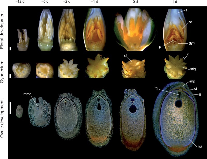
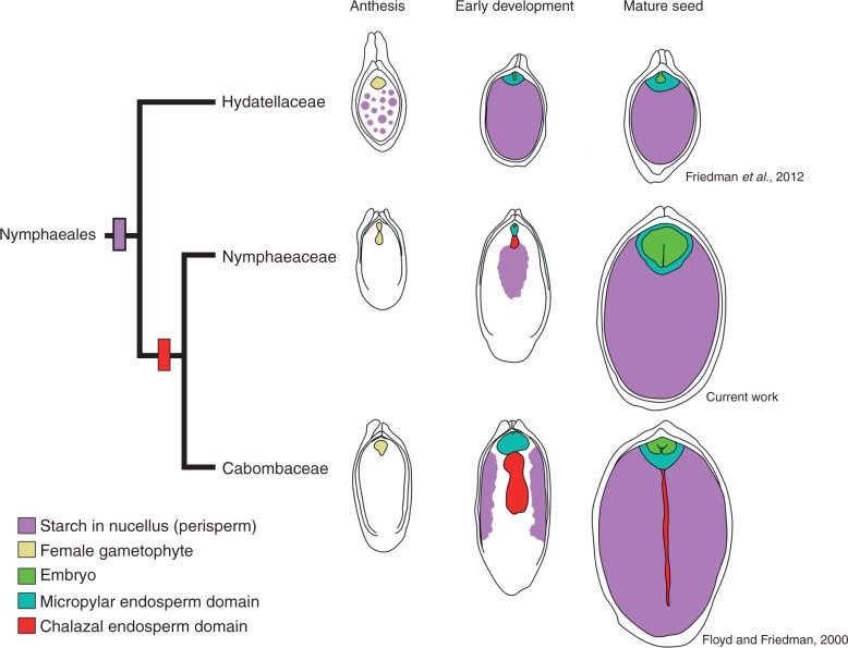
Background and aims: Nymphaea thermarum is a member of the Nymphaeales, of one of the most ancient lineages of flowering plants. This species was only recently described and then declared extinct in the wild, so little is known about its reproductive biology. In general, the complete ontogeny of ovules and seeds is not well documented among species of Nymphaea and has never been studied in the subgenus Brachyceras, the clade to which N. thermarum belongs.

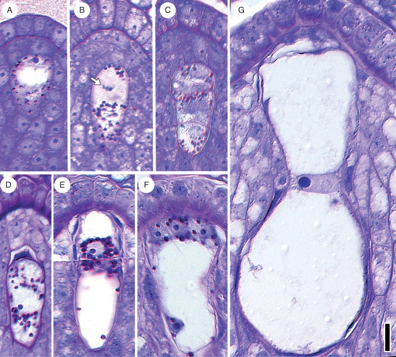
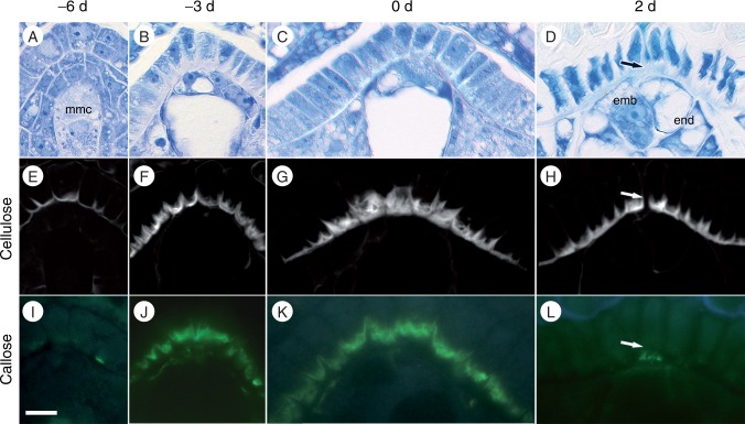
Methods: Flowers and fruits were processed for brightfield, epifluorescence and confocal microscopy. Flower morphology, with emphasis on the timing of male and female functions, was correlated with key developmental stages of the ovule and the female gametophyte. Development of the seed tissues and dynamics of polysaccharide reserves in the endosperm, perisperm and embryo were examined.

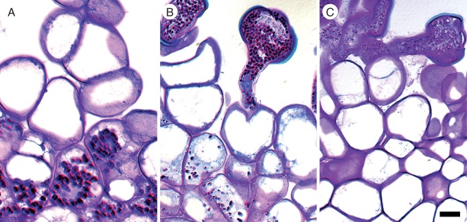
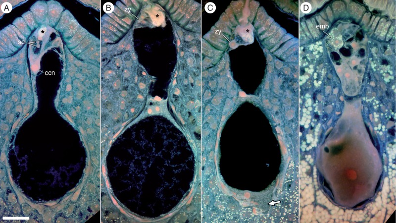
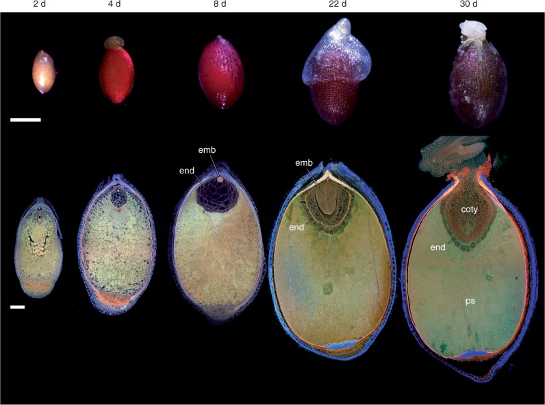
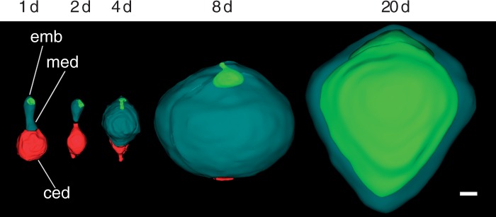
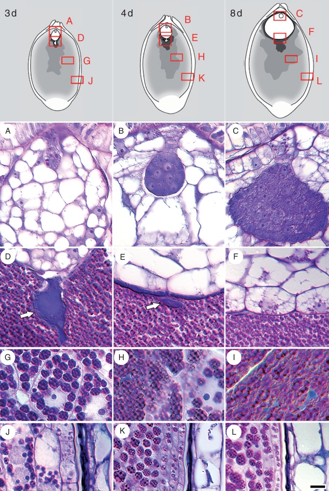
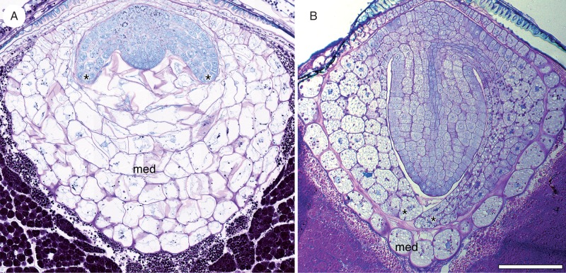
Key results: Pollen release in N. thermarum starts before the flower opens. Cell walls of the micropylar nucellus show layering of callose and cellulose in a manner reminiscent of transfer cell wall patterning. Endosperm development is ab initio cellular, with micropylar and chalazal domains that embark on distinct developmental trajectories. The surrounding maternal perisperm occupies the majority of seed volume and accumulates starch centrifugally. In mature seeds, a minute but fully developed embryo is surrounded by a single, persistent layer of endosperm.

Conclusions: Early male and female function indicate that N. thermarum is predisposed towards self-pollination, a phenomenon that is likely to have evolved multiple times within Nymphaea. While formation of distinct micropylar and chalazal developmental domains in the endosperm, along with a copious perisperm, characterize the seeds of most members of the Nymphaeales, seed ontogenies vary between and among the constituent families. Floral biology, life history traits and small genome size make N. thermarum uniquely promising as an early-diverging angiosperm model system for genetic and molecular studies.

Keywords: Early-diverging angiosperm; Nymphaea thermarum; Nymphaeales; embryo; endosperm; evo-devo; female gametophyte; flower biology; megagametogenesis; megasporogenesis; perisperm; protogyny; seed development; stigma.


Figures

F<sc>ig</sc>. 1.
Fig. 1.
Flower and ovule morphogenesis in Nymphaea thermarum. Stages in floral biology, gynoecium development and ovule morphogenesis are depicted at 12, 6, 2 and 1 days before anthesis, as well as the first and second day of anthesis (noted as -12 d, -6 d, -2 d, -1 d, 0 d and 1 d, respectively). Stereomicroscope images of floral buds (first row, some perianth removed to show all floral organs) and gynoecia (second row, all other floral organs removed), along with confocal optical sections of N. thermarum ovules pretreated for the Feulgen reaction and cleared (third row, whole ovules). (First row) Outer ranks of anthers dehisce and some stigmatic fluid is secreted at -1 d. Flowers then open for 2 consecutive days (0 d, 1 d), with a prominent drop of stigmatic fluid present at 0 d and fully dehisced anthers evident at 1 d. (Second row) Carpels begin to fuse at -6 d and the stigmatic surface is revealed by -2 d. Stigmatic fluid is present at -1 d and prominent at 0 d. (Third row) An endostomic micropyle is formed by -2 d. The mature female gametophyte, with an hour-glass shape, is present at -1 d. By 1 d, starch accumulation in nucellus (perisperm) is apparent. Abbreviations: c , carpel; fg, female gametophyte; gyn, gynoecium; ii, inner integument; mmc, megaspore mother cell; mp, micropyle; nu, nucellus (perisperm); oi, outer integument; p, pollen; sf, stigmatic fluid; st, stamen; stig, stigma; t, tepal. Scale bars = 100 µm.
F<sc>ig</sc>. 2.
Fig. 2.
Female gametophyte development in Nymphaea thermarum. Material was embedded in JB-4 resin, sectioned at 4 µm and stained with PAS reagent and toluidine blue. (A) Megaspore mother cell with starch granules (red/magenta) present in cytoplasm. (B) First meiotic division showing chromosomes (arrow) at the micropylar pole and starch granules at the chalazal pole. (C) Linear tetrad of four cellular megaspores. (D) Functional megaspore, formed from the chalazal-most megaspore, with degenerating micropylar megaspore. (E) Two-nucleate female gametophyte with starch surrounding the nuclei. This image represents a composite of two sequential sections. (F) Immature four-celled female gametophyte, with three cells at the micropylar end and one (the central cell) that occupies the rest of the female gametophyte. (G) Mature female gametophyte, devoid of starch and showing an hour-glass shape. Scale bar = 10 µm.
F<sc>ig</sc>. 3.
Fig. 3.
Micropylar nucellar epidermis in Nymphaea thermarum developing ovules. Ovules from 6 days before anthesis (-6 d), megaspore mother cell stage (A, E, I); 3 days before anthesis (-3 d, immature four-celled female gametophyte stage; B, F, J), first day of anthesis (0 d, mature female gametophyte stage: C, G, K) and 2 days after anthesis (2 d, post-fertilization: D, H, L). Material was embedded in JB-4 resin, sectioned at 4 µm and stained for general structure (toluidine blue: A–D), cellulose and other polysaccharides (calcofluor white: E–H) or callose (aniline blue: I–L). The inner tangential wall of the nucellar epidermis did not stain strongly with toluidine blue throughout ovule development (A–D), but did stain strongly for cellulose, starting at the megaspore mother cell stage (E) with cell wall elaboration reaching a maximum by 0 d (G). After fertilization, cellulose was absent at the site of pollen tube penetration (arrowhead in H). Callose did not accumulate until -3 d (I), but subsequently formed a discrete layer just interior to the cellulosic wall convolutions of the inner tangential wall (J–K). This callose layer was not present after pollination (L). Scale bar = 10 µm.
F<sc>ig</sc>. 4.
Fig. 4.
Pollen–stigma interactions in Nymphaea thermarum. Material was embedded in JB-4 resin, sectioned at 4 µm and stained with PAS reagent and toluidine blue. (A) Starch presence in subdermal the stigmatoid layer, prior to pollination (-2 d). (B) Pollen grains with high starch content 1 d before anthesis (-1 d), with reduction in stigmatoid starch reserves. (C) Absence of starch in stigmatic tissue, second day of anthesis (1 d). Scale bar = 10 µm.
F<sc>ig</sc>. 5.
Fig. 5.
Fertilization and endosperm differentiation in Nymphaea thermarum. Confocal images shown are maximum projections of ≥20 optical sections. Material was pretreated for the Feulgen reaction and embedded in JB-4 resin. (A) Pollen tube penetration of the nucellar cap and delivery of two sperm cells. The central cell nucleus is located next to the egg apparatus. (B) Primary endosperm nucleus migration and cellular division. Two endosperm domains are present: a micropylar endosperm domain and a chalazal endosperm domain. (C) Migration of chalazal endosperm domain nucleus to the chalazal pole and extension of a finger-like projection into the nucellus (arrow). The zygote and micropylar endosperm domain remain undivided. (D) The chalazal endosperm domain nucleus enlarges but does not divide. The micropylar endosperm domain has undergone several rounds of cell division, while the embryo is two-celled and has accumulated starch. Starch reserves are also present in the nucellus. The asterisk indicates pollen tube discharge. Abbreviations: ccn, central cell nucellus; emb, embryo; sc, sperm cells; zy, zygote. Scale bar = 10 µm.
F<sc>ig</sc>. 6.
Fig. 6.
Seed development in Nymphaea thermarum. Stereomicroscope images of seeds (top row) and single confocal optical sections or maximum projections of ≥20 optical sections; (bottom row) material treated for the Feulgen reaction. (Top row) External volumetric changes in developing seeds of N. thermarum accompanied a continued browning of the exotesta up to maturity (22 days after anthesis, noted as 22 d) and through germination (30 d). (Bottom row) Whole-mount imaging of seeds revealed that perisperm accounted for the majority of seed volume throughout development. The endosperm reached a maximum volume 8 d after anthesis (8 d). After this point, the growing embryo displaced most of the endosperm, leaving only the outermost layer of endosperm cells. This layer persisted through germination. Abbreviations: coty, cotyledons, end, endosperm; ps, perisperm. Scale bars = 100 µm.
F<sc>ig</sc>. 7.
Fig. 7.
Embryo–endosperm volumetric relationships in Nymphaea thermarum. Three-dimensional surface renderings of the developing offspring tissues reconstructed from z-stacks of whole-mount ovules. Offspring tissues are depicted on the second day of anthesis and at 2, 4, 8 and 20 days after anthesis (noted as 1 d, 2 d, 4 d, 8 d and 20 d, respectively). Immediately after fertilization, the chalazal endosperm domain was slightly larger than the micropylar domain. Growth of the micropylar domain occurred through 8 d, partly at the expense of the chalazal endosperm domain. The expansion of the embryo was comparatively negligible until after the micropylar endosperm reached its maximum volume. The chalazal domain was rarely evident after 8 d. By the time of seed maturity (20 d), the embryo had expanded into space previously occupied by the micropylar endosperm, displacing all but a single layer of peripheral endosperm cells. Abbreviations: ced, chalazal endosperm domain; emb, embryo; med, micropylar endosperm domain. Scale bar = 10 µm.
F<sc>ig</sc>. 8.
Fig. 8.
Insoluble polysaccharide zonation in Nymphaea thermarum developing seeds. Material was embedded in JB-4 resin, sectioned at 4 µm and stained with PAS reagent and toluidine blue. Seeds were examined at 3 (A, D, G, J), 4 (B, E, H, K) and 8 days (C, F, I, L) after anthesis (noted as 3 d, 4 d and 8 d, respectively). (A) Initially, starch is present throughout the filamentous embryo, but is later restricted to the suspensor and nascent root pole in the late-globular embryo (B). (C) Inception of the cotyledonary ridges. (D) Early and dense accumulation of starch in the endosperm-adjacent zone of the perisperm. (D–F) Starch is consistently absent from the chalazal endosperm (arrow), but is present in the peripheral layers of the micropylar endosperm 8 d after anthesis. (G–I) Perisperm transition zone, in which cells progressively accumulate starch. (J) Perisperm peripheral zone with some multinucleate cells. (J–K) Starch accumulation is delayed relative to other perisperm zones. (L) Starch aggregations remain discrete, even 8 d after anthesis. Scale bar = 10 µm
F<sc>ig</sc>. 9.
Fig. 9.
Seed maturation in Nymphaea thermarum. Material was embedded in JB-4 resin, sectioned at 4 µm and stained with PAS reagent and toluidine blue. (A) Prior to seed maturation, two cotyledons (asterisks) are initiated. The cells of the micropylar endosperm near the embryo become increasing vacuolate and distorted as the cotyledons expand. Starch is present in the micropylar endosperm domain. (B) In mature seeds, the embryo has expanded, displacing the majority of the micropylar endosperm. Starch is present in the persistent endosperm and throughout the cotyledons (asterisks) of the embryo. Scale bar = 100 µm.
F<sc>ig</sc>. 10.
Fig. 10.
Patterns of starch accumulation in the maternal tissues during seed development of Nymphaeales. While members of the Hydatellaceae show starch accumulation (pink) throughout the perisperm before fertilization, in Nymphaea (Nymphaeaceae) starch accumulates centrifugally in the perisperm after fertilization, and the chalazal endosperm (red) is ephemeral. In turn, Cabomba (Cabombaceae) shows centripetal starch accumulation in the perisperm, and the chalazal endosperm protrudes into the perisperm and persists at seed maturity. Teal, micropylar endosperm domain; green, embryo; beige, female gametophyte.

References

    1. Bachelier JB, Friedman WE. 2011. Female gamete competition in an ancient angiosperm lineage. Proceedings of the National Academy of Sciences of the USA 180: 12360–12365. - PMC - PubMed
    1. Baker HG, Baker I. 1979. Starch in angiosperm pollen grains and its evolutionary significance. American Journal of Botany 66: 591–600.
    1. Barrell P, Grossniklaus U. 2005. Confocal microscopy of whole ovules for analysis of reproductive development: the elongate1 mutant affects meiosis II. Plant Journal 43: 309–320. - PubMed
    1. Baskin CC, Baskin JM. 2007. Nymphaeaceae: a basal angiosperm family (ANITA grade) with a fully developed embryo. Seed Science Research 17: 293–296.
    1. Batygina TB, Kravtsova TI, Shamrov II. 1980. The comparative embryology of some representatives of the orders Nymphaeales and Nelumbonales. Botanicheskii Zhurnal 65: 1071–1086.

Publication types

MeSH terms