. 2015 Sep:81:76-92.
doi: 10.1016/j.nbd.2014.11.018. Epub 2014 Dec 8.

Overexpression of alpha-synuclein at non-toxic levels increases dopaminergic cell death induced by copper exposure via modulation of protein degradation pathways

Affiliations

Overexpression of alpha-synuclein at non-toxic levels increases dopaminergic cell death induced by copper exposure via modulation of protein degradation pathways

Annadurai Anandhan et al. Neurobiol Dis. 2015 Sep.

Abstract

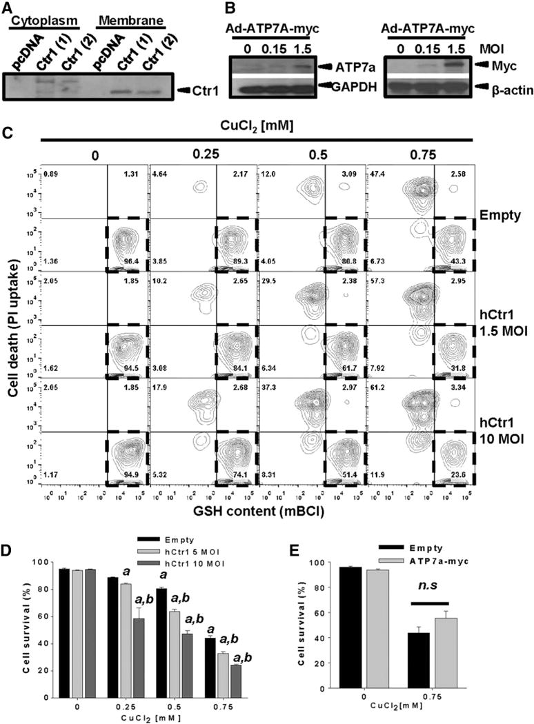
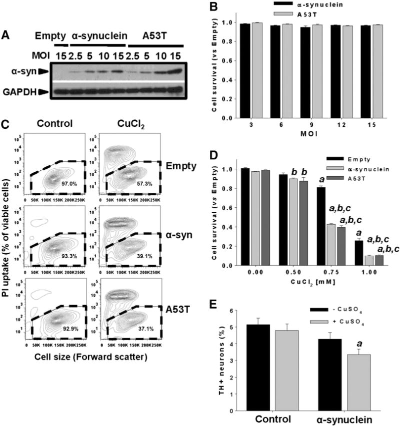
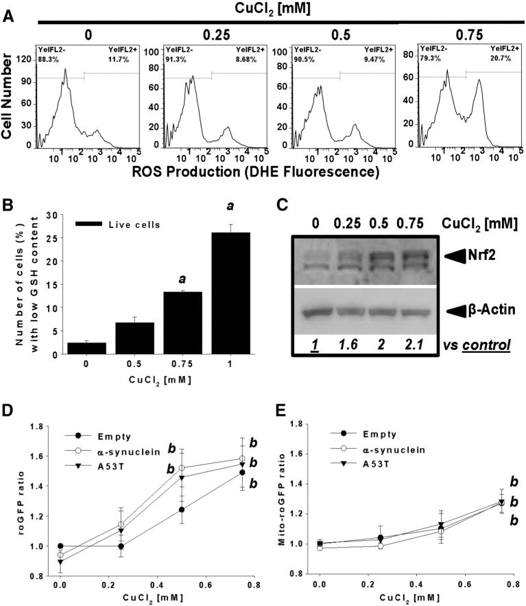
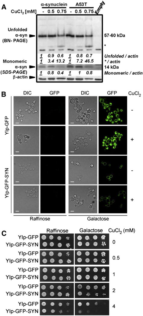
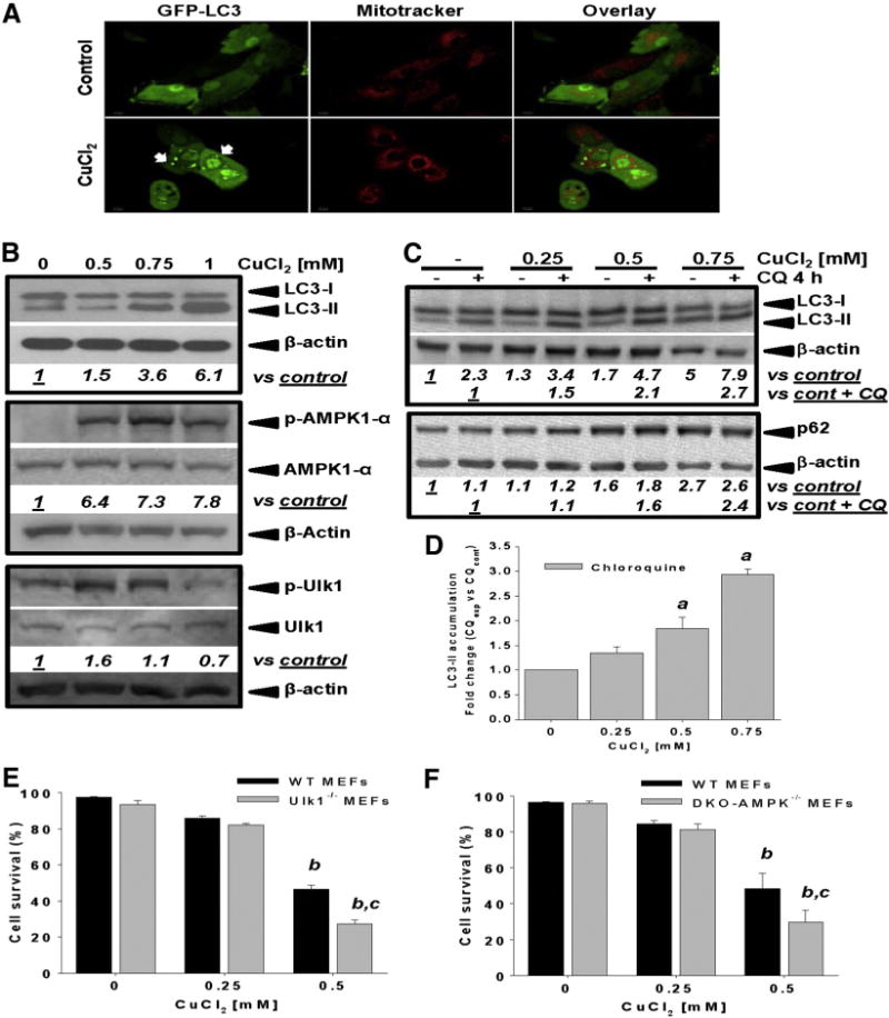
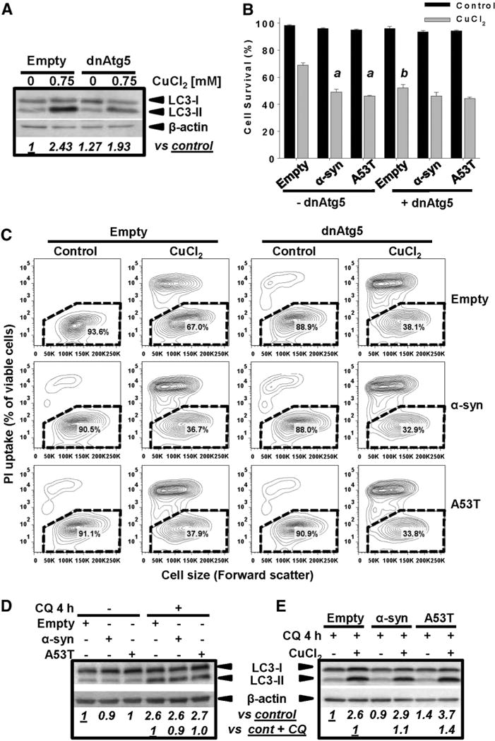
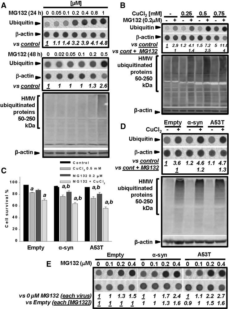
Gene multiplications or point mutations in alpha (α)-synuclein are associated with familial and sporadic Parkinson's disease (PD). An increase in copper (Cu) levels has been reported in the cerebrospinal fluid and blood of PD patients, while occupational exposure to Cu has been suggested to augment the risk to develop PD. We aimed to elucidate the mechanisms by which α-synuclein and Cu regulate dopaminergic cell death. Short-term overexpression of wild type (WT) or mutant A53T α-synuclein had no toxic effect in human dopaminergic cells and primary midbrain cultures, but it exerted a synergistic effect on Cu-induced cell death. Cell death induced by Cu was potentiated by overexpression of the Cu transporter protein 1 (Ctr1) and depletion of intracellular glutathione (GSH) indicating that the toxic effects of Cu are linked to alterations in its intracellular homeostasis. Using the redox sensor roGFP, we demonstrated that Cu-induced oxidative stress was primarily localized in the cytosol and not in the mitochondria. However, α-synuclein overexpression had no effect on Cu-induced oxidative stress. WT or A53T α-synuclein overexpression exacerbated Cu toxicity in dopaminergic and yeast cells in the absence of α-synuclein aggregation. Cu increased autophagic flux and protein ubiquitination. Impairment of autophagy by overexpression of a dominant negative Atg5 form or inhibition of the ubiquitin/proteasome system (UPS) with MG132 enhanced Cu-induced cell death. However, only inhibition of the UPS stimulated the synergistic toxic effects of Cu and α-synuclein overexpression. Our results demonstrate that α-synuclein stimulates Cu toxicity in dopaminergic cells independent from its aggregation via modulation of protein degradation pathways.

Keywords: A53T; AMPK; Autophagy; Copper and Parkinson's disease; Oxidative stress; Proteasome; Ubiquitination; α-Synuclein.


Conflict of interest statement

Conflict of interest statement

The authors certify that there is no conflict of interest.

Figures

Fig. 1
Fig. 1
Copper exposure induces apoptosis in dopaminergic cells. In A–B, cell survival after CuCl2 treatment (48h) was determined in cells co-stained with PI and mBCl. Two-dimensional 5% probability contour plots in A display cell death (PI uptake) vs GSH content (mBCl). Healthy cells were identified as PI- and high mBCl fluorescence (broken line regions in A) and quantified in the corresponding bar graphs in B. Numbers in quadrants (A) reflect number of cells in %. In C, well death was evaluated via Calcein retention assay and data was normalized vs control (DMSO). When indicated, cells were pre-treated with 50 μM pan-caspase (Z-VAD-FMK) or caspase 3 inhibitor (DMQD-CHO) for 1 h and inhibitors remained present throughout the experiment. In D, WB analysis of CuCl2-induced caspase 3 cleavage/activation. Numbers (italics) represent the densitometry analysis of cleaved caspase 3 normalized to β-actin signal with respect to control. The WB is cropped for simplification, but the unmodified version is found in Supplementary Fig. 1. Data in graphs represent means ± SE of at least n = 3. Two-way ANOVA, Holm-Sidak post hoc test, ap < 0.05 vs DMSO only; bp < 0.05 zVAD or Ac-DMQD vs DMSO within the corresponding [CuCl2] category.
Fig. 2
Fig. 2
Copper transport regulates cell death. In A, cytosolic and membrane fractions were extracted from two stable cell clones overexpressing hCtr1, and its expression levels were evaluated by WB. In B, whole cell lysates of cells transduced at the indicated MOI for 24 h with Ad-Empty or Ad-ATPA-myc were isolated, and the expression levelsof ATP7A were confirmed with anti-ATP7A (left panel) or anti-myc (right panel) antibodies. In C–E, cells were transduced with Ad-Empty, Ad-hCtr1 or Ad-ATPA-myc at the indicated MOI for 24 h. Then, cells were washed and treated with CuCl2 for 48 h. Cell death was determined by the PI uptake and the loss of in intracellular GSH as explained in Fig. 1A. In C, results were represented in two-dimensional 5%probability contour plots displaying cell death (PI uptake) vs GSH content (mBCl). Healthy cells were identified asPI- and high mBCl fluorescence (broken line regions). Numbers in quadrants reflect %s. Data in D and E is expressed as cell survival (i.e. % of cells with normal GSH content and PI- after treatments). Two-way ANOVA, Holm-Sidak post hoc test, ap < 0.05 vs Empty without CuCl2 supplementation, bp < 0.05 vs Empty within the corresponding [CuCl2] category. n.s. not significant.
Fig. 3
Fig. 3
Non-toxic overexpression of α-synuclein sensitizes dopaminergic neuroblastoma cells and primary midbrain cultures to Cu toxicity. In A and B, neuroblastoma cells were transduced with Ad-Empty, Ad-α-synuclein or Ad-α-synuclein A53T mutant (A53T) at the indicated MOI for 24 h. Cells were washed, and the expression levels of WT or A53T α-synuclein were measured by WB after 48 h of transduction (A), while cell survival was evaluated by Calcein retention (B). In C and D, cell death induced by CuCl2 treatment (48 h, 0.75 mM in C) was determined in neuroblastoma cells previously transduced with WT or A53T α-synuclein using PI uptake (C) or Calcein retention assay (D). In C, cell death is represented in two-dimensional 5% probability contour plots of PI fluorescence vs cell size (forward scatter) to depict cells with both compromised plasma membrane integrity (PI uptake) and decreased cell size. %s in contour plots represent the number of viable cells (PI- and normal cell size, broken line regions). Data in B and D represent means ± SE of n = 3. In E, primary midbrain cultures transduced, or not, with WT α-synuclein (MOI = 10, 72 h) were incubated in fresh media with or without CuSO4 (10 μM) for 24 h. Cells were stained with antibodies specific for MAP2 and TH and scored for dopaminergic cell viability. The data are presented as the mean ± SE of n = 7. Two-way ANOVA, Holm-Sidak post hoc test, ap < 0.05 vs Empty without CuCl2 or CuSO4 supplementation; bp < 0.05 vs 0 mM CuCl2 within the corresponding category of α-synuclein or A53T; cp < 0.05 vs Empty within the corresponding [CuCl2] tested.
Fig. 4
Fig. 4
The synergistic toxic effect of α-synuclein and CuCl2 is not mediated by increased oxidative stress. Cells were treated with increasing concentrations of CuCl2 for 48 h. ROS formation and GSH depletion were determined by DHE oxidation (A) and changesin mBCl fluorescence (B), respectively. InC, whole cell lysates of cells treated with CuCl2 were isolated to determine changes in Nrf2 levels by WB. Numbers (italics) represent the densitometry analysis normalized to β-actin with respect to control. In D–E, stable cells overexpressing roGFP and Mito-roGFP were transduced with Ad-Empty, Ad-α-synuclein or Ad-A53T for 24 h (3 MOI). Then, cells were washed and treated with CuCl2 (48 h) as indicated. Cells were stained with PI and only viable cells (PI−) were analyzed to determine alterations in the cytosolic (roGFP) or mitochondrial (Mito-roGFP) redox state by ratiometric analysis. Data in graphs were normalized with respect to Empty control values. Data in graphs represent means ± SE of n = 3 independent experiments. One-way ANOVA, Holm-Sidak post hoc test, ap < 0.05 vs 0 mM CuCl2. Two-way ANOVA, Holm-Sidak post hoc test, bp < 0.05 vs 0 mM CuCl2 within the corresponding virus category.
Fig. 5
Fig. 5
The synergistic toxic effect of α-synuclein and CuCl2 is not associated with changes in α-synuclein aggregation. In A, cells were transduced with Ad-Empty, Ad-α-synuclein or Ad-A53T for 24 h (3 MOI), washed and then treated with or without CuCl2 (48 h). TX-100-soluble (upper panel) and insoluble (lower panel) fractions were analyzed by BN- and SDS-PAGE, respectively, and α-synuclein was visualized by WB. Numbers (italics) represent the densitometry analysis normalized to β-actin with respect to the corresponding control. In B–C, W303 wild type S. cerevisiae strains containing genome-integrated, GAL1 promoter-driven GFP or α-synuclein-GFP expression cassettes were pre-grown in 2% glycerol (v/v)/ lactate (w/v) medium, serially diluted and spotted onto plates containing either 2% raffinose or 2% galactose (w/v) as a sole carbon source, and supplemented with the indicated amounts of CuCl2. Cells were incubated for 2 days at 28 °C. In B, visualization of GFP and α-synuclein-GFP distribution was assessed by confocal microscopy performed after 12 hours of cell growth in the indicated medium with or without 4 mM CuCl2.
Fig. 6
Fig. 6
Copper increases autophagy flux and its toxicity is regulated by AMPK-Ulk1 signaling. In A, autophagosome formation induced by 0.75 mM CuCl2 (48 h) was analyzed by confocal microscopy in cells transduced with Ad-GFP-LC3 (3 MOI, 24 h prior CuCl2 treatment). Mitochondria were labeled with Mitotracker. In B, accumulation of LC3-II, and phosphorylation of AMPKα1 and Ulk1 (Ser555) in response to increasing concentrations of CuCl2 (48 h) was evaluated by WB. In C and D, accumulation of LC3-II and p62 induced by CuCl2 (48 h) was determined in the presence or absence of chloroquine (CQ, 40 μM, 4 h prior sample harvesting). Numbers in B, C (italics) represent the densitometry analysis with respect to total AMPKα1, Ulk 1 or β-actin (for LC3-II) and normalized to the corresponding control ± CQ. Data in D represent the densitometry analysis of LC3-II with respect to control + CQ and are means ± SE of n = 3 independent experiments. Controls contained DMSO used as vehicle for CQ. In E–F, Ulk1−/−, DKO AMPK−/− or the corresponding WT MEFs were exposed to CuCl2 at the concentrations indicated (48 h). Cell viability was determined by simultaneous analysis of both PI uptake and changes in intracellular GSH (mBCl) by flow cytometry as exemplified in Fig. 1A. Viable cells were defined as cells with high intracellular glutathione levels (GSH) and PI-. %s represent means ± SE of n = 3 independent experiments. One-way ANOVA, Holm-Sidak post hoc test, ap < 0.05 vs control 0 mM CuCl2. Two-way ANOVA, Holm-Sidak post hoc test, bp < 0.05 vs WT 0 mM CuCl2 cp < 0.05 vs WT within the corresponding [CuCl2] tested.
Fig. 7
Fig. 7
Inhibition of Atg5-dependent autophagy increases Cu toxicity but not the synergistic toxicity of Cu/α-synuclein. Cells were transduced with Ad-dnAtg5 (1.5 MOI) and/or Ad-α-synuclein or Ad-A53T (3 MOI) for 24 h. Ad-Empty was used as control at the MOI required to normalized the MOI used for other treatments (4.5 MOI total), In A, D and E, autophagy (LC3-II accumulation) induced by 0.75 mM CuCl2 (48 h) was evaluated by WB. In D–E, autophagy flux was determined by incubation with CQ (40 μM) 4 h prior to sample isolation. Controls contained DMSO usedas vehicle for CQ. Numbersin WBs (italics) represent the densitometry analysis normalizedto β-actin with respect to controls± CQ. In C, cell death induced by 0.75 mM CuCl2 (48 h) is represented in two-dimensional 5% probability contour plots of PI fluorescence vs cell size (forward scatter) to depict cells with both compromised plasma membrane integrity (PI uptake) and decreased cell size. %s in the contour plots represent the population of viable cells (PI- and normal cell size, broken line regions). B: Two-way ANOVA, Holm-Sidak post hoc test was performed only on CuCl2 data, ap < 0.05 vs Empty + CuCl2; (with or without dnAtg5 transduction); bp < 0.05 vs -dnAtg5 within the corresponding virus category (empty, α-synuclein or A53T).
Fig. 8
Fig. 8
Inhibition of proteasomal activity increases Cu and α-synuclein toxicity. Accumulation of ubiquitin-bound proteins was evaluated by WB or dot blot. Numbers in dot blots (italics) represent the densitometry analysis normalized to β-actin with respect to the indicated controls. In A, cells were treated with MG132 as indicated. In B, cells were treated initially with CuCl2 for 24 h and then MG132 was added for 24 h (CuCl2 remained throughout the experiment). In C, cells were transduced with the indicated viral vectors for 24 h. Then cells were washed and treated with CuCl2 ± MG132 for 48 h. Cell viability was determined by simultaneous analysis of both PI uptake and changes in intracellular GSH (mBCl) by flow cytometry as exemplified in Fig. 1A. Viable cells were defined as cells with high intracellular GSH levels and PI−. %s represent means ± SE of n = 3 independent experiments. Three-way ANOVA, Holm-Sidak post hoc test, ap < 0.05 vs Control within the corresponding category of virus ± MG132;bp < 0.05 vs Empty within the corresponding category ± CuCl2 ± MG132. In D, cells were transduced with the indicated viral vectors for 24 h, washed, and then treated with CuCl2 for 48 h. In E, cells were transduced with the indicated viral vectors for 24 h, washed, and after 24 h, treated with MG132 for additional 24 h (72 h total). Controls contained DMSO used as vehicle for MG132.

Similar articles

Cited by

References

    1. Alvarez-Castelao B, et al. Mechanism of cleavage of alpha-synuclein by the 20S proteasome and modulation of its degradation by the RedOx state of the N-terminal methionines. Biochim Biophys Acta 2013 - PubMed
    1. Arnal N, et al. Clinical utility of copper, ceruloplasmin, and metallothionein plasma determinations in human neurodegenerative patients and their first-degree relatives. Brain Res. 2010;1319:118–130. - PubMed
    1. Arnesano F, et al. Copper-triggered aggregation of ubiquitin. PLoS One. 2009;4:e7052. - PMC - PubMed
    1. Auluck PK, et al. alpha-Synuclein: membrane interactions and toxicity in Parkinson’s disease. Annu Rev Cell Dev Biol. 2010;26:211–233. - PubMed
    1. Ayton S, et al. Ceruloplasmin dysfunction and therapeutic potential for parkinson disease. Ann Neurol 2012 - PubMed

Publication types

MeSH terms