Comparative Study
. 2014 Dec 11;3(6):e001491.
doi: 10.1161/JAHA.114.001491.

Mouse ERG K(+) channel clones reveal differences in protein trafficking and function

Affiliations
Comparative Study

Mouse ERG K(+) channel clones reveal differences in protein trafficking and function

Eric C Lin et al. J Am Heart Assoc. .

Abstract

Background: The mouse ether-a-go-go-related gene 1a (mERG1a, mKCNH2) encodes mERG K(+) channels in mouse cardiomyocytes. The mERG channels and their human analogue, hERG channels, conduct IKr. Mutations in hERG channels reduce IKr to cause congenital long-QT syndrome type 2, mostly by decreasing surface membrane expression of trafficking-deficient channels. Three cDNA sequences were originally reported for mERG channels that differ by 1 to 4 amino acid residues (mERG-London, mERG-Waterston, and mERG-Nie). We characterized these mERG channels to test the postulation that they would differ in their protein trafficking and biophysical function, based on previous findings in long-QT syndrome type 2.

Methods and results: The 3 mERG and hERG channels were expressed in HEK293 cells and neonatal mouse cardiomyocytes and were studied using Western blot and whole-cell patch clamp. We then compared our findings with the recent sequencing results in the Welcome Trust Sanger Institute Mouse Genomes Project (WTSIMGP).

Conclusions: First, the mERG-London channel with amino acid substitutions in regions of highly ordered structure is trafficking deficient and undergoes temperature-dependent and pharmacological correction of its trafficking deficiency. Second, the voltage dependence of channel gating would be different for the 3 mERG channels. Third, compared with the WTSIMGP data set, the mERG-Nie clone is likely to represent the wild-type mouse sequence and physiology. Fourth, the WTSIMGP analysis suggests that substrain-specific sequence differences in mERG are a common finding in mice. These findings with mERG channels support previous findings with hERG channel structure-function analyses in long-QT syndrome type 2, in which sequence changes in regions of highly ordered structure are likely to result in abnormal protein trafficking.

Keywords: genetic variability; hERG; long‐QT syndrome; mERG; mouse.


Figures

Figure 1.
Figure 1.
Western blot analysis of HEK293 cells expressing ERG channels. A, Empty vector and ERG clone transfected cells cultured at 37°C. B, ERG clone transfected cells cultured at 27°C. The table shows the ratio of upper (155‐kDa) band image density to the lower (135‐kDa) band image density. hERG indicates human ether‐a‐go‐go‐related gene; mERG, mouse ether‐a‐go‐go‐related gene; WT, wild type.
Figure 2.
Figure 2.
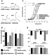
Electrophysiological properties of ERG channels in HEK293 cells. A, Representative current recordings of mERG‐London cultured at 37°C and 27°C (top) and mERG‐Waterston and mERG‐Nie cultured at 37°C (bottom). The arrows indicate peak tail current. Dotted line indicates 0 current. Scale is 200 pA by 1 second. B, The mean peak tail current is shown for WT hERG and the mERG channels cultured at 37°C and 27°C (n=6 for each recording). C, I‐to‐V relations for activation plotted for WT hERG and mERG channels cultured at 37°C and 27°C. Dotted line indicates 0 current. D, V1/2, and k values of the I‐to‐V activation relations for WT hERG and mERG channels cultured at 37°C and 27°C. hERG indicates human ether‐a‐go‐go‐related gene; mERG, mouse ether‐a‐go‐go‐related gene; WT, wild type.
Figure 3.
Figure 3.
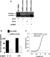
Pharmacological correction (ie, rescue) of mERG‐London. A, A representative Western blot analysis of HEK293 cells expressing WT hERG and the mERG‐London channel for different culture conditions. B, Mean peak tail current density of WT hERG and mERG‐London without culture (n=6) and with culture in E‐4031 (n=4). C, I‐to‐V relation for activation of mERG‐London cultured without and with E‐4031 followed by 1 to 2 hours of drug washout (n=4). Dotted line indicates 0 current. hERG indicates human ether‐a‐go‐go‐related gene; mERG, mouse ether‐a‐go‐go‐related gene; pS/pF, picoamperes per picofarad; WT, wild type.
Figure 4.
Figure 4.
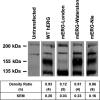
Western blot analysis of neonatal cardiomyocytes expressing WT hERG and mERG channels. All cardiomyocytes express 165‐ and 205‐kDa protein bands. Transfection with WT hERG, mERG‐Waterston, and mERG‐Nie resulted in 135‐ and 155‐kDa bands, whereas transfection with mERG‐London showed only the 135‐kDa band. The density ratio analysis of 155‐ to 135‐kDa bands is shown below the Western blots. hERG indicates human ether‐a‐go‐go‐related gene; mERG, mouse ether‐a‐go‐go‐related gene; WT, wild type.
Figure 5.
Figure 5.
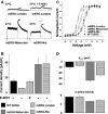
Electrophysiological properties of ERG channels in neonatal mouse cardiomyocytes. A, mERG‐London channel current traces at 37°C for control recordings and after 24 hours of culture in 10 μmol/L E‐4031 followed by drug washout for 1 to 2 hours (top traces) and control recordings for mERG‐Waterston and mERG‐Nie channels (bottom traces). The arrows indicate peak tail current. Dotted line indicates 0 current. Scale is 100 pA by 1 second. B, Peak tail current density for WT hERG, mERG‐London (without and with culture in E‐4031), mERG‐Waterston, and mERG‐Nie (n=6, 6, 8, 7, 4, respectively). C, I‐to‐V relations for activation plotted of WT hERG and mERG channels cultured at 37°C. Solid line indicates 0 current. Normalized peak tail I‐V plot for WT hERG, mERG‐London, mERG‐Waterston, and mERG‐Nie. Dotted line indicates 0 current. D, V1/2, and k values of the I‐to‐V activation relations for WT hERG and mERG channels cultured at 37°C. hERG indicates human ether‐a‐go‐go‐related gene; mERG, mouse ether‐a‐go‐go‐related gene; WT, wild type.
Figure 6.
Figure 6.
Putative topology map of a mERG1 α‐subunit. Modified from Anderson et al,. hERG indicates human ether‐a‐go‐go‐related gene; mERG, mouse ether‐a‐go‐go‐related gene.

References

    1. Sanguinetti MC, Jiang C, Curran ME, Keating MT. A mechanistic link between an inherited and an acquired cardiac arrhythmia: HERG encodes the IKr potassium channel. Cell. 1995; 81:299-307. - PubMed
    1. Trudeau MC, Warmke JW, Ganezky B, Robertson GA. HERG, a human inward rectifier in the voltage‐gated potassium channel family. Science. 1995; 269:92-95. - PubMed
    1. London B, Trudeau MC, Newton KP, Beyer AK, Copeland NG, Gilbert DJ, Jenkins NA, Satler CA, Robertson GA. Two isoforms of the mouse ether‐a‐go‐go‐related gene coassemble to form channels with properties similar to the rapidly activating component of the cardiac delayed rectifier K+ current. Circ Res. 1997; 81:870-878. - PubMed
    1. Anderson CL, Kuzmicki CE, Childs RR, Hintz CJ, Delisle BP, January CT. Large‐scale mutational analysis of Kv11.1 (hERG) reveals molecular insights into type 2 long QT syndrome. Nat Commun. 2014; 5:553510.1038/ncomms6535 - PMC - PubMed
    1. Zhou Z, Gong Q, Epstein M, January CT. HERG channel dysfunction in human long QT syndrome. Intracellular transport and functional defects. J Biol Chem. 1998; 273:21061-21066. - PubMed

Publication types

MeSH terms

Supplementary concepts

LinkOut - more resources