Comparative Study
. 2014 Dec 14:11:74.
doi: 10.1186/s12989-014-0074-0.

Diesel exhaust particle exposure in vitro impacts T lymphocyte phenotype and function

Affiliations
Comparative Study

Diesel exhaust particle exposure in vitro impacts T lymphocyte phenotype and function

Marina Pierdominici et al. Part Fibre Toxicol. .

Abstract

Background: Diesel exhaust particles (DEP) are major constituents of ambient air pollution and their adverse health effect is an area of intensive investigations. With respect to the immune system, DEP have attracted significant research attention as a factor that could influence allergic diseases interfering with cytokine production and chemokine expression. With this exception, scant data are available on the impact of DEP on lymphocyte homeostasis. Here, the effects of nanoparticles from Euro 4 (E4) and Euro 5 (E5) light duty diesel engines on the phenotype and function of T lymphocytes from healthy donors were evaluated.

Methods: T lymphocytes were isolated from peripheral blood obtained from healthy volunteers and subsequently stimulated with different concentration (from 0.15 to 60 μg/ml) and at different time points (from 24 h to 9 days) of either E4 or E5 particles. Immunological parameters, including apoptosis, autophagy, proliferation levels, mitochondrial function, expression of activation markers and cytokine production were evaluated by cellular and molecular analyses.

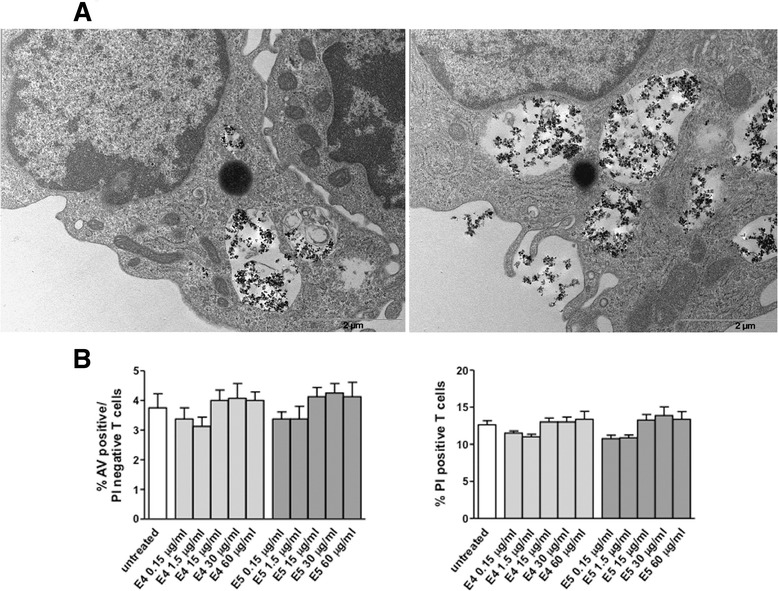
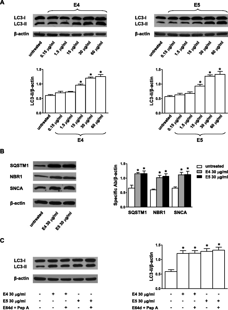
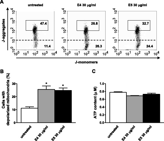
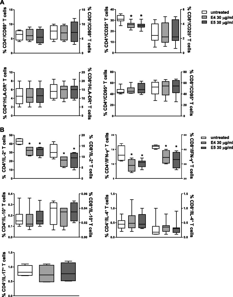
Results: DEP exposure caused a pronounced autophagic-lysosomal blockade, thus interfering with a key mechanism involved in the maintaining of T cell homeostasis. Moreover, DEP decreased mitochondrial membrane potential but, unexpectedly, this effect did not result in changes of the apoptosis and/or necrosis levels, as well as of intracellular content of adenosine triphosphate (ATP). Finally, a down-regulation of the expression of the alpha chain of the interleukin (IL)-2 receptor (i.e., the CD25 molecule) as well as an abnormal Th1 cytokine expression profile (i.e., a decrease of IL-2 and interferon (IFN)-γ production) were observed after DEP exposure. No differences between the two compounds were detected in all studied parameters.

Conclusions: Overall, our data identify functional and phenotypic T lymphocyte parameters as relevant targets for DEP cytotoxicity, whose impairment could be detrimental, at least in the long run, for human health, favouring the development or the progression of diseases such as autoimmunity and cancer.


Figures

Figure 1
Figure 1
Uptake of DEP by T lymphocytes and dose–response analysis of apoptosis/necrosis after nanoparticulate exposure. (A) TEM analysis was performed on T cells after 48 h incubation with E4 or E5 nanoparticles (both used at 30 μg/ml). DEP were found to be localized in membrane-surrounded vesicles in the cytoplasmic region (E4, left panel and E5, right panel). Note the integrity of ultrastructural features of mitochondria and the absence of signs of cell injury. (B) Apoptosis/necrosis assay involving dual staining with AV and PI was carried out using flow cytometry. Results of dose–response experiments performed at 48 h are shown. Data referred to both AV positive/PI negative and PI positive T lymphocytes are shown and are presented as mean ± SD of independent experiments performed in cells from 15 healthy donors.
Figure 2
Figure 2
DEP-induced autophagic-lysosomal blockade in human T lymphocytes. (A) LC3-II Western blot analysis of T-cell lysates (30 μg/lane) from one representative healthy donor (of the 15 analyzed) after treatment with different concentrations (0.15-60 μg/ml for 48 h) of E4 or E5 particles. Densitometry analysis of LC3-II levels relative to β-actin is also shown. Values are expressed as mean ± SD obtained from independent experiments performed in cells from 15 healthy donors. Statistically significant differences are indicated in the figure. *p < 0.05 versus untreated cells. (B) Western blot analysis of autophagic-lysosomal proteins (SQSTM1, NBR1, SNCA) in T-cell lysates from one representative healthy donor (of the 15 analyzed) after treatment with E4 or E5 (30 μg/ml for 48 h) particles. Densitometry analysis of specific protein levels relative to β-actin is also shown. Values are expressed as mean ± SD obtained from independent experiments performed in cells from 15 healthy donors. Statistically significant differences are indicated in the figure. *p < 0.05 versus untreated cells. (C) LC3-II Western blot analysis of T-cell lysates from one representative healthy donor (of the 15 analyzed) after treatment with E4 or E5 (30 μg/ml for 48 h) particles in the absence or presence of the lysosomal inhibitors E64d and pepstatin A. Densitometry analysis of LC3-II levels relative to β-actin is also shown. Values are expressed as mean ± SD obtained from independent experiments performed in cells from 15 healthy donors. Statistically significant differences are indicated in the figure. *p < 0.05 versus untreated cells. SQSTM1, sequestosome 1; NBR1, neighbor of BRCA1 gene 1; SNCA, α-synuclein; Pep A, pepstatin A.
Figure 3
Figure 3
Loss of ΔΨm but preserved ATP content after exposure of T lymphocytes to DEP. (A) Flow cytometry analysis of ΔΨm after staining with JC-1 in untreated T lymphocytes (left panel), T lymphocytes treated with E4 (middle panel) or E5 (right panel) particles (30 μg/ml for 24 h for both compounds). The results obtained in a representative experiment are shown. The numbers in the boxed areas represent the percentages of cells with hyperpolarized mitochondria. The percentages of cells with depolarized mitochondria are shown below the dashed line. (B) Mean percentage (and SD) of lymphocytes with depolarized mitochondria obtained from independent experiments performed in cells from 15 healthy donors is also shown. *p < 0.05 versus untreated cells. (C) ATP content detected by chemiluminescent assay in untreated and E4-and E5-treated T lymphocytes (30 μg/ml for 24 h for both compounds). Data are expressed as mean ± SD and are obtained from independent experiments performed in T lymphocytes from 5 out 15 randomly selected healthy donors.
Figure 4
Figure 4
Flow cytometry immunophenotyping of DEP-treated T lymphocytes. Flow cytometry analysis of T cell activation markers (A) and cytokine expression at the single cell level (B) carried out in CD4+ and CD8+ T lymphocytes from 15 healthy donors after treatment with 30 μg/ml of E4 or E5 particles for 48 h (activation markers) or 72 h (cytokine production). For CD4+ and CD8+ T lymphocyte subsets, data were expressed as the percentage of each subset within the CD4+ or CD8+ population considered as 100%. Data are represented as box plots displaying medians, 25th and 75th percentiles as boxes, and 10th and 90th percentiles as whiskers. *p < 0.05 versus untreated cells.

References

    1. Donaldson K, Poland CA. Inhaled nanoparticles and lung cancer - what we can learn from conventional particle toxicology. Swiss Med Wkly. 2012;142:w13547. - PubMed
    1. Donaldson K, Seaton A. A short history of the toxicology of inhaled particles. Part Fibre Toxicol. 2012;9:13. doi: 10.1186/1743-8977-9-13. - DOI - PMC - PubMed
    1. Kelly FJ, Fussell JC. Air pollution and airway disease. Clin Exp Allergy. 2011;41:1059–1071. doi: 10.1111/j.1365-2222.2011.03776.x. - DOI - PubMed
    1. Shah AS, Langrish JP, Nair H, McAllister DA, Hunter AL, Donaldson K, Newby DE, Mills NL. Global association of air pollution and heart failure: a systematic review and meta-analysis. Lancet. 2013;382:1039–1048. doi: 10.1016/S0140-6736(13)60898-3. - DOI - PMC - PubMed
    1. Nemmar A, Hoet PH, Vanquickenborne B, Dinsdale D, Thomeer M, Hoylaerts MF, Vanbilloen H, Mortelmans L, Nemery B. Passage of inhaled particles into the blood circulation in humans. Circulation. 2002;105:411–414. doi: 10.1161/hc0402.104118. - DOI - PubMed

Publication types

MeSH terms

LinkOut - more resources