. 2014 Dec 11:11:206.
doi: 10.1186/s12974-014-0206-3.

Antibodies to the inositol 1,4,5-trisphosphate receptor type 1 (ITPR1) in cerebellar ataxia

Affiliations

Antibodies to the inositol 1,4,5-trisphosphate receptor type 1 (ITPR1) in cerebellar ataxia

Sven Jarius et al. J Neuroinflammation. .

Abstract

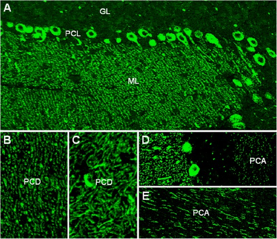
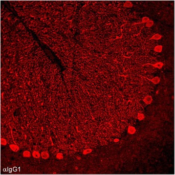
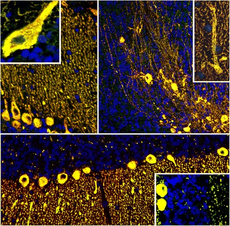
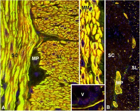
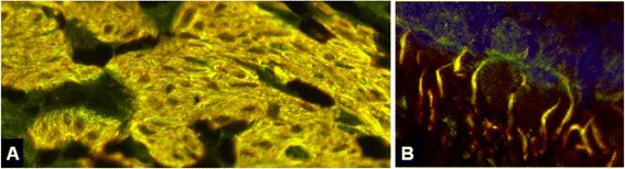
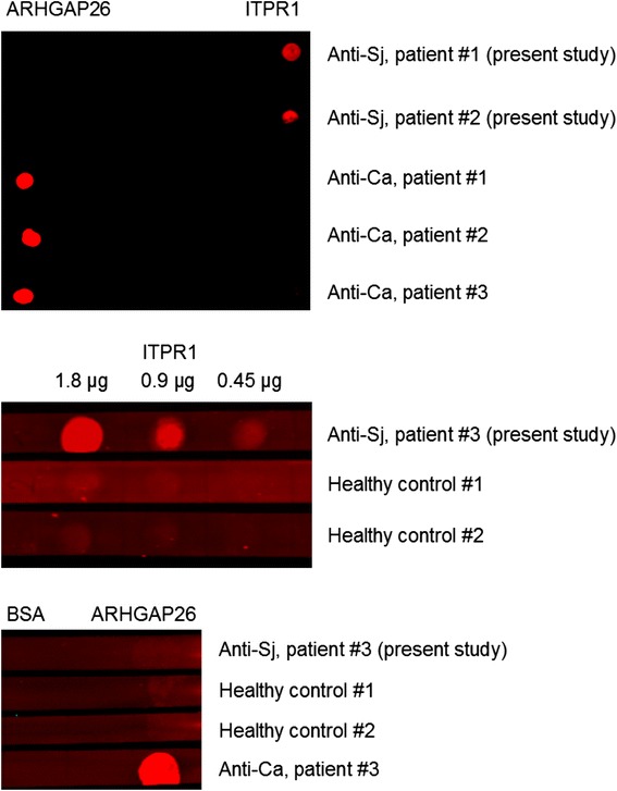
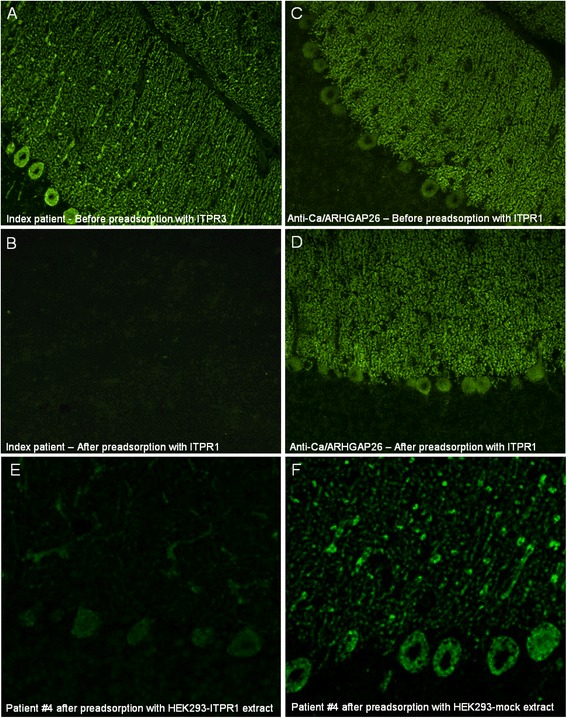
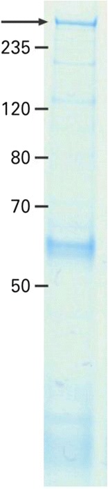
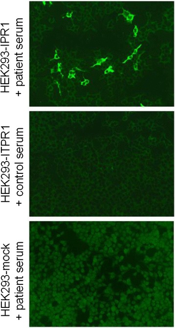
We report on a serum autoantibody associated with cerebellar ataxia. Immunohistochemical studies of sera from four patients referred for autoantibody testing revealed binding of high-titer (up to 1:5,000) IgG antibodies, mainly IgG1, to the molecular layer, Purkinje cell layer, and white matter on mouse, rat, porcine, and monkey cerebellum sections. The antibody bound to PC somata, dendrites, and axons, resulting in a binding pattern similar to that reported for anti-Ca/anti-ARHGAP26, but did not react with recombinant ARHGAP26. Extensive control studies were performed to rule out a broad panel of previously described paraneoplastic and non-paraneoplastic anti-neural autoantibodies. The characteristic binding pattern as well as double staining experiments suggested inositol 1,4,5-trisphosphate receptor type 1 (ITPR1) as the target antigen. Verification of the antigen included specific neutralization of the tissue reaction following preadsorption with ITPR1 (but not ARHGAP26) and a dot-blot assay with purified ITPR1 protein. By contrast, anti-ARHGAP26-positive sera did not bind to ITPR1. In a parallel approach, a combination of histoimmunoprecipitation and mass spectrometry also identified ITPR1 as the target antigen. Finally, a recombinant cell-based immunofluorescence assay using HEK293 cells expressing ITPR1 and ARHGAP26, respectively, confirmed the identification of ITPR1. Mutations of ITPR1 have previously been implicated in spinocerebellar ataxia with and without cognitive decline. Our findings suggest a role of autoimmunity against ITPR1 in the pathogenesis of autoimmune cerebellitis and extend the panel of diagnostic markers for this disease.


Figures

Figure 1
Figure 1
Binding of patient IgG to the Purkinje cell (PC) dendrites (PCD) in the molecular layer (ML), to the PC somata in the PC layer (PCL), and to the PC axons (PCA) in the white matter (WM) on murine cerebellum tissue sections. The observed staining pattern (A) is similar to that observed with anti-Ca/ARHGAP26 (see reference [30]). Note that PC nuclei, interneurons, and granular cells are spared. Also note that the PCD (B and C) and the PCA (D and E) staining patterns can differ significantly depending on sectional planes. Axonal staining may not be detectable on all sections. GL, Granular cell layer.
Figure 2
Figure 2
Analysis of the IgG subclasses of anti-Sj in the index patient. Subclass analysis revealed mainly IgG1 antibodies (depicted in red) with very few IgG2 and no IgG3 or IgG4 antibodies (not shown); no PC-specific antibodies to IgA or IgM were detectable (not shown). IgG1 was also the main anti-Sj IgG subclass in a second patient (not shown).
Figure 3
Figure 3
Perfect overlay of the binding pattern observed with patient IgG and that observed with a commercial antibody to ITPR1, a well-established specific marker of Purkinje cells (PCs). Anti-ITPR1 reactivity is depicted in red (Alexa Fluor® 568), the patient antibody in green (Alexa Fluor® 488), and yellow indicates overlay of the two antibodies. Nuclei are shown in blue (DAPI).
Figure 4
Figure 4
Double labeling of primate intestine sections with patient serum and with commercial antibodies to anti-ITPR1 or ARHGAP26, respectively. The anti-Sj index serum and the commercial antibody to ITPR1 (A) stained both the stratum circulare (SC) and the stratum longitudinale (SL) of the tunica muscularis as well as the muscularis mucosae (MM) and structures adjacent to the enteric villi (V), with a perfect overlay, but spared the plexus myentericus Auerbach (MP), which is located between SC and SL. Conversely, the anti-Ca index serum [30] and the commercial antibody to ARHGAP26 (B) both stained the MP (and the plexus submucosus Meissner; not shown) but spared the enteric muscle cells. Anti-ITPR1 or anti-ARHGAP26 reactivity is depicted in red (Alexa Fluor® 568), the patient antibody in green (Alexa Fluor® 488), and yellow indicates overlay of the two antibodies. Nuclei are shown in blue (DAPI).
Figure 5
Figure 5
Double labeling of primate bulbus oculi tissue sections with patient serum and a commercial antibody to anti-ITPR1. An overlay of the index patient’s IgG and a commercial antibody to ITPR1 was also observed outside the CNS and the intestine, e.g., in the eye bulb (A: ciliary muscle, B: retina with rod and cone processes), confirming the specificity of the patient antibody for ITPR1. Anti-ITPR1 reactivity is depicted in red (Alexa Fluor® 568), the patient antibody in green (Alexa Fluor® 488), and yellow indicates overlay of the two antibodies. Nuclei are shown in blue (DAPI).
Figure 6
Figure 6
Results from an dot-blot assay employing purified ITPR1 and ARHGAP26 as test substrates. IgG from the patients’ serum bound to full-length ITPR1 but not to human full-length ARHGAP26; contrarywise, anti-Ca-positive control sera bound to ARHGAP26 but not to ITPR1 (upper panel). The minimum amount of ITPR1 that resulted in a significant staining intensity was 0.45 μg/μL (middle panel). Healthy control sera bound neither to ITPR1 nor to ARHGAP26 (middle and lower panel).
Figure 7
Figure 7
Results from preadsorption experiments. Preadsorption of the patient serum with purified rat ITPR1 or an extract of HEK293 cells expressing murine ITPR1 resulted in complete loss of binding to cerebellar tissue sections in an indirect immunofluorescence assay (A, B, E). In contrast, binding of anti-Ca/ARHGAP26-positive sera was not affected by preadsorption with ITPR1 (C, D). Preadsorption of anti-ITPR1-positive patient serum with full-length human ARHGAP26 (not shown) or with an extract of mock-transfected HEK293 (F) did not affect binding to PCs.
Figure 8
Figure 8
Histoimmunoprecipitation with a reference patient serum revealed a band at around 300 kDa (arrow).
Figure 9
Figure 9
Confirmation of ITPR1 as the target antigen by a recombinant cell-based indirect immunofluorescence assay employing HEK293 cells transfected with full-length human ITPR1 and mock-transfected HEK293 cells as control substrates.

References

    1. Graus F, Delattre JY, Antoine JC, Dalmau J, Giometto B, Grisold W, Honnorat J, Smitt PS, Vedeler C, Verschuuren JJ, Vincent A, Voltz R. Recommended diagnostic criteria for paraneoplastic neurological syndromes. J Neurol Neurosurg Psychiatry. 2004;75:1135–1140. doi: 10.1136/jnnp.2003.034447. - DOI - PMC - PubMed
    1. Voltz R. Paraneoplastic neurological syndromes: an update on diagnosis, pathogenesis, and therapy. Lancet Neurol. 2002;1:294–305. doi: 10.1016/S1474-4422(02)00135-7. - DOI - PubMed
    1. Graus F, Keime-Guibert F, Rene R, Benyahia B, Ribalta T, Ascaso C, Escaramis G, Delattre JY. Anti-Hu-associated paraneoplastic encephalomyelitis: analysis of 200 patients. Brain. 2001;124:1138–1148. doi: 10.1093/brain/124.6.1138. - DOI - PubMed
    1. Pittock SJ, Lucchinetti CF, Lennon VA. Anti-neuronal nuclear autoantibody type 2: paraneoplastic accompaniments. Ann Neurol. 2003;53:580–587. doi: 10.1002/ana.10518. - DOI - PubMed
    1. Peterson K, Rosenblum MK, Kotanides H, Posner JB. Paraneoplastic cerebellar degeneration. I. A clinical analysis of 55 anti-Yo antibody-positive patients. Neurology. 1992;42:1931–1937. doi: 10.1212/WNL.42.10.1931. - DOI - PubMed

Publication types

Substances