

Reproducible extracellular vesicle size and concentration determination with tunable resistive pulse sensing
- PMID: 25498889
- PMCID: PMC4263901
- DOI: 10.3402/jev.v3.25922
Reproducible extracellular vesicle size and concentration determination with tunable resistive pulse sensing
Abstract
Introduction: The size of extracellular vesicles (EVs) can be determined with a tunable resistive pulse sensor (TRPS). Because the sensing pore diameter varies from pore to pore, the minimum detectable diameter also varies. The aim of this study is to determine and improve the reproducibility of TRPS measurements.
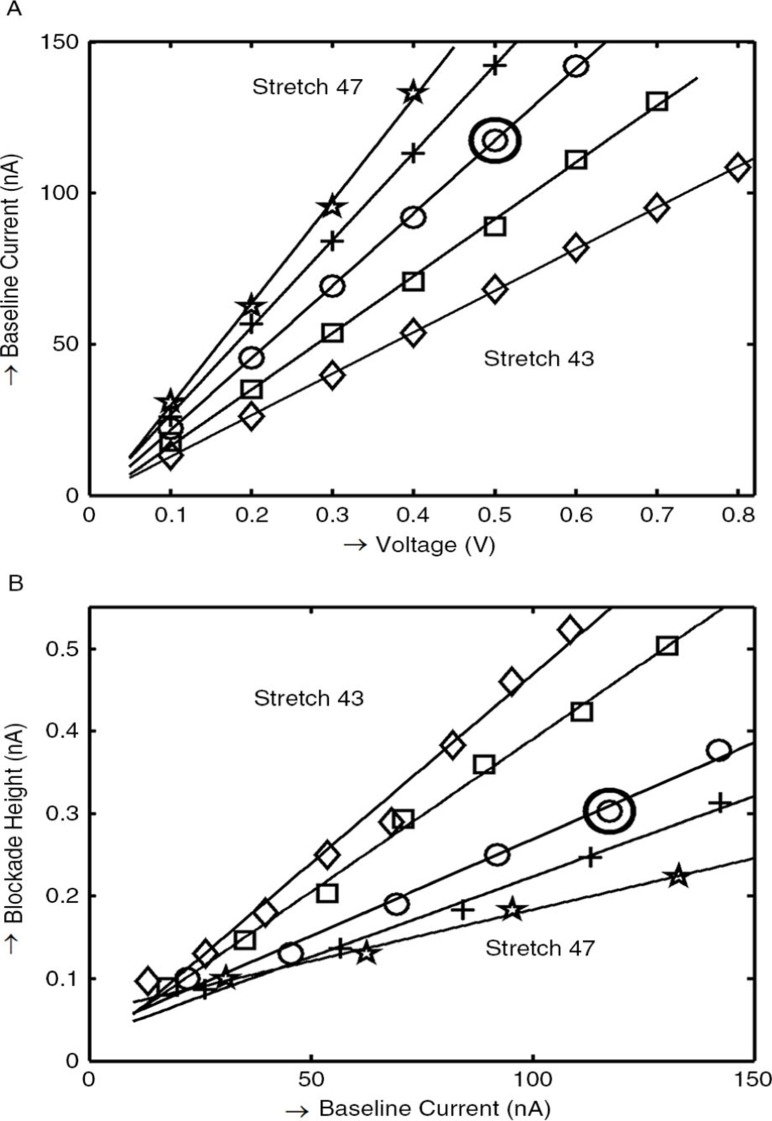
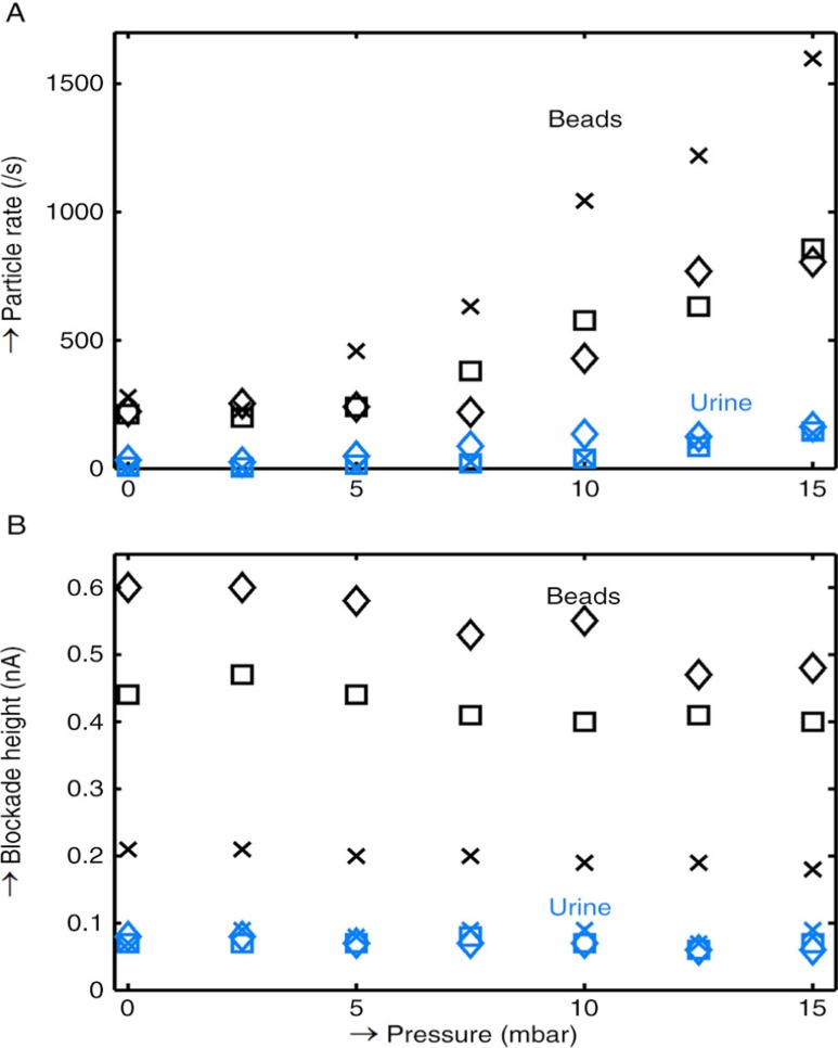
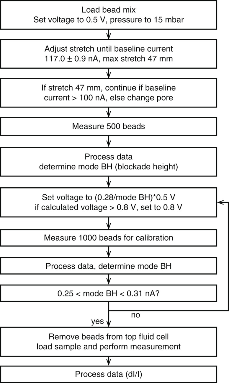
Methods: Experiments were performed with the qNano system (Izon) using beads and a standard urine vesicle sample. With a combination of voltage and stretch that yields a high blockade height, we investigate whether the minimum detected diameter is more reproducible when we configure the instrument targeting (a) fixed stretch and voltage, or (b) fixed blockade height.
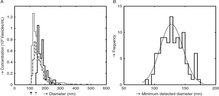
Results: Daily measurements with a fixed stretch and voltage (n=102) on a standard urine sample show a minimum detected vesicle diameter of 128±19 nm [mean±standard deviation; coefficient of variation (CV) 14.8%]. The vesicle concentration was 2.4·109±3.8·109 vesicles/mL (range 1.4·108-1.8·1010). When we compared setting a fixed stretch and voltage to setting a fixed blockade height on 3 different pores, we found a minimum detected vesicle diameter of 118 nm (CV 15.5%, stretch), and 123 nm (CV 4.5%, blockade height). The detected vesicle concentration was 3.2-8.2·108 vesicles/mL with fixed stretch and 6.4-7.8·108 vesicles/mL with fixed blockade height. Summary/conclusion: Pore-to-pore variability is the cause of the variation in minimum detected size when setting a fixed stretch and voltage. The reproducibility of the minimum detectable diameter is much improved by setting a fixed blockade height.
Keywords: exosomes; extracellular vesicles; microparticles; nanoparticles; resistive pulse sensing; size determination.
Figures
References
-
- van der Pol E, Coumans FAW, Gardiner C, Sargent IL, Harrison P, Sturk A. Particle size distribution of exosomes and microvesicles by transmission electron microscopy, flow cytometry, nanoparticle tracking analysis, and resistive pulse sensing. J Thromb Haemostasis. 2014;12:1182–92. - PubMed
-
- Ito T, Sun L, Bevan MA, Crooks RM. Comparison of nanoparticle size and electrophoretic mobility measurements using a carbon-nanotube-based coulter counter, dynamic light scattering, transmission electron microscopy, and phase analysis light scattering. Langmuir. 2004;20:6940–5. - PubMed
-
- DeBlois RW, Bean CP, Wesley RK. Electrokinetic measurements with submicron particles and pores by the resistive pulse technique. J Colloid Interface Sci. 1977;61:323–35.
-
- Willmott GR, Bauerfeind LH. Detection of polystyrene sphere translocations using resizable elastomeric nanopores; 2010. arXiv preprint; 1002.0611.
-
- Kozak D, Anderson W, Vogel R, Chen S, Antaw F, Trau M. Simultaneous size and ζ-potential measurements of individual nanoparticles in dispersion using size-tunable pore sensors. ACS Nano. 2012;6:6990–7. - PubMed
LinkOut - more resources
Full Text Sources
Other Literature Sources
