Review
. 2015:4:60-73.
doi: 10.1016/j.redox.2014.11.008. Epub 2014 Nov 28.

Molecular mechanisms of the microsomal mixed function oxidases and biological and pathological implications

Affiliations
Review

Molecular mechanisms of the microsomal mixed function oxidases and biological and pathological implications

Arthur I Cederbaum. Redox Biol. 2015.

Abstract

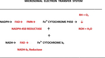
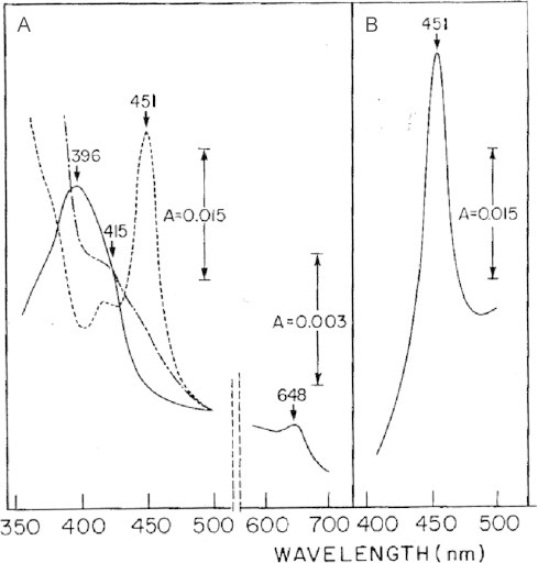
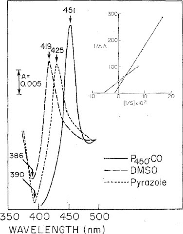
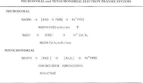
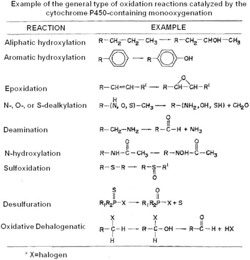
The cytochrome P450 mixed function oxidase enzymes play a major role in the metabolism of important endogenous substrates as well as in the biotransformation of xenobiotics. The liver P450 system is the most active in metabolism of exogenous substrates. This review briefly describes the liver P450 (CYP) mixed function oxidase system with respect to its enzymatic components and functions. Electron transfer by the NADPH-P450 oxidoreductase is required for reduction of the heme of P450, necessary for binding of molecular oxygen. Binding of substrates to P450 produce substrate binding spectra. The P450 catalytic cycle is complex and rate-limiting steps are not clear. Many types of chemical reactions can be catalyzed by P450 enzymes, making this family among the most diverse catalysts known. There are multiple forms of P450s arranged into families based on structural homology. The major drug metabolizing CYPs are discussed with respect to typical substrates, inducers and inhibitors and their polymorphic forms. The composition of CYPs in humans varies considerably among individuals because of sex and age differences, the influence of diet, liver disease, presence of potential inducers and/or inhibitors. Because of such factors and CYP polymorphisms, and overlapping drug specificity, there is a large variability in the content and composition of P450 enzymes among individuals. This can result in large variations in drug metabolism by humans and often can contribute to drug-drug interactions and adverse drug reactions. Because of many of the above factors, especially CYP polymorphisms, there has been much interest in personalized medicine especially with respect to which CYPs and which of their polymorphic forms are present in order to attempt to determine what drug therapy and what dosage would reflect the best therapeutic strategy in treating individual patients.

Keywords: Cytochrome P450; Liver microsomal drug metabolism system; NADPH-cytochrome P450 reductase; P450 catalytic cycle; P450 induction; P450 multiple forms; P450 polymorphisms; Personalized medicine.


Figures

None
Graphical abstract
Fig. 1
Fig. 1
Spectral characteristics of cytochrome P450. A: Absolute spectra of CYP2E1 purified from pyrazole-treated rats . The sample cuvette contained 0.39 nmol of CYP2E1 in 0.1 M KPi, pH 7.4 buffer, 20% glycerol, 0.1 mM EDTA, 0.2% Emulgen 911 and 0.5% sodium cholate in a final volume of 1 ml. The reference cuvette was identical except for the omission of the CYP2E1. The scanned spectra were: oxidized CYP2E1 (solid line); dithionite reduced CYP2E1 (dot/dashed lines -.-.-.-.-.); carbon monoxide-bound reduced CYP2E1 (dashed line - - - -). B: The CO-CYP2E1 difference spectrum. Dithionite-reduced CYP2E1 (0.39 nmol/ml) was present in the sample and reference cuvettes. The sample cuvette was saturated with CO and spectra recorded over the indicated wavelengths.
Fig. 2
Fig. 2
Cartoon of the membrane topography of cytochrome P450 and of NADPH-cytochrome P450 reductase. Electrons for the reduction of molecular oxygen are provided by the reductant/cofactor NADPH which is generated by the pentose phosphate pathway or mitochondrial transhydrogenase, and transferred via NADPH-cytochrome P450 reductase to the heme of cytochrome P450. Both the NADPH-P450 reductase and P450 are microsomal membrane associated, large parts of these enzymes protrude into the cytosol and are attached to the membrane via their amino terminus.
Fig. 3
Fig. 3
Substrate binding spectra to CYP2E1. CYP2E1 and various concentrations of the CYP2E1 ligands DMSO (14–364 mM) or pyrazole (6–51 µM) were added and the spectrum was recorded over the indicated wavelengths until no further changes were observed. A CO-CYP2E1 binding spectrum is shown by the solid lines with a peak at 451 nm. DMSO produces a type II substrate binding spectrum with CYP2E1 with a peak at 419 nm and trough at 386 nm. Pyrazole also produces a type II binding spectrum with a peak at 425 nm and trough at 390 nm. A Lineweaver–Burk plot of the concentration dependence was linear (insert) and the spectral dissociation constants were calculated to be 21 mM for DMSO and 0.04 mM for pyrazole. Results are from Ref. .
Fig. 4
Fig. 4
Electron transfer to cytochrome P450. NADPH is the preferred reductant for microsomal cytochrome P450. Electrons are passed from NADPH to the FAD cofactor of the reductase which passes electrons to the FMN cofactor which than reduces the heme of P450. NADH is less effective than NADPH in reacting with the P450 reductase and reducing P450. NADH effectively reduces the FAD component of the NADH-cytochrome b5 reductase which then reduces the second major heme enzyme in microsomes, cytochrome b5. The system is important in fatty acid desaturation. However, it is not very efficient, relative to the NADPH cytochrome P450 reductase in reducing the heme of cytochrome P450. For reduction of mitochondrial cytochrome P450, iron sulfur proteins are required. The scheme depicts the roles of the FAD adrenodoxin reductase in reducing the adrenodoxin iron–sulfur protein and subsequently, mitochondrial cytochrome P450.
Fig. 5
Fig. 5
Absorption spectrum of microsomal cytochrome b5. Solid line is the spectrum for oxidized cytochrome b5 with a peak at 412 nm. The dashed line is the absorption spectrum after reduction with dithionite, with a peak at 423 nm. Reduced cytochrome b5 does not bind CO.
Fig. 6
Fig. 6
Phase 2 conjugation enzymes.
Fig. 7
Fig. 7
Phases 1 and 2 acetaminophen metabolism. Acetaminophen consumed in “normal” amounts can be removed by direct conjugation by sulfotransferase or glucuronidation by glucuronyl transferases to produce acetaminophen sulfate or acetaminophen glucuronide, which are excreted. When present at high levels, the conjugation reactions become limiting and acetaminophen can be oxidized by several CYPS, especially CYP2E1 to form the reactive N-acetyl-p-benzoquinone imine. The quinone imine can be removed by conjugation with GSH. However, when formed in high amounts at high levels of acetaminophen or when GSH levels are low because of liver disease or alcohol intake, the reactive quinone imine forms covalent adducts with proteins, especially mitochondrial proteins. Impairment of mitochondrial bioenergetics leads to liver necrosis. Acetaminophen toxicity is one of the leading causes of liver damage and emergency room visits.
Fig. 8
Fig. 8
General type of reactions catalyzed by cytochrome P450 enzymes. Other reactions, not shown, include aryl migration, ring contraction, ring formation, ring coupling, dimer formation, group migration (shown in Ref. [33]).
Fig. 9
Fig. 9
The CYP catalytic cycle. Please see text for discussion. Oxygen surrogates such as hydroperoxides can replace NADPH, oxygen and the NADPH-cytochrome P450 reductase in promoting certain CYP-catalyzed reactions by forming active oxygenated–CYP complexes. For example: RH+Fe3+ (CYP)→RH-Fe3+ (CYP) RH-Fe3+ (CYP)+XOOH→XOH+R (Fe–OH)3+ (CYP) R∙ (Fe–OH)3+ (CYP)→ROH-Fe3+ (CYP)→ROH+Fe3+ CYP.
Fig. 10
Fig. 10
Microsomal production of superoxide anion radical. Microsomes were isolated from chronic ethanol-fed rats and dextrose pair-fed controls. Generation of superoxide was assayed by determining the superoxide dismutase-sensitive production of the nitroxyl radical formed from the interaction of superoxide with 1-hydroxy-2,2,6,6-tetramethyl-4-oxo-piperidine. Either NADPH (spectra b and c) or NADH (spectra d and e) were used as cofactors. ESR measurements were carried out at room temperature in a flat quartz cuvette using a Bruker E-300 spectrometer. Spectrum a is that of 0.04 mM of a standard nitroxyl radical. Splitting constants for the resulting triplet were AN=16.0 G and g=2.005. Results are from Refs. .
Fig. 11
Fig. 11
Chlormethiazole (CMZ) lowers alcohol-induced oxidant stress. SV129 female mice were fed the Lieber-DeCarli ethanol or dextrose liquid diets for 4 weeks. At 2 weeks, some of the ethanol-fed mice were also treated with the CYP2E1 inhibitor, CMZ, at a dose of 50 mg/kg body wt, IP, every other day and fed ethanol for the remaining 2 weeks. Lipid peroxidation in liver homogenates was determined by a thiobarbituric acid (TBARs) assay. Immunohistochemical staining for 3-nitrotyrosine (3-NT) protein adducts or 4-hydroxynonenal (4-HNE) protein adducts was carried out using anti-3-NT or 4-HNE antibodies, followed by a rabbit ABC staining system (Santa Cruz). Results are from Ref. .
Fig. 12
Fig. 12
Alcohol-induced oxidant stress is lower in CYP2E1 knockout (KO) mice. CYP2E1 KO mice and CYP2E1 knockin (KI) mice in which the human 2E1 transgene was introduced into the corresponding mouse null mice to produce a humanized CYP2E1 in the mouse background in the absence of the mouse CYP2E1, were kindly provided by Dr. Frank Gonzalez, NCI, NIH . SV129 female wild type (WT) and the KO and the KI mice were fed the ethanol or dextrose diets for 4 weeks. Liver homogenates (1:10) were prepared in 150 mM KCl and assayed for TBARs and for total GSH levels. Results are from Ref. .

References

    1. Nebert D.W., Nelson D.R., Coon M.J., Estabrook R.W., Feyereisen R., Fujii-Kuriyama Y., Gonzalez F.J., Guengerich F.P., Gunsalus I.C., Johnson E.F., Loper J.C., Sato R., Waterman M.R., Waxman D.J. The P450 superfamily: update on new sequences, gene mapping, and recommended nomenclature. DNA and Cell Biology. 1991;10:1–4. - PubMed
    1. McKinnon R.A., Sorich M.J., Ward M.B. Cytochrome P450 Part 1: multiplicity and function. Journal of Research in Pharmacy Practice. 2008;38:55–57.
    1. Kalyanaraman B. Teaching the basics of redox biology to medical and graduate students: oxidants, antioxidants and disease mechanisms. Redox Biology. 2013;1:244–257. 24024158 - PMC - PubMed
    1. Klingenberg M. Pigments of rat liver microsomes. Archives of Biochemistry and Biophysics. 1958;75(2):376–386. 13534720 - PubMed
    1. Garfinkel D. Studies on pig liver microsomes. I. Enzymic and pigment composition of different microsomal fractions. Archives of Biochemistry and Biophysics. 1958;77:493–509. - PubMed

Publication types

LinkOut - more resources