. 2015 Mar;36(3):1602.e17-27.
doi: 10.1016/j.neurobiolaging.2014.10.032. Epub 2014 Oct 31.

Novel mutations support a role for Profilin 1 in the pathogenesis of ALS

Affiliations

Novel mutations support a role for Profilin 1 in the pathogenesis of ALS

Bradley N Smith et al. Neurobiol Aging. 2015 Mar.

Abstract

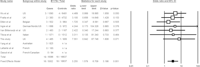
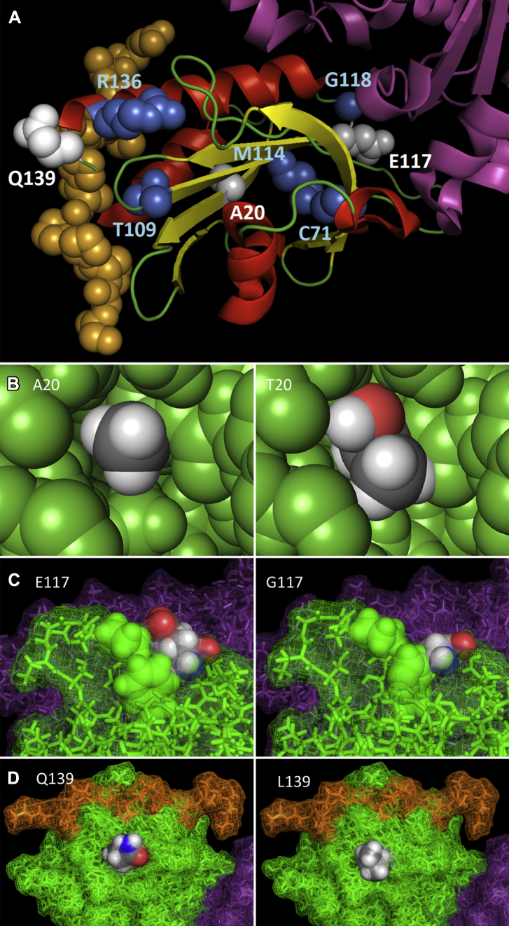
Mutations in the gene encoding profilin 1 (PFN1) have recently been shown to cause amyotrophic lateral sclerosis (ALS), a fatal neurodegenerative disorder. We sequenced the PFN1 gene in a cohort of ALS patients (n = 485) and detected 2 novel variants (A20T and Q139L), as well as 4 cases with the previously identified E117G rare variant (∼ 1.2%). A case-control meta-analysis of all published E117G ALS+/- frontotemporal dementia cases including those identified in this report was significant p = 0.001, odds ratio = 3.26 (95% confidence interval, 1.6-6.7), demonstrating this variant to be a susceptibility allele. Postmortem tissue from available patients displayed classic TAR DNA-binding protein 43 pathology. In both transient transfections and in fibroblasts from a patient with the A20T change, we showed that this novel PFN1 mutation causes protein aggregation and the formation of insoluble high molecular weight species which is a hallmark of ALS pathology. Our findings show that PFN1 is a rare cause of ALS and adds further weight to the underlying genetic heterogeneity of this disease.

Keywords: Amyotrophic lateral sclerosis; Profilin 1; TDP-43 proteinopathy.


Figures

Fig. 1
Fig. 1
Residue conservation of mammals and viper showing mutations identified in this study. The residues, indicated with a red star, for A20, E117, and Q139 are fully conserved in all mammalian species and viper. Non-mammalian species were not included because of the lack of a clear one to one orthologous relationship in the profilin family.
Fig. 2
Fig. 2
Meta-analysis of published E117G case-control frequencies and those identified in this study. Data are presented as a forest plot. Squares and lines correspond to the study specific odds ratios (ORs) and 95% confidence intervals (CIs), respectively. The bottom row (filled square) shows the meta-analysis OR and CIs under fixed and random effects models. The fixed effect model is preferred as the studies showed minimal heterogeneity (p = 0.962, χ2-based Q-test), although there was no difference between the two methods. The area of the squares reflects the study specific weights (inverse of the variance). Variant counts from 4300 ESP European Americans and the 1000 Genomes project have been incorporated into the Wu et al. (2012) control data. Studies by Yang et al. (2013), Daoud et al., 2013 and Lattante et al. (2012) did not screen local control cohorts and so did not contribute to the final meta-analysis calculation.
Fig. 3
Fig. 3
Familial gene mutations in PFN1. Pedigrees of PFN1 mutation positive cases identified in this study are shown for (A) A20T - Family 1 and (B) E117G - Family 2. Gender information has been withheld and denoted by diamonds. The arrow denotes the proband of each kindred. DNA was unavailable from other affected and unaffected members to test segregation of PFN1 mutations.
Fig. 4
Fig. 4
Pymol rendering of novel mutations identified in this study and all published PFN1 reports. (A) A schematic diagram of PFN1 bound to VASP and actin. VASP is shown as orange spheres, actin as purple ribbons, and PFN1 as red helices, yellow beta sheets, and green loops. Mutations identified in this study are indicated by white spheres, whereas those described elsewhere are shown as blue spheres. B–D show the reference amino acid (left diagram) and variant amino acid (right diagram) for the A20T, E117G, and Q139L mutations, respectively. The A20T mutation in the core of the protein potentially destabilizes a beta sheet because of steric hindrance, the E117G mutation potentially destabilizes an adjacent surface loop, whereas the surface Q139L mutation has no apparent structural impact. All PFN1 atoms are colored green, actin purple, and VASP orange, except those belonging to the highlighted residue side chain which are colored according to atom (gray = carbon, white = hydrogen, red = oxygen, blue = nitrogen). All diagrams are derived from PDB: 2PAV and rendered with PyMol (The PyMOL Molecular Graphics System, Version 1.5.0.4 Schrödinger, LLC.). Abbreviations: PDB, protein data bank; PFN1, profilin 1; VASP, vasodilator-stimulated phosphoprotein.
Fig. 5
Fig. 5
TDP-43 immunohistochemistry in 3 cases showing PFN1 mutations. (A) TDP-43 immunopositive skein-like neuronal cytoplasmic inclusion (NCI) seen in the XIIth nerve nucleus of the medulla (patient 2; E117G). (B) A globular TDP-43 immunopositive NCI seen in the anterior horn of the spinal cord (patient 3; E117G). (C) A TDP-43 immunopositive glial cytoplasmic inclusion (GCI) in the spinal cord (patient 6; Q139L). No PFN1 immunopositivity was seen in any of the regions examined in any of the 3 cases. Scale bar: (A) 40 μm, (B) 40 μm, and (C) 15 μm. Abbreviation: PFN1, profilin 1.
Fig. 6
Fig. 6
Immunocytochemistry of transfected cells shows granule accumulation of mutant PFN1 in the cytoplasm. (A) Immunofluorescent images of HEK293T cells expressing V5-PFN1WT, V5-PFN1E117G, and V5-PFN1Q139L show diffuse anti-V5 staining (green). The cells expressing V5-PFN1C71G and V5-PFN1A20T show distinct punctate granular structures in the cytoplasm. Nuclei are counterstained with DAPI (blue) in the merged image. Scale bar, 10 μm. (B) Quantification of cells containing granules shows that the V5-PFN1C71G and V5-PFN1A20T constructs have an increased percentage of cells with granules compared with V5-PFN1WT, whereas V5-PFN1E117G and V5-PFN1Q139L are similar to the wild type (*p < 0.05). Bars represent means ± SEM. Abbreviations: PFN1, profilin 1; SEM, standard error of the mean.
Fig. 7
Fig. 7
Fractionation of cell lysates shows that mutant PFN1 is more insoluble than the wild type. (A–C) Solubility fractionation of HEK293T cell transfected with V5-PFN1 constructs showing lysate (L), NP-40 soluble (S), and urea soluble/NP-40 insoluble (I) fractions. The WT protein was predominantly soluble as were the E117G and Q139L proteins. Both the C71G and A20T constructs showed significant decreases in the soluble fraction compared with the WT (***p < 0.001) alongside significant increases in the insoluble fraction (*p < 0.05). Bars represent means ± SEM. There was also an increase in high molecular weight detergent resistant oligomers in the C71G and A20T insoluble fractions. (D–F) Solubility fractionation of fibroblasts from a patient with the A20T mutation and a control individual showing lysate (L), NP-40 soluble (S), and urea soluble/NP-40 insoluble (I) fractions. PFN1 was predominantly found in the soluble fraction in both cell lines. Treatment with MG132 resulted in a significant shift of PFN1 from the soluble to the insoluble fraction in the A20T cell line (***p < 0.001). There was no change to the solubility of PFN1 in the control line (ns = nonsignificant). Bars represent means ± SEM. Abbreviations: PFN1, profilin 1; SEM, standard error of the mean; WT, wild type.

References

    1. Abramzon Y., Johnson J.O., Scholz S.W., Taylor J.P., Brunetti M., Calvo A., Mandrioli J., Benatar M., Mora G., Restagno G., Chio A., Traynor B.J. Valosin-containing protein (VCP) mutations in sporadic amyotrophic lateral sclerosis. Neurobiol. Aging. 2012;33:2231.e1–2231.e6. - PMC - PubMed
    1. Adzhubei I.A., Schmidt S., Peshkin L., Ramensky V.E., Gerasimova A., Bork P., Kondrashov A.S., Sunyaev S.R. A method and server for predicting damaging missense mutations. Nat. Methods. 2010;7:248–249. - PMC - PubMed
    1. Al-Chalabi A., Jones A., Troakes C., King A., Al-Sarraj S., van den Berg L.H. The genetics and neuropathology of amyotrophic lateral sclerosis. Acta Neuropathol. 2012;124:339–352. - PubMed
    1. Arnold K., Bordoli L., Kopp J., Schwede T. The SWISS-MODEL workspace: a web-based environment for protein structure homology modelling. Bioinformatics. 2006;22:195–201. - PubMed
    1. Brooks B.R. Versailles minimal dataset for diagnosis of ALS: a distillate of the 2nd Consensus Conference on accelerating the diagnosis of ALS. Versailles 2nd Consensus Conference participants. Amyotroph. Lateral Scler. Other Mot. Neuron Disord. 2000;1(Suppl 1):S79–S81. - PubMed

Publication types

MeSH terms