. 2014 Dec;16(12):993-1006.
doi: 10.1016/j.neo.2014.10.001.

In vivo Mn-enhanced MRI for early tumor detection and growth rate analysis in a mouse medulloblastoma model

Affiliations

In vivo Mn-enhanced MRI for early tumor detection and growth rate analysis in a mouse medulloblastoma model

Giselle A Suero-Abreu et al. Neoplasia. 2014 Dec.

Abstract

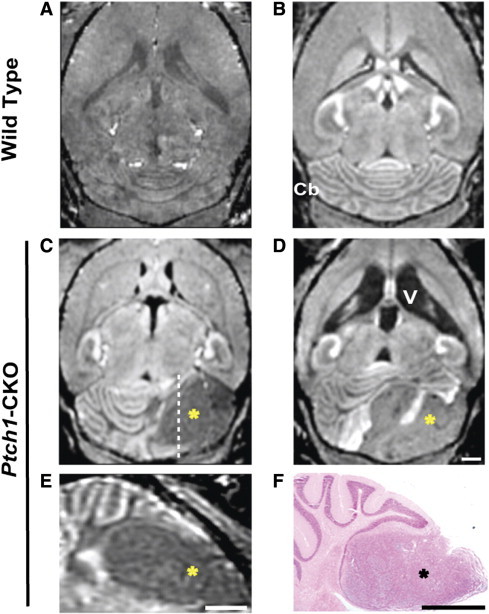
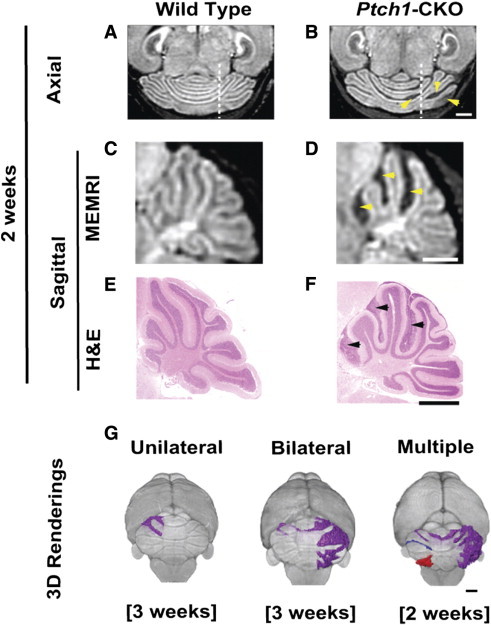
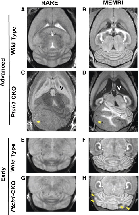
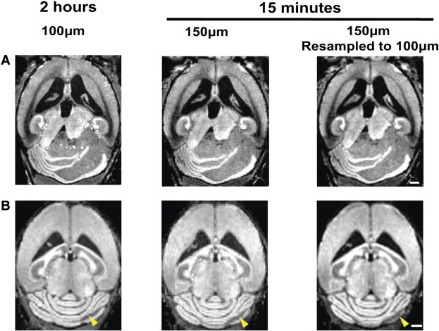
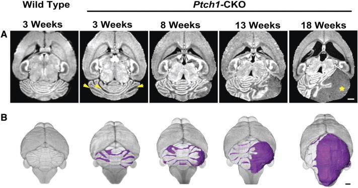
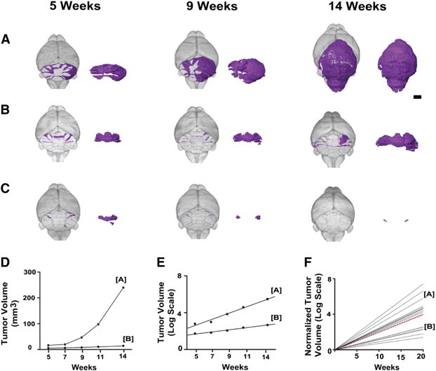
Mouse models have increased our understanding of the pathogenesis of medulloblastoma (MB), the most common malignant pediatric brain tumor that often forms in the cerebellum. A major goal of ongoing research is to better understand the early stages of tumorigenesis and to establish the genetic and environmental changes that underlie MB initiation and growth. However, studies of MB progression in mouse models are difficult due to the heterogeneity of tumor onset times and growth patterns and the lack of clinical symptoms at early stages. Magnetic resonance imaging (MRI) is critical for noninvasive, longitudinal, three-dimensional (3D) brain tumor imaging in the clinic but is limited in resolution and sensitivity for imaging early MBs in mice. In this study, high-resolution (100 μm in 2 hours) and high-throughput (150 μm in 15 minutes) manganese-enhanced MRI (MEMRI) protocols were optimized for early detection and monitoring of MBs in a Patched-1 (Ptch1) conditional knockout (CKO) model. The high tissue contrast obtained with MEMRI revealed detailed cerebellar morphology and enabled detection of MBs over a wide range of stages including pretumoral lesions as early as 2 to 3 weeks postnatal with volumes close to 0.1 mm(3). Furthermore, longitudinal MEMRI allowed noninvasive monitoring of tumors and demonstrated that lesions within and between individuals have different tumorigenic potentials. 3D volumetric studies allowed quantitative analysis of MB tumor morphology and growth rates in individual Ptch1-CKO mice. These results show that MEMRI provides a powerful method for early in vivo detection and longitudinal imaging of MB progression in the mouse brain.


Figures

Figure 1
Figure 1
In vivo MEMRI detection of MB tumors in Ptch1-CKO mice. Compared to non–Mn-enhanced T1w MRI (A), post-contrast signal enhancement in axial MEMRI image (B) enabled visualization of normal brain and Cb anatomy in wild-type mice. MEMRI also allowed detection of advanced MB tumors as hypointense areas (*) within the Cb of Ptch1-CKO mice, typically located in the Cb hemispheres (C and D), and demonstrated severely enlarged ventricles (V) in some mice (D). Panel E shows a sagittal section from the mouse shown in C (dashed line indicates the approximate location of the section) and highlights the MB tumor (*). A matched histologic section (F), stained with H&E, showed good correlation of tumor location with MEMRI (E). Scale bars, 1 mm for A to F (scale on D for A–D).
Figure 2
Figure 2
MEMRI allowed detection of early-stage MB lesions in Ptch1-CKO mice. Axial (A and B) and sagittal (C and D) MEMRI images (dashed lines on A and B show the approximate location of the sagittal sections shown in C and D, respectively) revealed multiple hypointense areas (yellow arrowheads) in the cerebella of Ptch1-CKO mice (B and D) compared to wild-type controls (A and C) at 2 to 3 weeks of age. Matched sagittal H&E sections (E and F) demonstrated accurate histologic correlation between areas of negative contrast in MEMRI (D; yellow arrowheads) and early pretumoral lesions (F; black arrowheads). Representative 3D volume renderings from MEMRI data demonstrate distinct patterns of Cb lesions seen in individual Ptch1-CKO mice at 2 to 3 weeks of age. These early pretumoral lesions were detected as unilateral, bilateral, and multiple (separated lesions shown in different colors; see Supplemental Video 1 for more detailed visualization), as demonstrated in G. Scale bars, 1 mm.
Figure 3
Figure 3
MEMRI was more sensitive for detecting early-stage MB than conventional T2w MRI. Representative examples are shown for wild-type (A, B, E, and F) and Ptch1-CKO mice (C, D, G, and H) scanned with both non-contrast T2w RARE (left panels; A, C, E, and G) and post-contrast T1w MEMRI sequences (right panels; B, D, F, and H). These images show improved tissue contrast in MEMRI compared to RARE images and show higher sensitivity for visualization of tumor tissue (*) using MEMRI (D) compared to RARE (C), especially for detection of early pretumoral lesions with MEMRI (H, arrowheads), which could not be detected with RARE (G). Scale bars, 1 mm.
Figure 4
Figure 4
MEMRI detection of advanced and early MB lesions with both high-throughput and high-resolution protocols. MEMRI images of Ptch1-CKO mice demonstrated that both advanced-stage tumors (A) and early pretumoral lesions (B, arrowheads) were detected with either the high-resolution (100 μm; 2 hours) or high-throughput (150 μm; 15 minutes) sequences. High-throughput images were improved by resampling the 150-μm images to 100 μm in post-processing (right panels). Scale bars, 1 mm.
Figure 5
Figure 5
In vivo longitudinal MEMRI enabled imaging tumor progression from early to advanced stages. Axial MEMRI images (A) and 3D volume renderings (B) of a representative individual Ptch1-CKO mouse at sequential time points (3-18 weeks) demonstrate the feasibility of longitudinal imaging using MEMRI. Note also that bilateral pretumoral lesions were observed at early time points (arrowheads), but at later times, only one area exponentially progressed to an advanced MB (*). Representative examples of an axial MEMRI and 3D rendering of a control mouse are shown at the first time point as a reference. Scale bars, 1 mm.
Figure 6
Figure 6
Longitudinal MEMRI revealed variability in MB tumor growth patterns. 3D renderings from longitudinal MEMRI images of representative Ptch1-CKO mice showed a range of MB progression rates from fast (A) to slow (B). In each panel, the segmented MB lesions are shown in color within the whole brain (left) and separately (right). Several examples of early lesions that regressed below detectability with MEMRI were also observed (C; N = 5/21). Quantitative analysis of MB lesion volume (D) demonstrated the distinct growth rate patterns of the mice shown in A and B and also showed excellent fit to an exponential growth model, plotted in semi-log format (E). To compare MB growth rates between mice, lesion volumes were normalized to their initial volumes and again plotted in semi-log format (F; N = 16), demonstrating the variability observed in MB growth rates in the Ptch1-CKO mouse model. Dashed red line represents the mean Td = 4.3 weeks. Scale bar in A, 2 mm.
Figure 7
Figure 7
IHC analysis of MB tumors in Ptch1-CKO mice. MB tumors were analyzed using IHC for Ki67 and p27, as markers of proliferation and differentiation, respectively (N = 12). DAB staining for Ki67 at low (× 3; A and B) and high (× 20; C and D) magnification showed the presence of multiple Ki67-negative areas in some tumors (B and D) but in not others (A and C). (Note: the red boxes show the approximate location of C and D on A and B, respectively.) Within the same two tumors, Ki67 (red; E and F)/p27 (green; G and H) double fluorescent staining revealed that the Ki67-negative areas (F) were regions of differentiation with high p27-expression (H and J). Conversely, tumors with uniform Ki67 expression (E) had scattered p27-expressing cells (G) mixed among the Ki67-expressing cells (I). Scale bars, 1 mm in A and B; 50 μm in C to J.
Figure 8
Figure 8
IHC analysis of early pretumoral and later non-progressing lesions. Cerebellar sections show examples of pretumoral lesions (PTL) in two different 3-week-old Ptch1-CKO mice (A and B and C and D). H&E-stained sections (A and C) show the cerebellar morphology and the approximate locations (yellow boxes) of the immunofluorescent sections (B and D) stained for Ki67 (red), p27 (green), and TUNEL (white) and the overlay of all three stains. Examples are also provided for tumors and non-progressing lesions (*) in 15-week-old (E–H) and 13-week-old (I and J) Ptch1-CKO mice. Dashed lines demarcate the PTLs and non-progressing lesions, as well as the molecular layer (ML) and internal granule layer (IGL). Early PTLs (B and D), tumors (E and I), and lesions that were not progressing (F–H and J) all showed high levels of Ki67, with variable p27 and TUNEL staining. p27 expression was more widespread and clustered in the PTLs and tumors when compared to non-progressing lesions. Notably, sections of both PTLs and non-progressed lesions showed some p27-positive cells in the ML between the lesion and the internal granule layer (yellow arrows). Magnification: × 1.5 (A and C) and × 20 (B, D, and E–J). Scale bars, 1 mm in A and C; 50 μm in B, D, and E–J.

References

    1. Siegel R, Naishadham D, Jemal A. Cancer statistics, 2012. CA Cancer J Clin. 2012;62:10–29. - PubMed
    1. Pui C-H, Gajjar AJ, Kane JR, Qaddoumi IA, Pappo AS. Challenging issues in pediatric oncology. Nat Rev Clin Oncol. 2011;8:540–549. - PMC - PubMed
    1. Northcott PA, Jones DT, Kool M, Robinson GW, Gilbertson RJ, Cho YJ, Pomeroy SL, Korshunov A, Lichter P, Taylor MD. Medulloblastomics: the end of the beginning. Nat Rev Cancer. 2012;12:818–834. - PMC - PubMed
    1. Rutkowski S, von Hoff K, Emser A, Zwiener I, Pietsch T, Figarella-Branger D, Giangaspero F, Ellison DW, Garre ML, Biassoni V. Survival and prognostic factors of early childhood medulloblastoma: an international meta-analysis. J Clin Oncol. 2010;28:4961–4968. - PubMed
    1. Gajjar A, Chintagumpala M, Ashley D, Kellie S, Kun LE, Merchant TE, Woo S, Wheeler G, Ahern V, Krasin MJ. Risk-adapted craniospinal radiotherapy followed by high-dose chemotherapy and stem-cell rescue in children with newly diagnosed medulloblastoma (St Jude Medulloblastoma-96): long-term results from a prospective, multicentre trial. Lancet Oncol. 2006;7:813–820. - PubMed

Publication types