Review
. 2014 Dec;41(6):848-59.
doi: 10.1053/j.seminoncol.2014.09.013. Epub 2014 Oct 7.

Altering the response to radiation: sensitizers and protectors

Affiliations
Review

Altering the response to radiation: sensitizers and protectors

Deborah E Citrin et al. Semin Oncol. 2014 Dec.

Abstract

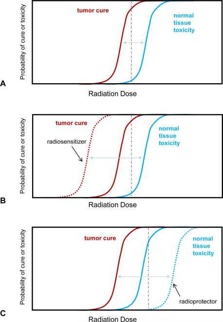
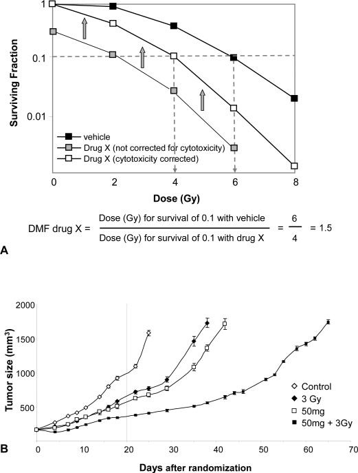
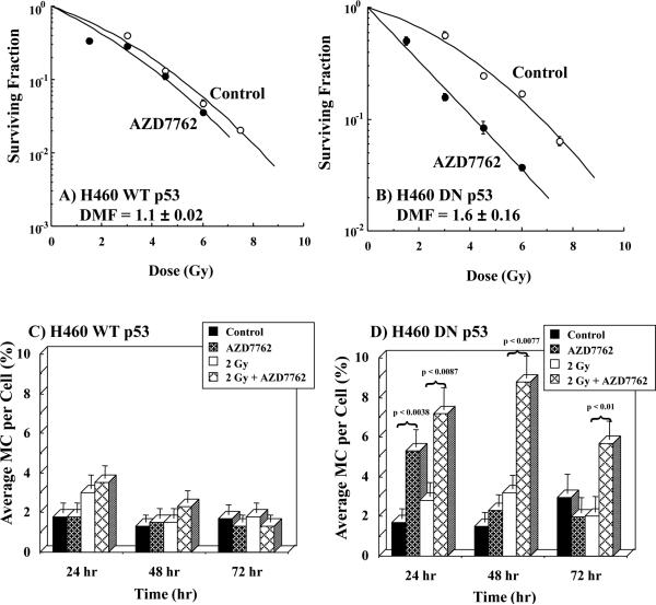
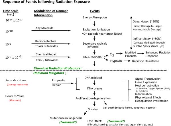
A number of agents are used clinically to enhance the efficacy of radiotherapy today, many of which are cytotoxic chemotherapies. Agents that enhance radiation induced tumor cell killing or protect normal tissues from the deleterious effects of ionizing radiation are collectively termed radiation modifiers. A significant effort in radiobiological research is geared towards describing and testing radiation modifiers with the intent of enhancing the therapeutic effects of radiation while minimizing normal tissue toxicity. In this review, we discuss the characteristics of these agents, the testing required to translate these agents into clinical trials, and highlight some challenges in these efforts.


Figures

Figure 1
Figure 1. Therapeutic ratio and radiation modifiers
A) The probability of tumor cure (red) and normal tissue toxicity (blue) can be plotted as a factor of radiation dose. Note that these are parallel idealized sigmoidal curves which may not be representative of the clinical condition. A therapeutic dose may be selected that delivers a high chance of tumor cure with an acceptable rate of toxicity (black dashed line). B) A tumor selective radiation sensitizer increases the probability of tumor cure for a given dose (red dashed line), hence the tumor control curve is shifted to the left. This results in a higher chance of cure for the same radiation dose as delivered in A) (black dashed line). Alternatively, a lower dose may be delivered to yield the same rate of cure with less normal tissue toxicity. C) A normal tissue radioprotector decreases the probability of normal tissue toxicity for a given dose of radiation, thus pushing the toxicity curve to the right (blue dashed line). This allows a higher dose to be given to obtain a higher rate of cure with less or equivalent normal tissue injury.
Figure 2
Figure 2. Evaluation of radiation dose modification in vitro and in vivo
A) Cells are plated at varying densities after exposure to Drug X or vehicle followed by exposure to radiation. Following incubation for 10-14 days, colonies of 50 or more cells are counted. The number of colonies that are counted is compared to the number of cells plated and a surviving fraction is generated. Plotting the surviving fraction over a range of radiation doses allows for the generation of a survival curve. The survival of the cell line when treated with vehicle and radiation is compared to that obtained when cells are treated with Drug X and radiation. The surviving fraction of the drug and radiation combination must include a correction for the cytotoxicity of the drug alone (open squares). If this correction is not made, a dose modifying factor cannot be calculated and the ability to sensitize or protect cannot be accurately determined. In this example, lack of correction for cytotoxicity (grey squares) appears to make the drug much more effective as a sensitizer than it actually is. B) A dose modifying factor may also be calculated for tumor regrowth. Mice are implanted with a tumor xenograft. Once tumors reach a measurable and consistent size, mice are randomized to treatment with vehicle, vehicle with radiation (in this example 3Gy), drug (in this example 50 mg of AZD6244), or radiation with drug. The dose modifying factor is calculated by determining the number of days it takes tumors in each group triple in volume. Comparing volumes at only one time point (dotted line at 20 days) does not allow a calculation of DMF and may over or underestimate the efficacy of the investigational agent. Figure 2B reproduced from () with permission.
Figure 3
Figure 3
The effects of radiation with and without the Chk1/2 inhibitor AZD7762 (100 nM, added 1 hr prior to and 24 hr post-radiation) on radiation-induced cell killing and mitotic catastrophe. In this study the influence of p53 on Chk1/2 inhibition/radiation response was evaluated using a H460 lung cancer cell line (wild type p53) as opposed to H460 cells containing a dominant negative p53 construct (H460 DN p53). Wild type p53 cells (A & C) were not sensitized by the combination of AZD7762 and radiation; whereas, the radiosensitivity H460 DN p53 cells were significantly enhanced by AZD7762 (B,) as well as DNA damage as measured by mitotic catastrophe (D). (Adapted from reference () with permission)
Figure 4
Figure 4. Sequence of events following radiation exposure
The chart is divided into three parts by dashed lines suggesting events and reactions that might be modified by radiation protectors (top), radiation mitigators, and treatment (bottom). Reproduced from ().
Figure 5
Figure 5. Serum, tissue, and tumor levels of WR-2721 (Amifostine) in Fischer 344 rats as a function of time after an i.p. injection
Notice that normal tissue levels rapidly achieve mM concentrations of WR-2721; whereas, tumor levels are quite low initially. With time tumor levels gradually increase, while normal tissue levels begin to decrease. This study demonstrates that chemical radioprotector tissue concentrations in the mM range can be achieved quickly. Further the study underscores the importance of the timing of radiation exposure with within 15-30 min after injection to take advantage of the differential concentration of WR-2721 in normal as opposed to tumor tissue. (Adapted from reference (), with permission)
Figure 6
Figure 6. The effects of selumetinib and 5-FU on tumor cell and xenograft radiosensitivity
A) The HCT116 cell line was exposed to 15 μmol/L of 5-FU (or vehicle) for 18 hours and selumetinib (or vehicle) for 2 hours and irradiated with graded doses of X-rays. Colony forming efficiency was determined 10 to 12 days later and survival curves generated after normalizing for cell killing by selumetinib, 5-FU or selumetinib + 5FU in the absence of IR. B) Mice harboring HCT116 xenografts were randomized into four groups: vehicle, selumetinib, radiation + 5-FU, or selumetinib + radiation + 5-FU. Each drug was given as a single dose prior to a single dose of radiation. The study demonstrates that radiation modifiers can be evaluated in the context of chemoradiotherapy and highlights the need to account for the toxicity of both the investigational agent and chemotherapy in calculations of modifying capability. Adapted from reference () with permission.
Figure 7
Figure 7. The effect of the radioprotector Tempol on oral mucositis resulting from fractionated irradiation and cisplatin (CDDP)
To measure ulceration tongues were removed from the various groups and stained with Toluidine blue. Ulcerations were clearly present in tongues from mice treated with the combination of CDDP and radiation (Panel A, second row, marked by arrows). Tempol either delivered systemically or topically afforded near complete protection (A, third and fourth rows). Using the same Tempol/CDDP/radiation protocol as used in A, SCCVII tumor regrowth for the various groups is shown in B. Tempol did not interfere with CDDP mediated enhancement of radiation. (Adapted from reference (), with permission).

References

    1. Gilbert M. Radiotherapy in Hodgkin's Disease (malignant granulomatosis); anatomic and clinical foundations; governing principles, results. Am J Roentgenol. 1939;41:198–241.
    1. Peters M. A study in survivals in Hodgkin's disease treated radiologically. Am J Roentgenol. 1950;63:299–311.
    1. Coutard H. Roentgen therapy of epitheliomas of the tonsillar region, hypopharynx, anf larynx from 1920 to 1926. AJR Am J Roentgenol 1932. 28:313.
    1. Minchinton AI, Tannock IF. Drug penetration in solid tumours. Nat Rev Cancer. 2006;6:583–592. - PubMed
    1. Kuban DA, Tucker SL, Dong L, Starkschall G, Huang EH, Cheung MR, et al. Long-term results of the M. D. Anderson randomized dose-escalation trial for prostate cancer. Int J Radiat Oncol Biol Phys. 2008;70:67–74. - PubMed

Publication types

Substances