Review
. 2015 Mar;25(3):158-70.
doi: 10.1016/j.tcb.2014.11.002. Epub 2014 Dec 11.

Protecting the mitochondrial powerhouse

Affiliations
Review

Protecting the mitochondrial powerhouse

Morten Scheibye-Knudsen et al. Trends Cell Biol. 2015 Mar.

Abstract

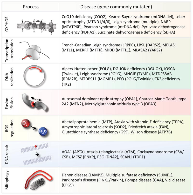
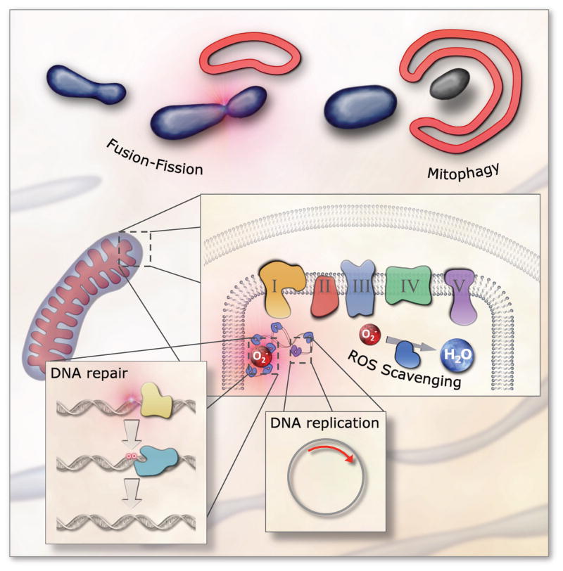
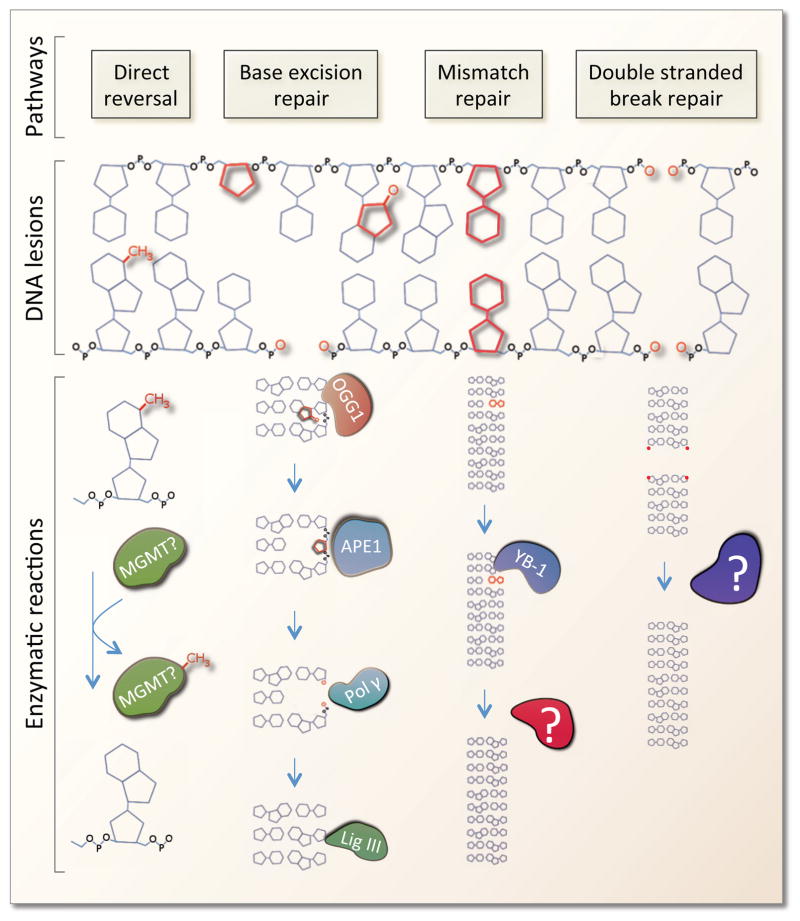
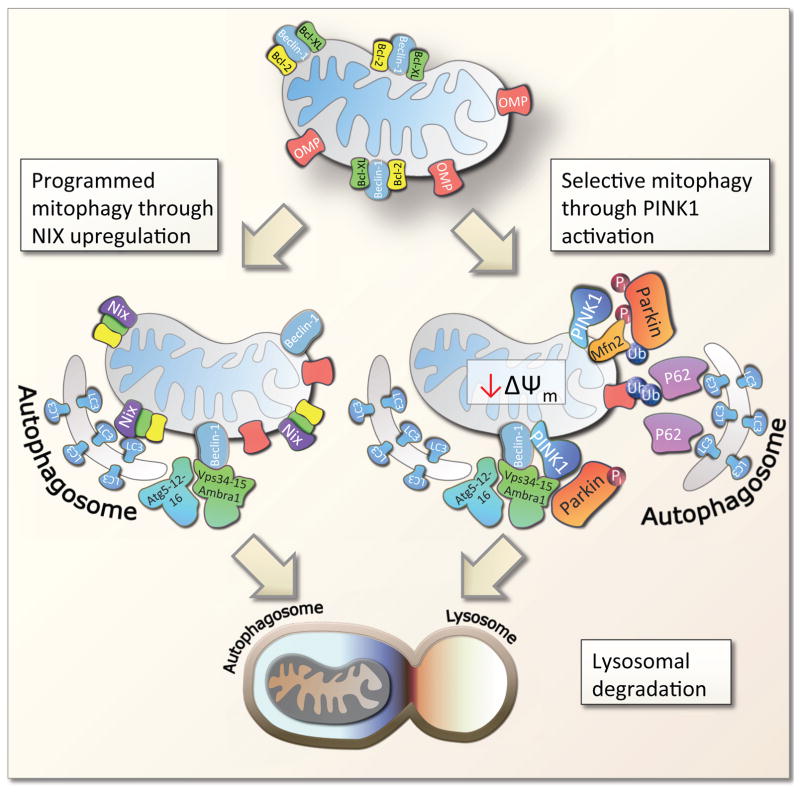
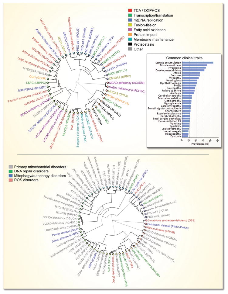
Mitochondria are the oxygen-consuming power plants of cells. They provide a critical milieu for the synthesis of many essential molecules and allow for highly efficient energy production through oxidative phosphorylation. The use of oxygen is, however, a double-edged sword that on the one hand supplies ATP for cellular survival, and on the other leads to the formation of damaging reactive oxygen species (ROS). Different quality control pathways maintain mitochondria function including mitochondrial DNA (mtDNA) replication and repair, fusion-fission dynamics, free radical scavenging, and mitophagy. Further, failure of these pathways may lead to human disease. We review these pathways and propose a strategy towards a treatment for these often untreatable disorders.

Keywords: DNA repair; disease; mitochondria; mitophagy; reactive oxygen species.


Figures

Figure 1
Figure 1
Examples of mitochondrial pathways that are associated with mitochondrial diseases.
Figure 2
Figure 2. Mitochondrial maintenance pathways
Several pathways have evolved that maintain mitochondrial integrity. These include faithful DNA replication; efficient DNA repair; enzymes and molecules that remove reactive oxygen species (ROS); mitochondrial morphology regulation through fission and fusion; and whole organelle removal through mitophagy.
Figure 3
Figure 3. Mitochondrial DNA repair pathways
A number of different DNA lesions have been shown to be reparable in the mitochondria. Direct reversal of spurious methylation has been shown to occur, however, the enzyme(s) involved is not currently known. Base excision repair is the major DNA repair pathway in mitochondria and most proteins involved in this pathway have been shown to be present in mitochondria. Base excision repair consists of multiple steps. The lesion is initially identified by a number of different glycosylases (eg. OGG1, NEIL1 etc.) that remove the damaged base leaving an abasic site. The abasic site is then recognized by APE1 that removes the ribose allowing for final repair by DNA polymerase γ and ligase III. Mismatch repair (MMR) has been shown in human mitochondria where Y-box-binding protein 1 (YB-1) is proposed to be a key player. The downstream enzymes in mitochondrial MMR are, however, still not known. DNA double stranded break repair (DSBR) may occur in the mitochondria by blunt end ligation, although the enzymes involved in this pathway are not known.
Figure 4
Figure 4. The mechanism of mitophagy
Mammalian mitophagy is believed to occur through at least two pathways, programmed mitophagy and selective mitophagy, although significant cross talk between these has been found. Programmed mitophagy was discovered in maturing red blood cells through upregulation of the protein NIX that may facilitate the dissociation of the anti-mitophagic proteins Bcl-2 and Bcl-XL from the pro-mitophagic protein Beclin-1. This potentially derepresses Beclin-1 allowing recruitment of the Vps34-15-AMBRA complex that in turn will associate with the autophagosome elongation machinery atg5-12/atg16 leading to the formation of an autophagosome. NIX coats the mitochondria and associates directly with LC3 which binds to the growing autophagosome. Mitophagy is completed by the fusion of the mitophagosome with a lysosome. In selective mitophagy the initiating event is believed to be mitochondrial inner membrane depolarization leading to PINK1 accumulation at the outer membrane. PINK1 phosphorylates Parkin, Mfn2 and other proteins leading to the activation of Parkin. Parkin is a E3-ubiquitin ligase that upon activation ubiquitinates outer mitochondrial membrane proteins (OMPs), possibly VDAC, are ubiquitinated. Vps34-15-AMBRA complex and the Atg5-12/atg16 complex is recruited by activated PINK1/Parkin. Outer membrane ubiquitination leads to the recruitment of p62 which will associate with LC3 on the growing autophagosome. When the autophagosome is completed, fusion with a lysosome will lead to complete degradation of the mitochondria.
Figure 5
Figure 5. The complex clinical relationship between mitochondrial disorders
The dendrograms show the association between diseases based on the prevalence of the clinical traits in the diseases. The closer two diseases are the more signs and symptoms are shared ei. the more similar the diseases are. Genes commonly mutated in these disorders are shown in the parentheses. The top dendrogram shows primary mitochondrial diseases and the colors represent the specific putative pathway that may be affected in that disease. The insert shows the average prevalence of commonly altered clinical traits in the mitochondrial disorders depicted in the dendrogram. The bottom dendrogram show how diseases with dysregulation of ROS, DNA repair and mitophagy associate with the primary mitochondrial disorders.

References

    1. Prachar J. Mouse and human mitochondrial nucleoid--detailed structure in relation to function. Gen Physiol Biophys. 2010;29:160–174. - PubMed
    1. Becker T, et al. Mitochondrial protein import: from transport pathways to an integrated network. Trends Biochem Sci. 2012;37:85–91. - PubMed
    1. Wallace DC. A mitochondrial paradigm of metabolic and degenerative diseases, aging, and cancer: a dawn for evolutionary medicine. Annu Rev Genet. 2005;39:359–407. - PMC - PubMed
    1. Poole JC, et al. Multiplex analysis of mitochondrial DNA pathogenic and polymorphic sequence variants. Biol Chem. 2010;391:1115–1130. - PMC - PubMed
    1. Dunbar DR, et al. Different cellular backgrounds confer a marked advantage to either mutant or wild-type mitochondrial genomes. Proc Natl Acad Sci U S A. 1995;92:6562–6566. - PMC - PubMed

Publication types