. 2015 Jun;9(6):1333-51.
doi: 10.1038/ismej.2014.219. Epub 2014 Dec 12.

Is there a common water-activity limit for the three domains of life?

Affiliations

Is there a common water-activity limit for the three domains of life?

Andrew Stevenson et al. ISME J. 2015 Jun.

Abstract

Archaea and Bacteria constitute a majority of life systems on Earth but have long been considered inferior to Eukarya in terms of solute tolerance. Whereas the most halophilic prokaryotes are known for an ability to multiply at saturated NaCl (water activity (a(w)) 0.755) some xerophilic fungi can germinate, usually at high-sugar concentrations, at values as low as 0.650-0.605 a(w). Here, we present evidence that halophilic prokayotes can grow down to water activities of <0.755 for Halanaerobium lacusrosei (0.748), Halobacterium strain 004.1 (0.728), Halobacterium sp. NRC-1 and Halococcus morrhuae (0.717), Haloquadratum walsbyi (0.709), Halococcus salifodinae (0.693), Halobacterium noricense (0.687), Natrinema pallidum (0.681) and haloarchaeal strains GN-2 and GN-5 (0.635 a(w)). Furthermore, extrapolation of growth curves (prone to giving conservative estimates) indicated theoretical minima down to 0.611 aw for extreme, obligately halophilic Archaea and Bacteria. These were compared with minima for the most solute-tolerant Bacteria in high-sugar (or other non-saline) media (Mycobacterium spp., Tetragenococcus halophilus, Saccharibacter floricola, Staphylococcus aureus and so on) and eukaryotic microbes in saline (Wallemia spp., Basipetospora halophila, Dunaliella spp. and so on) and high-sugar substrates (for example, Xeromyces bisporus, Zygosaccharomyces rouxii, Aspergillus and Eurotium spp.). We also manipulated the balance of chaotropic and kosmotropic stressors for the extreme, xerophilic fungi Aspergillus penicilloides and X. bisporus and, via this approach, their established water-activity limits for mycelial growth (∼0.65) were reduced to 0.640. Furthermore, extrapolations indicated theoretical limits of 0.632 and 0.636 a(w) for A. penicilloides and X. bisporus, respectively. Collectively, these findings suggest that there is a common water-activity limit that is determined by physicochemical constraints for the three domains of life.


Figures
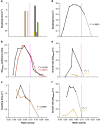
Figure 1
Figure 1
Growth in relation to water activity and/or lower water-activity values at which growth was observed for halophilic Bacteria and Archaea cultured in high-magnesium saline substrates (except for data for H. saccharovorum and Salinicola strain LC26 that were obtained in high-NaCl media (see Table 1 and Supplementary Table S1), and for S. ruber and S. longa on NaCl-supplemented media indicated by black lines in (e) and (f) respectively). (a) Halophilic Archaea Halorubrum saccharovorum (strain NCIMB 2081T; shown in brown), Halobacterium sp. NRC-1 and Halococcus morrhuae (strain NCIMB 787T; both represented in yellow), Haloquadratum walsbyi (strain DSM 16790; black), Halococcus salifodinae (strain DSM 13046; purple), Halobacterium noricense (strain DSM 15987T; hashed) and Natrinema pallidum (strain NCIMB 777T; green), and Bacteria Salinicola strain LC26 and Pontibacillus strain AS2 (both represented in grey) cultured in media supplemented with various concentrations of NaCl and MgCl2 and/or glycerol and ethylene glycol to give a range of water-activity values and incubated at 20 °C for Pontibacillus strain AS2 and Salinicola strain LC26 or 37 °C for all other species (see Table 1 and Supplementary Table S1). (b) Haloarchaeal strains GN-2 and GN-5 shown in orange and black, respectively, cultured in bittern brines supplemented with peptone at 37 °C for 6 days (calculated and replotted against water activity using data from Javor, 1984). (c) A mixed halophile community (identified using DAPI; red line) and Bacteria within this community (quantified using molecular probes; black line) by inoculating a synthetic seawater medium with supplemented NaCl using crystalliser brine and incubating at 37 °C (calculated and replotted against water activity using data from Antón et al., 2000). (d) The archaeon Halobacterium strain 004.1 in a synthetic seawater medium supplemented with NaCl, MgCl2, Na2SO4 and KCl at 37 °C (see Materials and methods and Supplementary Table S2). (e) The bacterium Salinibacter ruber strain DSM 13855T in complex media supplemented with addition of water from the Dead Sea (0.812 to 0.777 aw) and without (0.840 aw) at 35 °C (yellow line) or complex media supplemented with NaCl and incubated at 37 °C (black line; calculated and replotted against water activity using data from Sher et al., 2004 and Peña et al., 2010; see also Materials and methods and Supplementary Table S6). (f) The bacterium Salisaeta longa strain DSM 21114T in complex media supplemented with addition of water from the Dead Sea (0.926 to 0.792 aw) at 35 °C (yellow line) or complex media supplemented with NaCl (black line; Materials and methods and Supplementary Table S7). For all media, water-activity values were determined as described in the Materials and methods and at the same temperature as incubation was carried out for each set of media. Curves were extrapolated via regression analyses (dotted lines; for details see Supplementary Table S1) in order to determine the theoretical water-activity minima for growth. Pink dashed lines indicate the previously accepted water-activity limit for extremely halophilic Bacteria and Archaea (see Brown, 1990; Grant, 2004; Kminek et al., 2010).
Figure 2
Figure 2
Growth curves for halophilic Bacteria and Archaea cultured in high-NaCl substrates and plotted in relation to water activity. (a) The bacterium Halorhodospira halochloris (strain and incubation temperature not specified) cultured in a defined medium supplemented with NaCl (calculated and replotted against water activity using data from Deole et al., 2013). (b) The bacterium Halorhodospira halophila (strain DSM 244T; incubation temperature not specified) cultured in a defined medium supplemented with NaCl (calculated and replotted against water activity using data from Deole et al., 2013). (c) The bacterium Halanaerobium lacusrosei (strain DSM 10165T) cultured in a complex medium supplemented with NaCl and incubated at 37 °C (calculated and replotted against water activity using data from Cayol et al., 1995). (d) The Aacterium Actinopolyspora halophila (strain ATCC 27976T) cultured in a complex medium supplemented with NaCl, after a 14-day incubation at 37 °C (calculated and replotted against water activity using data from Yoshida et al., 1991). (e) The Archaea ‘Haloarcula californiae' (strain DSM 8905; black line) and ‘Haloarcula sinaiiensis' (strain DSM 8928; orange line) cultured in a complex medium supplemented with NaCl and incubated at 37 °C (calculated and replotted against water activity using data from Javor et al., 1982). (f) The archaeon Halorhabdus utahensis (strain DSM 12940T) in a defined medium supplemented with NaCl and incubated at 30 °C (calculated and re-plotted against water activity using data from Wainø et al., 2000). For all media, water activity values were determined as described in the Materials and methods and at the same temperature as incubation was carried out for each set of media. Curves were extrapolated via regression analyses (dotted lines; for details see Supplementary Table S1) in order to determine the theoretical water activity minima for growth. Pink dashed lines indicate the previously accepted water activity limit for extremely halophilic Bacteria and Archaea (see Brown, 1990; Grant, 2004; Kminek et al., 2010).
Figure 3
Figure 3
Radial extension rates for two strains of the xerophilic ascomycete Aspergillus penicillioides on solid media (MYPiA) supplemented with glycerol and other solutes over a range of concentrations, buffered at various pH values and incubated at different temperatures (Supplementary Table S5) and plotted in relation to water activity: strains JH06THH (black bars) and JH06THJ (red bars). For A. penicilliodes strain JH06THH, data relating to the following media were replotted from Williams and Hallsworth (2009): 0.647, 0.656, 0.667 and 0.670 aw. Medium composition and incubation temperatures for several treatments with common water-activity values differed (that is, 0.655i, 0.655ii, 0.702i, 0.702ii 0.714i, 0.714ii, 0.795i and 0.795ii; for details see Supplementary Table S8). The black arrow indicates the lowest water-activity at which growth of each strain was observed during an incubation period of six months. The line graph shows extrapolated growth curves plotted using data obtained on the biologically permissive media only in order to determine the theoretical extent of the water-activity windows for growth of each species; the yellow dashed line indicates the original water-activity limit for hyphal growth of the most xerophilic fungi (Pitt and Christian, 1968). For growth rate values of >0.75 mm per day, variation was ±0.10 mm per day, and for those of <0.75 mm per day, variation was ±0.040 mm per day (see Williams and Hallsworth, 2009).
Figure 4
Figure 4
Radial extension rates for three strains of the xerophilic ascomycete Xeromyces bisporus on solid media (MYPiA) supplemented with glycerol and other solutes over a range of concentrations, buffered at various pH values and incubated at different temperatures (Supplementary Table S5) and plotted in relation to water activity: strains FRR 0025 (blue bars), FRR 2347 (black bars) and FRR 3443 (pink bars). For all three strains of X. bisporus, data relating to the following media were replotted from Williams and Hallsworth (2009): 0.647, 0.653, 0.655ii, 0.656, 0.665, 0.670, 0.702ii and 0.714ii aw. Medium composition and incubation temperatures for several treatments with common water-activity values differed (that is, 0.655i, 0.655ii, 0.702i, 0.702ii, 0.714i, 0.714ii, 0.795i and 0.795ii; for details see Supplementary Table S8). The black arrow indicates the lowest water-activity at which growth of each strain was observed during an incubation period of six months. The line graph shows extrapolated growth curves plotted using data obtained on the biologically permissive media only in order to determine the theoretical extent of the water-activity windows for growth of each species; the yellow dashed line indicates the original water-activity limit for hyphal growth of the most xerophilic fungi (Pitt and Christian, 1968). For growth rate values of >4.0 mm per day, variation was ±0.20 mm per day, for those between 0.75 and 4.0 mm per day, variation was ±0.10 mm per day, and for those of <0.75 mm per day, variation was ±0.040 mm per day (see Williams and Hallsworth, 2009).
Figure 5
Figure 5
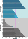
Lower water-activity limits for cell division of the most xerophilic eukaryotic microbes (upper pale-blue panel) and Bacteria and Archaea thus far identified (lower pale-grey panel) on salt-supplemented substrates (mid-blue and dark-grey bars, respectively) and sugar- or polyol-supplemented substrates (dark-blue and mid-grey bars, respectively): (i) haloarchaeal strain GN-5 (Figure 1b), (ii) haloarchaeal strain GN-2 (Figure 1b), (iii) Halorhodospira halophila (strain DSM 244T; Figure 2b), (iv) Halorhabdus utahensis (strain DSM 12940T; Figure 2f), (v) Halobacterium strain 004.1 (Figure 1d), (vi) Actinopolyspora halophila (strain ATCC 27976T; Figure 2d), (vii) Halanaerobium lacusrosei (strain DSM 10165T; Figure 2c), (viii) Halorhodospira halochloris (strain not specified; Figure 2a), (ix) Bacteria within a mixed halophile community (Figure 1c), (x) Natrinema pallidum (strain NCIMB 777T; Figure 1a and Table 1), (xi) Halobacterium noricense (strain DSM 15987T; Figure 1a and Table 1), (xii) Halcococcus salifodinae (strain DSM 13046; Figure 1a and Table 1), (xiii) ‘Haloarcula californiae' (strain DSM 8905; Figure 2e), (xiv) Haloquadratum walsbyi (strain DSM 16790; Figure 1a and Table 1), (xv) ‘Haloarcula sinaiiensis' (strain DSM 8928; Figure 2e), [xvi) Mycobacterium parascrofulaceum (strain LAIST_NPS017), (xvii) Mycobacterium smegmatis (strain ATCC 10143), (xviii) Tetragenococcus halophilus (strains T11 and T15), (xix) Saccharibacter floricola (strain DSM 15669T), (xx) Staphylococcus aureus (strains ATCC 6538P, NA and FM1), (xxi) Asaia bogorensis (strain JCM 10569T), (xxii) Gluconacetobacter diazotrophicus (strain DSM 5601T), (xxiii) Streptomyces albidoflavus (strain JCM 4198T), (xxiv) Staphylococcus epidermidis, (xxv) Halotalea alkalilenta (strain AW-7T), (xxvi) Streptomyces rectiviolaceus (strain JCM 9092T), (xxvii) Micromonospora grisea (strain JCM 3182), (xxviii) Sarcina sp. (strain 2b), (xxix) Lactococcus lactis (strain not specified), (xxx) Micromonospora sp. (strain JCM 3050), (see Supplementary Table S3 for (xvi) to (xxx); Stevenson and Hallsworth, 2014 for (xxiii; xxvi; xxvii; xxx)), (xxxi) Basipetospora halophila (strain FRR 2787), (xxxii) Wallemia ichthyophaga (strain EXF-994), (xxxiii) Dunaliella salina (strain UTEX 200), (xxxiv) Polypaecilum pisce (strain FRR 2733), (xxxv) Dunaliella peircei (strain UTEX 2192), (xxxvi) Aspergillus penicilliodes (strain FRR 2612), (xxxvii) germination of Wallemia sebi (strain FRR 1473), (xxxviii) Eurotium halotolerans (strain EXF-4356), (xxxix) Halocafeteria seosinensis (strain EHF34), (xl) Dunaliella parva (strain UTEX 1983), (xli) Pleurostomum flabellatum (strain CCAP 1959/1), (xlii) Hortaea werneckii (strain EXF-225), (xliii) Euplaesiobystra hypersalinica (strain CCAP 1528/1), (xliv) Wallemia muriae (strain EXF-951), (xlv) Debaryomyces hansenii (strain DSM 70590); see Supplementary Table S2 for entries (xxxi) to (xlv), (xlvi) germination of Xeromyces bisporus (strain FRR 0025) on a watchglass in a humidity-controlled atmosphere (Pitt and Christian, 1968), (xlvii) Zygosaccharomyces rouxii (strain not specified) on a high-sugar medium (von Schelhorn, 1950), (xlviii) germination of Aspergillus echinulatus (strain not specified) on a watchglass in a humidity-controlled atmosphere (Snow, 1949), (xlix) A. penicillioides (strain JH06THJ) (Figure 3), (l) X. bisporus (strain FRR 3443) (Figure 4), (li) Eurotium amstelodami (strains FRR 2792 and FRR 0475) on media supplemented with glycerol and other solutes (Williams and Hallsworth, 2009), (lii) Eurotium chevalieri strain JH06THI (Williams and Hallsworth, 2009), (liii) Xerochrysium xerophilium (formerly Chyrsosporium xerophilum Pitt et al., 2013) (strain CBS 153.67T) on a medium supplemented with glucose and fructose (Leong et al., 2011), (liv) Eurotium repens (strain JH06JPD) on a medium supplemented with glycerol and other solutes (Williams and Hallsworth, 2009), (lv) germination and growth of Eurotium halophilicum (strain FRR 2471) on a medium supplemented with glucose and fructose (Andrews and Pitt, 1987), (lvi) germination of Aspergillus penicillioides (strain not specified) in complex substrates (Pitt and Hocking, 1977); (lvii) germination of Bettsia fastidia (formerly Chrysosporium fastidium, Pitt et al., 2013) (strain FRR 77) on a watchglass in a humidity-controlled atmosphere (Pitt and Christian, 1968), (lviii) germination of W. sebi (strain FRR 1473) on a medium supplemented with glucose and fructose (Pitt and Hocking, 1977), (lix) hyphal growth of B. fastidia (strain FRR 77) (Pitt and Christian, 1968; Williams and Hallsworth, 2009), (lx) germination of Eurotium rubrum (strain FRR 0326) (Gock et al., 2003). For each bar, the shaded region extends to the lowest empirically determined water-activity value (see also Supplementary Tables S1–S3 and Figures 1, 2, 3, 4). Only lower water-activity limits for growth are indicated (unless spore germination is indicated); note that some of these species may be unable to grow close to a water activity of 1 (for examples, see Figure 1). The pink dashed line indicates the previously accepted water-activity limit for growth of the most halophilic Bacteria and Archaea (see Brown, 1990; Grant, 2004; Kminek et al., 2010); the yellow dashed line indicates the original water-activity limit for hyphal growth of the most xerophilic fungi (Pitt and Christian, 1968).

References

    1. Anderson H. The reddening of salted hides and fish. Appl Microbiol. 1954;2:64–69. - PMC - PubMed
    1. Andrews S, Pitt JI. Further studies on the water relations of xerophilic fungi, including some halophiles. J Gen Microbiol. 1987;133:233–238.
    1. Antón J, Rosselló-Mora R, Rodríguez-Valera F, Amann R. Extremely halophilic Bacteria in crystallizer ponds from solar salterns. Appl Environ Microbiol. 2000;66:3052–3057. - PMC - PubMed
    1. Arai H. Foxing caused by fungi: twenty-five years of study. Int Biodeter Biodegr. 2000;46:181–188.
    1. Arino J, Ramos J, Sychrova H. Alkali metal cation transport and homeostasis in yeasts. Microbiol Mol Biol Rev. 2010;74:95–120. - PMC - PubMed

Publication types

LinkOut - more resources